DNA Profiling & CODIS Forensic Analysis
 Call Kai
 Learn
 Practice Test
 Spaced Repetition
 Match
 Flashcards
 Knowt Play
1/61

There's no tags or description
Looks like no tags are added yet.
62 Terms
What are prenatal screening tests?
screening tests are routine procedures that focus on testing for anatomical and physiological abnormalities in a fetus during pregnancy.
What type of screenings are done?
ultrasounds and fetal heartbeat tests, testing of cell-free genetic material from the fetus found in maternal blood has become a popular screening method, especially in pregnant women who are 35 or older.
How is testing of cell-free genetic material from the fetus found in maternal blood?
This screening method requires a blood sample to be taken from the mother. The fetus's genetic material can be analyzed in multiple assays from the mothers blood.
What exactly is often tested?
sex discernment, paternity testing, identification of single-gene disorders (such as sickle-cell disease and cystic fibrosis), and diagnosis of Down syndrome.
Genetic Material in cells is encoded as what?
deoxyribonucleic acid (DNA
What are the purpose of DNA?
negatively charged nucleic acid molecule that functions to store biological information and transmit that information to the next generation.
DNA contains genes? What does that mean?
segments of a DNA molecule that contain the information required to synthesize a functional gene product that is either a protein or a ribonucleic acid (RNA) with its own biological functions.
How is DNA physically structured?
DNA has two strands of nucleic acid, biopolymers composed of nucleotides. Each nucleotide building block contains the five-carbon sugar deoxyribose, a phosphate group, and a specific nitrogenous base.
What is the nitrogenous base in DNA?
In DNA, the major nitrogenous bases are adenine (A), guanine (G), cytosine (C), and thymine (T). The two DNA strands are complementary: adenine always pairs with thymine and cytosine always bonds with guanine.
How many hydrogen bonds are between the major nitrogenous bases?
Three hydrogen bonds form between G and C and two form between A and T.
What are the pairs of nucleotides often referred as?
The pairs of nucleotides are called base pairs (bp)
What is base pairs of bp often used for?
and the bp number is used to measure DNA length.
Select the statements that correctly describe properties of DNA.
DNA is a negatively charged molecule.
DNA molecules vary in length, measured in base pairs.
What are the nucleotides wound together as?
The two strands of nucleotides wound together in a spiral, called a double helix, resemble a twisted ladder (Phosphate backbone with base pairs in the middle of helix held together by hydrogen bonds)
What are the phosphate backs made of?
The phosphate backbones, which form the two sides of the ladder, are composed of alternating deoxyribose (Sugar)and phosphate molecules.
What bonds hold the phosphate backbones with their nucleotides?
Covalent bonds, known as phosphodiester linkages, join successive nucleotides
Picture of the structure of the DNA double helix:

How does DNA create daughter cell during cell division?
The two strands of DNA can be separated and then each strand can be made into a complete double-stranded DNA molecule by synthesizing a new complementary DNA strand. DNA replication can also occur outside the cell (in vitro)
There are several molecular biology techniques used to study DNA such as?
DNA extraction, restriction enzyme digestion, polymerase chain reaction (PCR), and gel electrophoresis.
What is the purpose of DNA extraction?
Before a scientist can study DNA, they must first remove the DNA from cells through a process called DNA extraction. Isolate DNA from the Sample.
How does DNA extraction work?
1.When cells are broken open or lysed, all the inner contents are released.
2.An extraction process, such as a phenol-chloroform extraction, can separate DNA from other intracellular components, such as proteins and lipids.
3.Following the extraction process, DNA is dissolved in an aqueous solution containing contaminants such as salts and sugars.
4.DNA can then be purified through ethanol precipitation. When ethanol is added to the mixture, the strong attraction of the DNA molecules to the water molecules is disrupted, allowing the DNA to precipitate out of the solution and become isolated.
What do restrictive enzymes do?
Restriction enzymes cleave (or cut) DNA only at a specific sequence of bases recognized by that enzyme
Examples of restrictive enzymes?
the restriction enzyme SmaI cleaves DNA only when it encounters the base sequence CCCGGG.
2.The restriction enzyme EcoRI cuts DNA only at the base sequence GAATTC.
What is the function of a restriction enzyme?
It cuts DNA only at a specific base sequence and is used to cut large DNA molecules into small pieces.
What happens when you cut DNA with restrictive enzymes?
Cutting cellular DNA with restriction enzymes produces smaller DNA segments for use in downstream applications
What are some downstream applications with restrictive enzymes?
recombinant DNA can be created by digesting genetic material from different sources with the same restriction enzymes and joining the fragments together.
How is digestive DNA amplified?
Digested DNA can also be amplified using polymerase chain reaction (PCR).
What does the PCR process do to DNA?
PCR can exponentially amplify a specific segment of DNA. The PCR process generates millions to billions of copies of a particular DNA sequence.
What three steps are required by PCR?
DNA sequence by repeating the following three steps for a total of 20-40 cycles:
1.Denaturation. In the denaturation step, the sample is heated to break the hydrogen bonds between the complementary bases, separating the strands of the double-stranded DNA molecule into two single-stranded DNA molecules.
2.Annealing. In the annealing step, the sample is cooled so that primers, short, known sequences of DNA that are complementary to the DNA sequence of interest and serve as a starting point for DNA synthesis, can bind to each of the single strands of DNA.
3.Elongation (extension). In the elongation step, the sample is heated again and DNA polymerase, an enzyme that catalyzes the synthesis of DNA, synthesizes new copies of the original DNA.
How many DNA samples are for each sample of PCR?
The first cycle duplicates the original amount of DNA. The next cycle quadruples the original amount of DNA, and so on. Most PCR reactions are set to cycle through a total of 20-40 cycles, which means that millions to billions of new copies of the original DNA are produced when all the cycles are completed.
What is Gel electrophoresis?
Gel electrophoresis separates macromolecules, such as DNA, by size and electric charge using a semipermeable agarose electrophoresis gel and an electrophoresis chamber that is composed of an empty plastic chamber and a power supply with leads connected to the two ends of the chamber.
How does gel electrophoresis work?
1.Samples containing the DNA to be analyzed are added to wells in the gel and the electrophoresis chamber is filled with a conducting buffer, such as Tris/Borate/EDTA (TBE)
2.Loading buffers are combined with the DNA samples prior to the sample being added to the well.
3.A DNA ladder is also added to one of the wells of the gel.
4.Once the samples have been loaded into the wells, an electric current is run through the agarose gel. A negative electrode, often colored black, is located at the top of the gel, near the wells, and a positive electrode, often colored red, is at the end of the gel.
5.The agarose gel has many microscopic holes and channels that allow it to act as a filter to separate the DNA molecules based on size.
6.The negatively charged DNA will migrate through the gel toward the positive electrode. Smaller fragments of DNA move through the agarose gel faster than larger fragments.
7.The dye in the loading buffer in each well is not bound to the DNA and is slightly smaller than any of the DNA fragments. The electric current is stopped once the dye, the smallest molecule in each sample, reaches the end of the agarose gel.
What is a loading buffer?
These loading buffers color the transparent DNA sample and increase its density, allowing the DNA to sink into the well instead of floating away into the conducting buffer.
What is a DNA ladder?
This ladder is a reference that contains DNA fragments of known length that can be used to estimate the size of the DNA in a sample. A typical DNA ladder is a 100 bp DNA ladder. It yields eight bands, ranging from 50 bp to 1,000 bp.
What is the goal of gel electrophoresis?
The goal of gel electrophoresis is to separate the DNA fragments as much as possible to differentiate the fragments while ensuring that the smallest DNA fragments in the sample do not run off the gel.
How exactly is the gel added to gel electrophoresis and what type of gel is used?
there are fluorescent dyes, such as ethidium bromide, that bind to DNA. The fluorescent dye can be added to the gel directly or the gel can be incubated in a solution containing the dye after the DNA has run through the gel. Once the fluorescent dye binds DNA, the DNA fragments can be visualized through the application of UV light
What is a locus?
A locus (plural: loci) refers to a fixed location on a chromosome where a gene or specific DNA sequence is positioned.
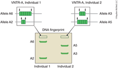
What are Variable number tandem repeats (VNTRs)?
loci where short sequences of DNA (typically 10-100 bp) are repeated in tandem (next to each other). Each individual has VNTR loci containing the same repeated sequences, but the number of repeats is highly variable from person to person.
How are VNTRs distributed to offspring?
Individuals receive one copy of a particular VNTR from their mother and one copy from their father. Given the high variability in the number of repeats, the number of repeats is most likely different for the two copies within an individual.
What are VNTR's useful for?
DNA fingerprinting
What is DNA fingerprinting?
a method used to identify whether two individuals are related
How is a VNTR done?
1.DNA is cut with restriction enzymes on either side of the VNTRs to generate two different-size DNA fragments, one of which contains the maternal VNTR and one of which contains the paternal VNTR.
2.The fragments are then amplified through PCR.
3. Finally, the fragments are selectively separated by size and electrical charge to obtain a DNA fingerprint for a specific individual (Figure 11.4).
4.The fingerprint can then be compared to a fingerprint from another individual that was obtained using the same restriction enzyme. People that are related will have fragments that are the same in size and charge.
Picture of DNA fingerprint:
Select the objectives of the DNA lab:
Analyze DNA samples using gel electrophoresis.
Determine a child's paternity.
Memorize this question:
In gel electrophoresis,
an electrical current
causes DNA fragments to move through a gelatin‑like matrix where they separate by their
size.
The
smallest
fragment is the one that travels the greatest distance through the gel.
What purpose does a DNA ladder serve in gel electrophoresis?
A DNA ladder contains DNA fragments of known length that can be used as a reference in order to estimate the size of DNA in a sample. A typical DNA ladder is 100 bp100 bp and yields 11 bands, ranging from 50 to 1000 bp1000 bp
In the lab, you will employ DNA fingerprinting by using VNTR fragments to identify the biological father of a child. Select the people from which DNA samples were collected and will be analyzed in the lab.
mother, child, two paternal fathers that could be the dad.
In this lab, how long should gel electrophoresis of the DNA be carried out in the electrophoresis chamber to obtain fully interpretable results?
1 minute
How can the DNA fragments that are separated in the gel be visualized?
Dye the DNA fragments with a fluorescent dye, such as ethidium bromide, and view the fragments under UV light.
Select the actions that should be taken when the gel is in the UV viewer.
taking a screenshot of the gel
measuring the distance each band traveled
Select the statements that correctly describe variable number tandem repeats (VNTRs).
A VNTR only cleaves DNA at a specific sequence of bases recognized by the VNTR.
VNTRs are loci where short sequences of DNA (typically 1010-100 bp100 bp) are repeated in tandem.
Each individual has VNTR loci with the same repeated sequences, but the number of repeats varies greatly.
DNA Results for Experiment:

Complete the statement.
In gel electrophoresis, DNA fragments travel at a rate
inversely proportional
to their length, thereby creating a pattern of fragments ordered from
longest to shortest
in size from the well to the opposite edge of the gel.
Unknown DNA fragment length can be estimated using the equation of a straight line in a standard curve. This curve is formed from the log of known fragment lengths of a DNA ladder in base pairs plotted against the distances the DNA ladder fragments traveled in the gel in millimeters. The known fragment lengths are called standards. Plotting them against their measured value, distance traveled in this case, creates a standard curve.
The standard curve for DNA is semilogarithmic with a negative slope. This is because smaller DNA fragments move faster than larger DNA fragments do, all at a rate inversely proportional to the logarithm of their length.
Calculate the sizes of the DNA fragments at 6 mm6 mm and 16 mm16 mm using the standard curve. Report your answers to the nearest whole number.
6 MM-11437
16MM-3929

Use the data from the experiment to estimate the sizes, within 20 base pairs, of each person's DNA fragments. The mother, the child, alleged father #1 (A.F.#1), and alleged father #2 (A.F.#2) each have two DNA fragments.
mother's longer fragment:1000
base pairs
mother's shorter fragment:240
base pairs
child's longer fragment:1000
base pairs
child's shorter fragment:475
base pairs
A.F.#1's longer fragment:770
base pairs
A.F.#1's shorter fragment:600
base pairs
A.F.#2's longer fragment:475
base pairs
A.F.#2's shorter fragment:350
base pairs
Using the gel from the lab, identify the child's father.
A.F.#2
Select the statements that helped identify the child's father.
None of the child's fragments matched any of A.F.#1's fragments.
The child's 475 bp fragment matched one of A.F.#2's fragments.
Match different types of gel loading or running errors with the description of what would be observed on the ethidium bromide‑stained gel.
Gel run too long
smaller bands not observed
Gel not run long enough
poorly separated bands clustered near well end
Loading dye not added
no bands observed
Identify the suspect that is guilty of a crime by analyzing the gel bands of DNA left at the crime scene and from two different suspects.
Suspect 2

The FBI and other law enforcement agencies make use of a system called the Combined DNA Index System (CODIS). CODIS records the genetic profiles of people from all 50 states. This allows for comparisons between DNA found at crime scenes and DNA profiles from known offenders. CODIS includes a core of 13 variable loci for each included individual.
The likelihood of someone's DNA sample matching 13 loci is extremely small, unless the DNA is from the same person.
A man received his results from a DNA testing company that traces family histories. He is surprised to see that he has a 25% match to a woman that he and his extended family have never met and have no prior knowledge of. This degree of matching is expected for aunt/uncle with a niece or nephew, or for half‑siblings. Thinking he has discovered a half‑sister, the man arranges to meet the woman. During their meeting he learns that the woman's birth parents gave her up for adoption as a newborn.
The man wants to know if he shares the same biological mother or the same biological father with this woman, but cannot ask his parents because they are both deceased. However, he has a locket with some of his mother's hair. He arranges to have DNA testing on his own DNA, the adopted woman's DNA, and his mother's DNA from the hair.
Man and A.H.S. are half‑siblings. His mother is not related to A.H.S.

When some students conducted gel electrophoresis, they obtained gel photos with uninterpretable results. Their lab instructor told them to propose the most likely cause for the error in their results.
Match each description of a gel pattern to the most likely cause of the observed pattern.
Ethidium bromide was left out of the gel.
No bands are seen in the entire gel.
The gel was run a little too long.
Smaller DNA fragments are missing.
The gel was not run long enough.
The bands are not well separated.
Sample did not get into the gel.
No bands are seen in one lane of the gel.