BMOL30040 L2/L1 JR ionotropic receptors
 Call Kai
 Learn
 Practice Test
 Spaced Repetition
 Match
 Flashcards
 Knowt Play
1/37

There's no tags or description
Looks like no tags are added yet.
38 Terms
learning objectives

RECAP: signalling pathways

3 classes of cell-surface receptors
ion-channel-coupled Rs aka:
ionotropic Rs
ligand-gated ion channels
ion-channel-associated Rs
G-protein couples Rs (GPCRs)
enzyme-coupled Rs
3 classes of cell-surface receptors image

RECAP: memb transport of small molecules
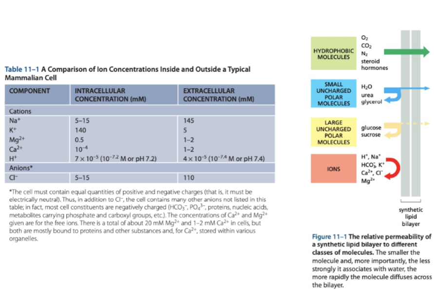
RECAP: passive vs. active transport

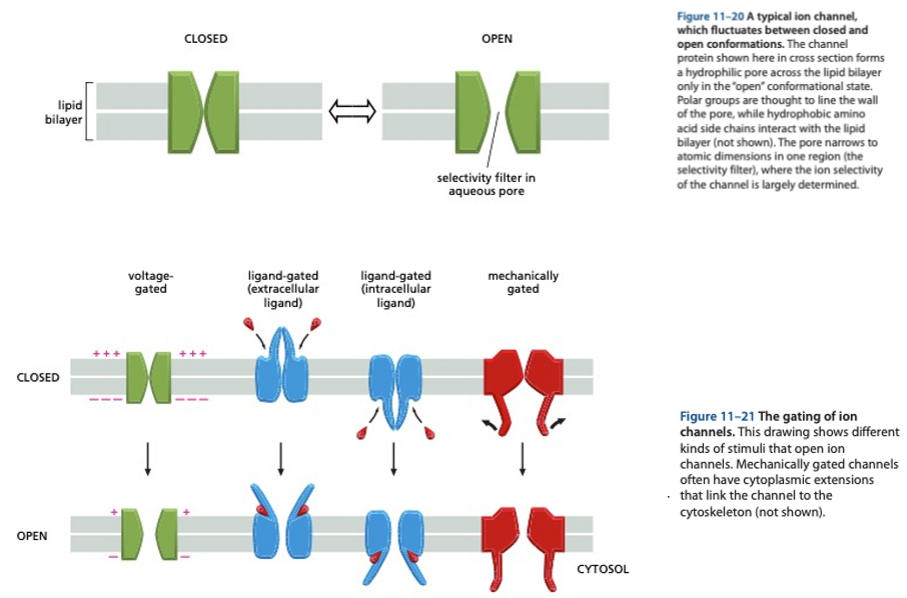
RECAP: ion channels
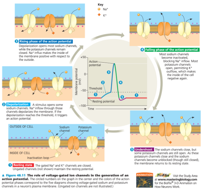
RECAP: action potential
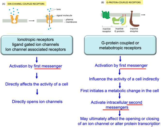
ionotropic Rs vs. GPCRs
ionotropic:
activation by first messenger
directly affects activity of cell
directly opens ion channels
GPCRs:
activation by first messenger
influence the activity of the cell indirectly
first initiates a metabolic change in the cell
activates IC second messengers
may ultimately affect the opening/closing of an ion channel or alter protein transcription
ionotropic Rs vs. GPCRs image
ion-channel-coupled receptors mediate fast responses image

ICCRs mediate fast responses
assoc. with nervous system
rapidly convert EC signals into electrical signals at chemical synapse
conc in specialised region on post-syn plasma memb
open transiently in response to NT binding
this produces brief permeability change in the memb
NTs: Ach, Glut, GABA
different types of ICCRs image

ligand-gated ion channels open in response to ___
first messenger binding to the outside of the memb
voltage-gated ion channels open in response to ___
a difference in local memb potential
stretch-activated ion channels open in response to ___
a mechanical deformation of the plasma memb
the response generated by activation of ligand-gated ion channel depends on the ion channel selectivity
excitatory NTs: open cation (+) channels → Na+ (& sometimes Ca2+) influx → depol. of post-syn memb
inhibitory NTs: open Cl- or K+ channels → reduce firing, harder for memb to be depol
the response generated by activation of ligand-gated ion channel depends on the ion channel selectivity
image

note about excit/inhib NTs
many can be either excit or inhib, depending on:
where they are released
what Rs they bind to
ionic conditions that they encounter
Na+ ions
opening an ion-channel selective for Na+ ions causes an influx of Na+
membrane becomes depolarised (positively charged)
once memb potential reached -40 mV, voltage-sensitive Na+ channels open causing an AP
membrane depolarisation initiates opening of voltage-sensitive Ca2+ channels that results in NT release

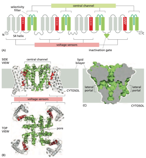
structure of voltage-gated Na+ channels
single polypeptide chain
contains 4 homologous domains
S4 helices are unique that they contain an abundance of arginines (+)
the inactivation gate is part of a flexible loop connecting the 3rd and 4th domains and acts as a plug that obstructs the pore in inactivated state
lateral portals are large enough to allow entry of small, hydrophobic, pore-blocking drugs (anaesthetics) that block ionic conductance
structure of voltage-gated Na+ channels image
Na+ channels and AP image

3 states of Na+ channel relating to AP
membrane at rest (highly polarised): closed conformation of the channel has the lowest free energy and therefore most stable
membrane is depolarised: the energy of the open conformation is lower, so the channel has high probability of opening. But the free energy of the inactivated conformation is lower still
therefore, after a random variable period spent in the open state, the channel becomes inactivated. thus, the open conformation corresponds to a metastable state that can exist only transiently when the memb depolarises
Cl- ion channels hyperpol the memb
opening ion channel selective for Cl- causes influx of Cl-
causes the inside of the memb to become hyperpolarised (more negative)
it is now less likely that voltage-sensitive Na+ or Ca2+ channels will open, thus NT release is inhibited

nicotinic acetylcholine receptor (nAChR)
muscle contraction and learning, memory and processes of cognition
first receptor to be isolated at molecular level
nAChR image

acetylcholine (ACh)
ACh is the NT at the NMJ
also has functions in autonomic NS
internal transmitter
and as final product released from parasympathetic NS
primary NT of parasympathetic pathway
ACh image

nicotine structure image

nAChR structure
many subunit structures:
5 subunits arranged in circular shape that spans the memb
alpha, beta, gamma etc. (usually 2 alpha)
ACh binds to 2 alpha subunits
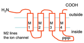
each sunubit is singly polypeptide chain that crosses the memb a number of times
two domains
EC domain: binds the NT
Transmemb. domain: contains the ion channel. Called M1, M2, M3, M4
nAChR structure image

nAChR subunit structure image
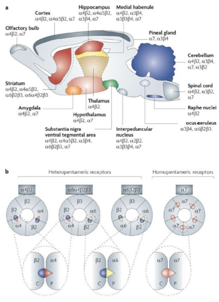
variation of nAChR structure across NS
vary in combination of subunits across brain and NS
can be either hetero- or homopentameric
the numbers refer to variations in the AA sequence in each type of subunit
Rs named according to tje subunits that for the ACh binding site - the a4b2 receptor is widely distributed in brain
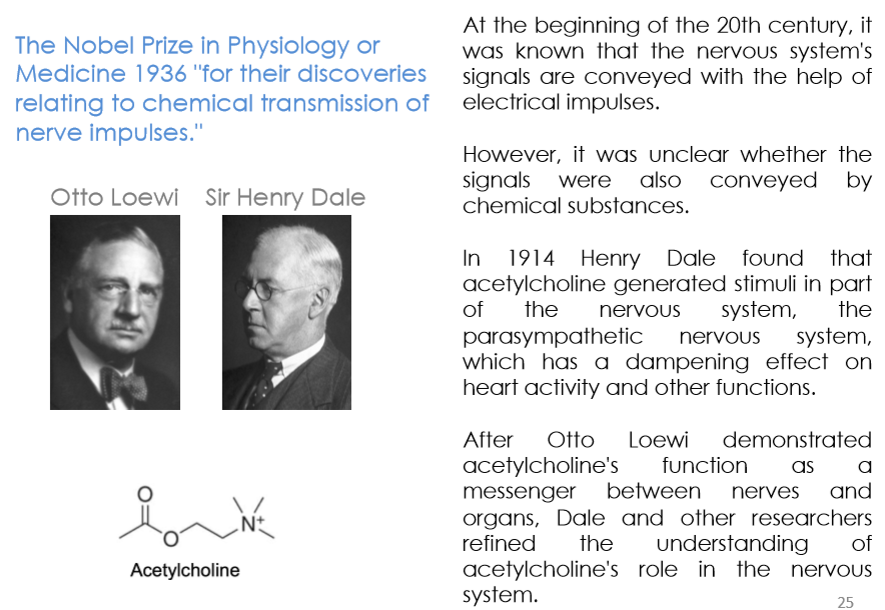
ACh discovery
structure of nAChR discovery

tools for nAChR research
fish electric organs
have many nAChRs
similar to NMJ
400 g fresh tissue yields only mgs of nAChR protein
snake toxins (venoms)
indian cobra: contains component that blocks NT and causes paralysis
bungarus multicinctus: purified polypeptide alpha-bungarotoxin binds to end-plate of NMJ with high affinity for irreversible blockade of nAChR
***neuromuscular transmission involves the sequential activation of 5 different sets of ion channels
