CT head anatomy cross sectional pics (WIP)
 Call Kai
 Learn
 Practice Test
 Spaced Repetition
 Match
 Flashcards
 Knowt Play
1/188

Earn XP
Description and Tags
CT protocl questions are from Mosbys book, pics are from CT tutor website (for now, 11/16/22)
189 Terms

A =
Falx Cerebri
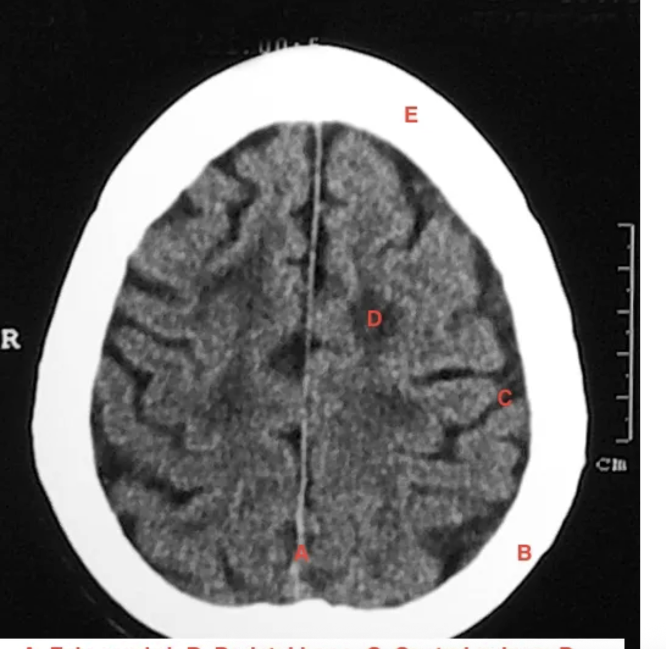
B=
parietal bone

C is
Central sulcus

D =
corona radiata

E=
frontal bone

a=
falux cerbri
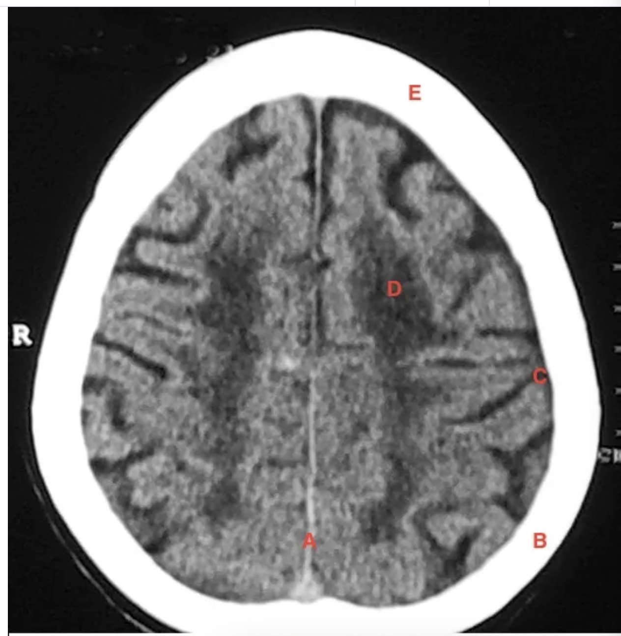
b =
parietal bone
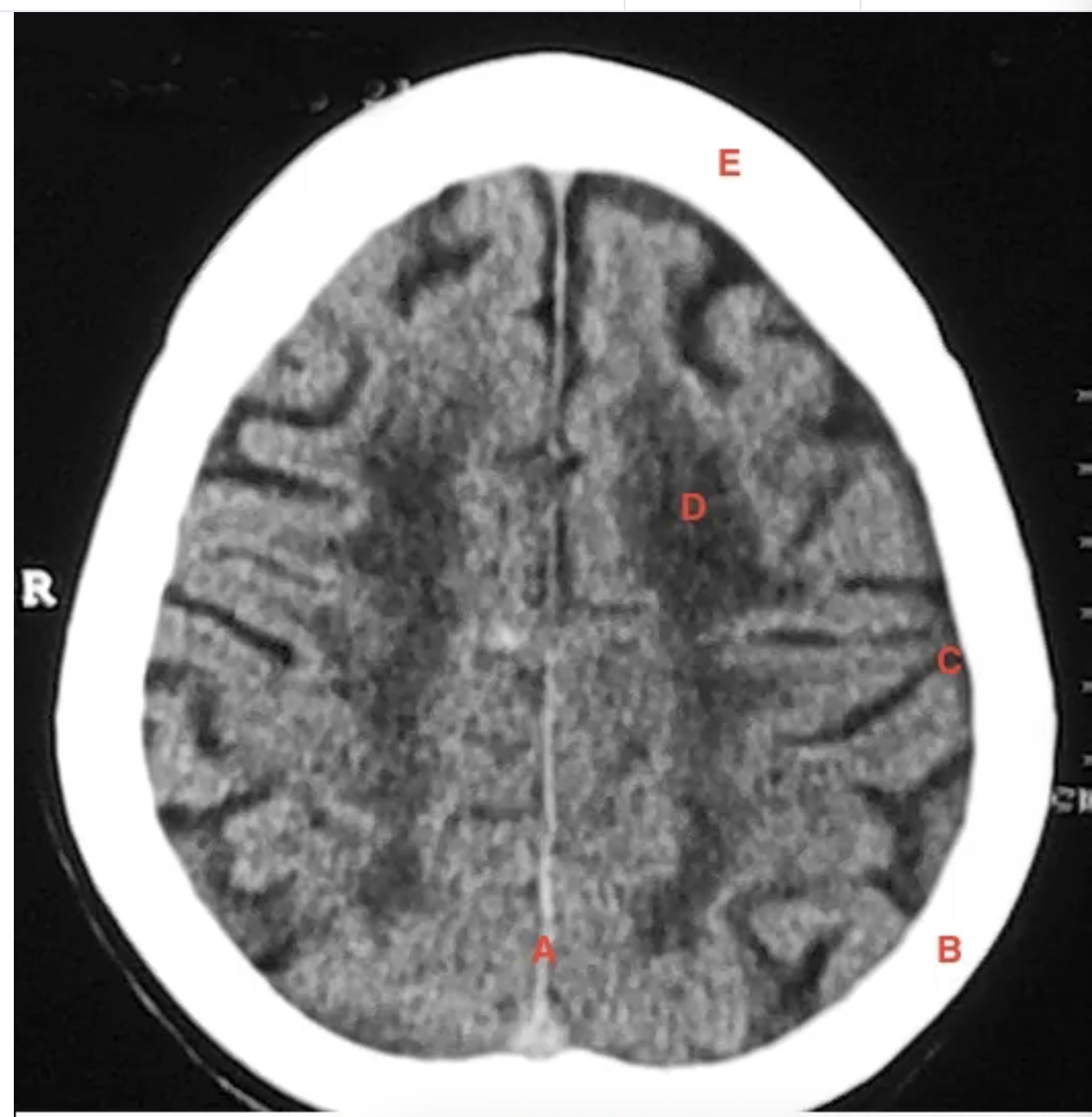
c =
central sulcus

d=
white matter

e=
frontal bone
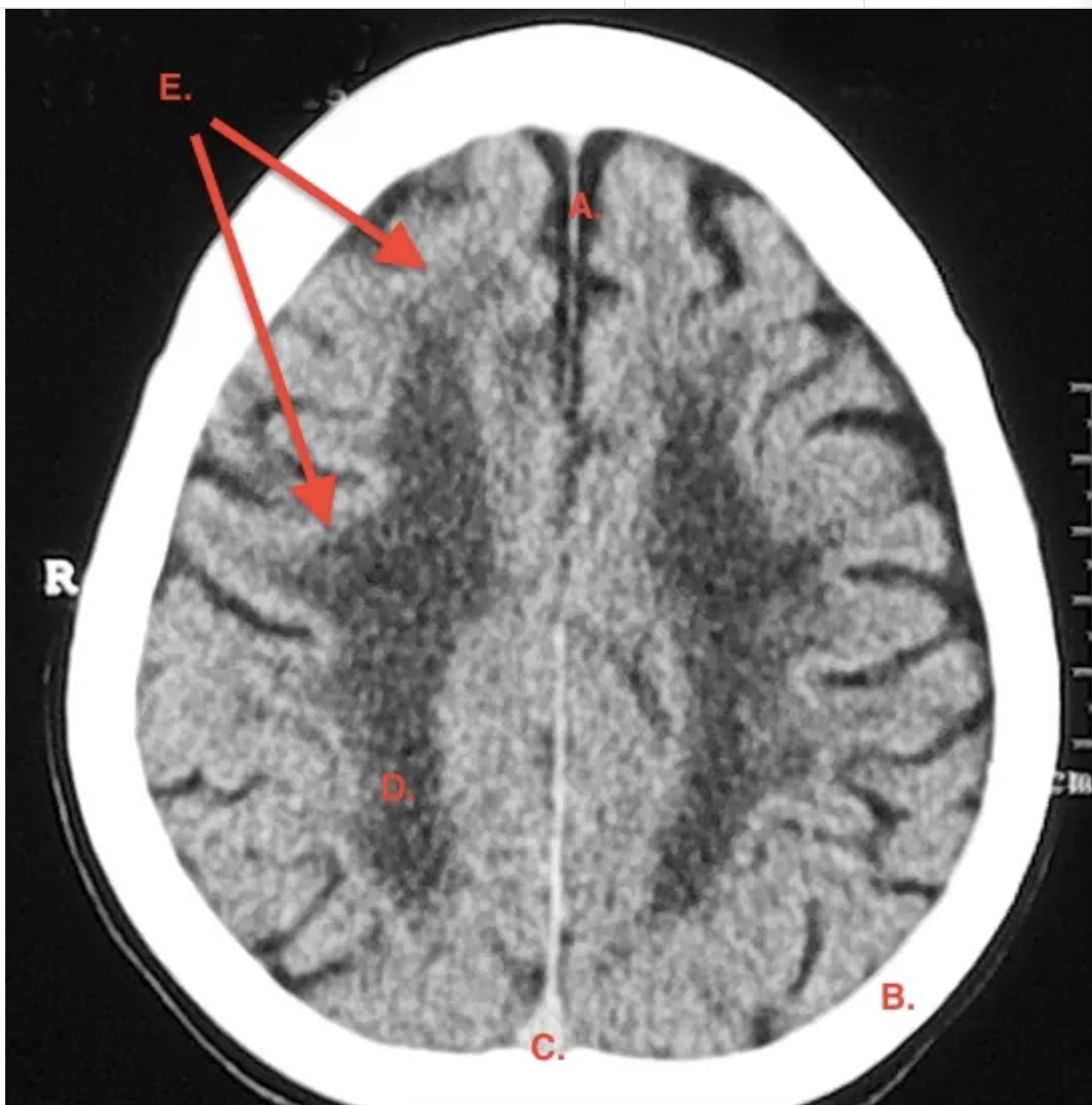
a =
falx cerebri

b =
parietal bone

c =
superior sagital sinus

d =
parietal lobe

e =
frontal lobe

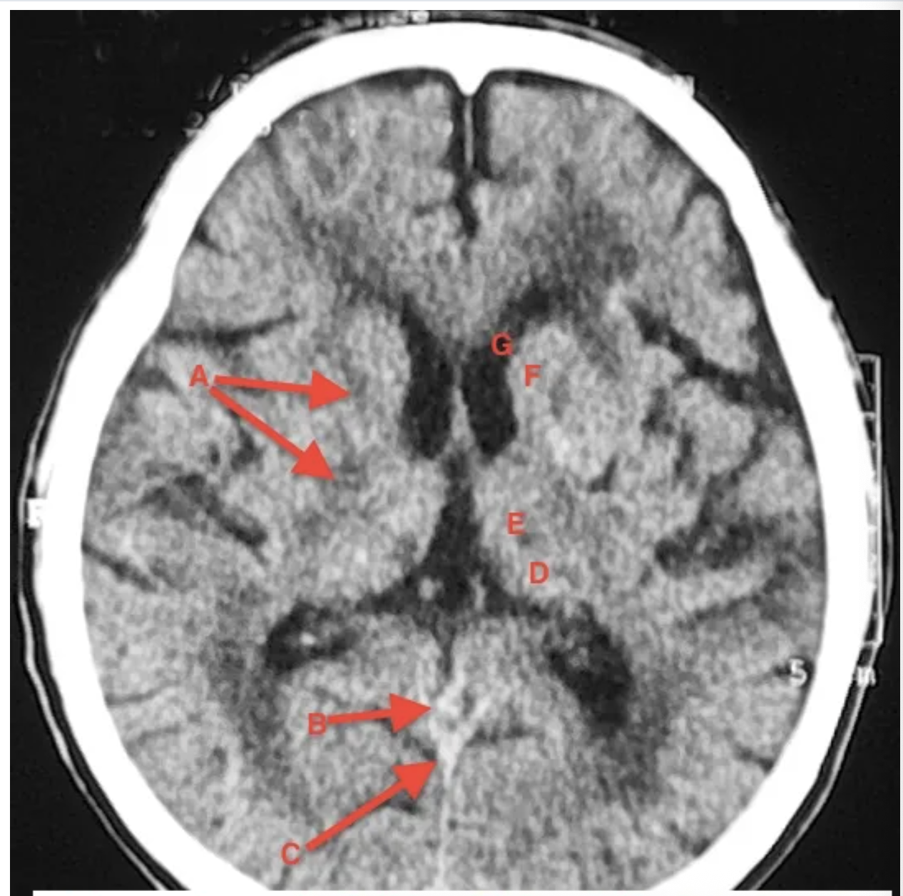
a
corpus callosum
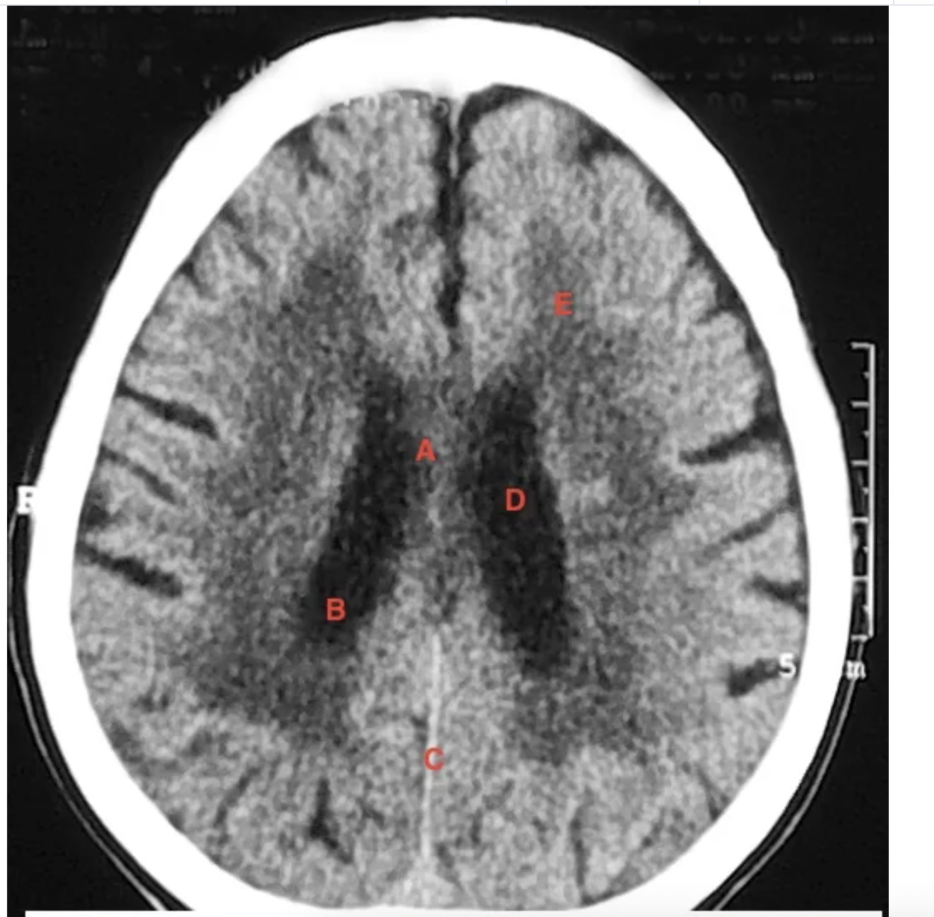
b
collateral trigone of lateral ventricle

c =
Falx Cerebri

d
body of lateral ventricle

e
white matter

a
septum pelucidum

b
splenium of corpus callosum
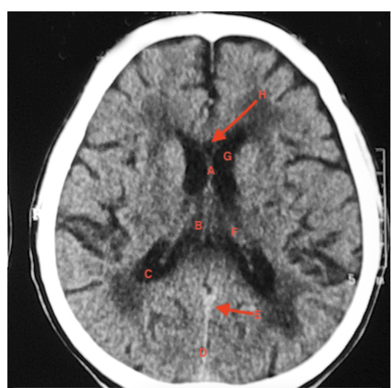
c
posterior horn of lateral ventricle
d
straight sinus

e
vein of galen

f
choroid plexus

g
anterior horn of lateral ventricle
h
genu of corpus colusum
a
internal capsule

b
vein of galen

c
straight sinus
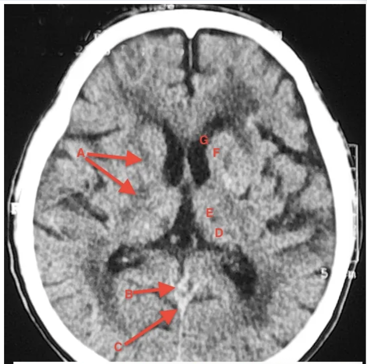
d
tail of caudate nucleus

e
thalamus nucleus

f
head of caudate nucleus

g
anterior horn of lateral ventricle

a
thalamus

c
co influences of cinuses

d
straight sinus

e
superior cistern
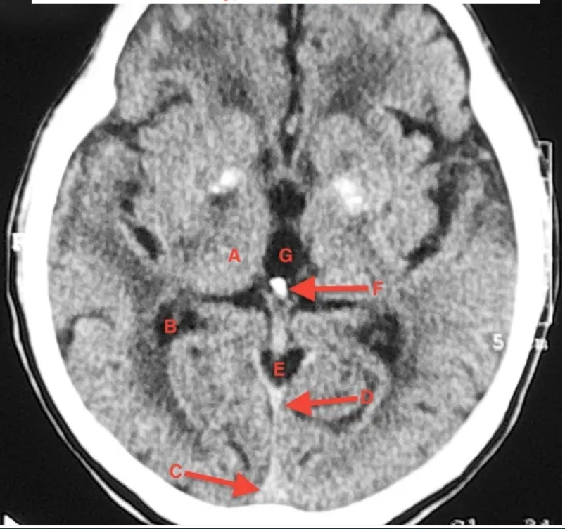
F
pineal body

G
third ventricle

A
third ventricle

b
cerebral aqueduct

c
cerebellum

d
co influence of sinuses

e
quadrigeminal plate
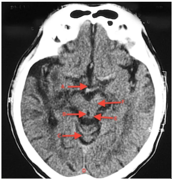
F
cerebral peduncle
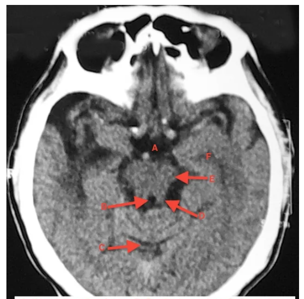
a
intrapeduncular cistern

b
Cerebral aquaduct

c
cerebellum

F
lateral ventricle (temporal horn)

A
pons
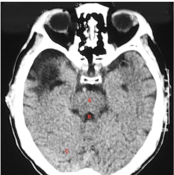
B
Fourth Ventricle

C
Cerebellum

a
sphenoid Sinus

b
Dorsum Sallae

C
cerebellum

D
Fourth Ventricle

F
mastoid air cells
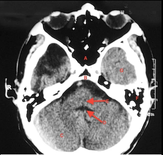
G
temporal lobe
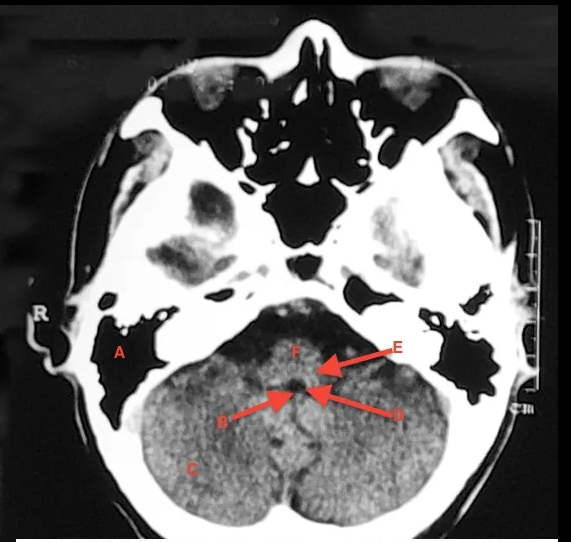
A
Mastoid air cells

B
Choroid plexus

c
cerebellum

D
fourth ventricle

E
medulla oblongata`

F
pons

A
Nasal Septum

B
Nasal Concha
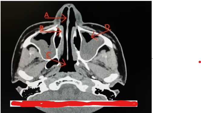
C
nasopharynx

D
Maxillary SInus
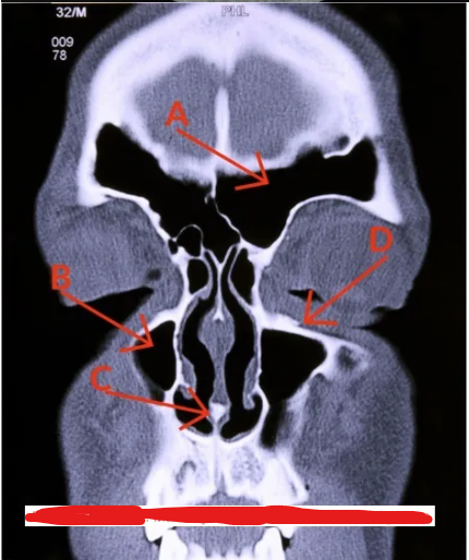
A
frontal sinus

b
maxillary sinus

C
Vomer

D
Maxillary Bone
T/F: When performing a CT head exam, it is common for there to be two separate slice thicknesses utilized throughout.
true
During a CT head exam, when acquiring images from the skull base through the posterior fosa, ____ sections (____ mm) may be used to reduce beam hardening artifacts
Thin (2-5mm)
During a head CT, if two slice thicknesses are utilized, ____ slice thickness (__mm) may be acquired through the posterior fossa to vertex
Thick (5-10mm)
During a CT brain exam, axial plane images are acquired paralllel to the
infraorbital-meatal line
Indications for a CT head without contrast
Trauma
Hematoma
intracranial hemorrhage
infarction
dementia
hydrocephalus
stroke
metastatic lesions/tumors/mass
Unless contraindicated, what CT head protocols require IV contrast
arteriovenous malformation
suspected neoplasm
attention to the pituitary gland
If trying to look at the pituitary gland, images can be acquired on the axial plane or the ________ plane
coronal
CT studies of the temporal bones and internal auditory canal require a high resolution imaging technique, which consists of the following:
Thin slices
Small targeted display field of view (DFOV)
High-resolution reconstruction algorithm

A
incus

b
temporal lobe

c
cochlea

D
vestibule

e
external auditory meatus

F
Carotid canal

G
mastoid air cells

H
cerebellum
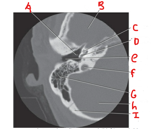
i
occipital bone

A
incus
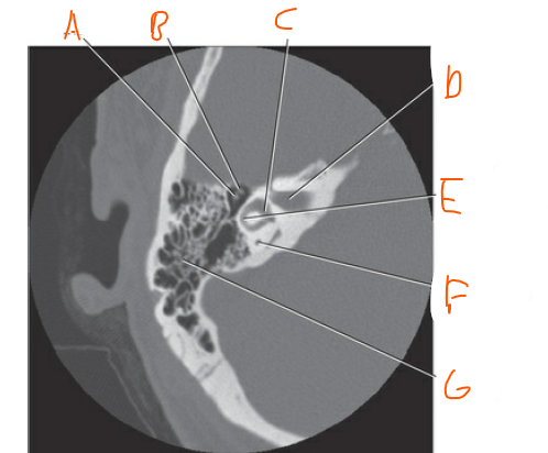
b
malleus

C
vestibule
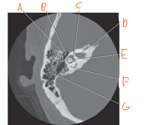
D
Internal auditory meatus
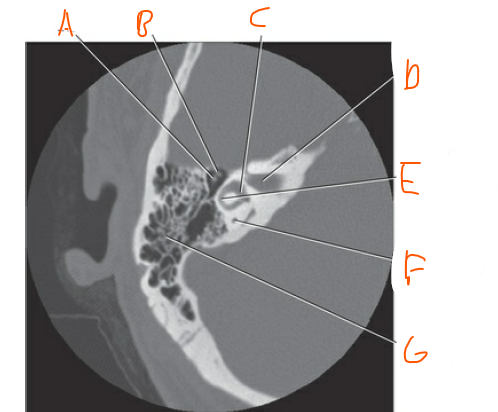
E
lateral semicircular canal

F
Posterior semicircular canal

G
mastoid air cells
When imaging the Internal auditory canals, images should be obtained/reconstructed in the ____ and _____ planes
axial and coronal
T/F: CT examinations of the temporal bones are typically performed without contrast
true