Biol 1110- Circulation
 Call Kai
 Learn
 Practice Test
 Spaced Repetition
 Match
 Flashcards
 Knowt Play
1/46

There's no tags or description
Looks like no tags are added yet.
47 Terms
What do the two sides of the heart do?
Right side: receives deoxygenated blood from body and sends it to lungs for oxygenation (pulmonary circuit)
Left side: receives oxygenated blood from the lungs and sends it out to body (systemic circuit)
Heart structure
Epicardium: outer layer, secretes serous fluid
Myocardium: muscular bulk, composed of cardiac muscle fibres
Endocardium: inner lining, composed of squamous epithelial tissue
Pericardium: thick sack of connective tissue which surrounds heart, secretes serous fluid to lubricate exterior of heart
What do “leaky” valves cause?
They can cause heart murmurs due to the backward flow of blood through the valve
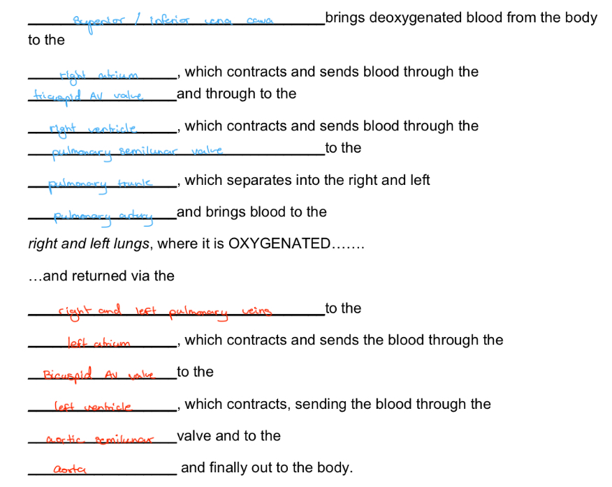
What is the path of blood flow through the heart?
.
Extrinsic control
Regulated by the autonomic NS
Sympathetic and Parasympathetic divisions work to increase and decrease heart rate as needed:
sympathetic: flight or fight response, increase heart rate due to adrenaline and epinephrine
Parasympathetic: brings heart rate down through action of acetylcholine
Cardiovascular system and Homeostasis
Respiratory: CVS picks up oxygen at the lung and distributes it to all areas of body., delivers CO2 to the lungs to be exhaled.
Digestive: The bloodstream circulates nutrients from intestinal walls, liver processes nutrients via hepatic portal system.
Urinary: bloodstream brings wastes to the kidneys to be filtered, removed as urine. Erythropoietin, hormone produced by the kidney, increases red blood cell production in red bone marrow.
Skeletal: CVS delivers calcium + phosphates to bone tissue, promoting growth.
Muscular: bloodstream delivers glucose to muscle tissue.
Nervous: medulla oblongata regulates heart rate and areas of brain control artery diameter—> controlling blood pressure.
Lymphatic: White blood cells produced by red bone marrow fight pathogens.
Why do unicellular organisms not need a circulatory system?
Materials can diffuse directly across the plasma membrane between the cell and external environment.
Why do multicellular organisms require adaptations for material exchange?
Most cells are too far from the external environment for efficient direct diffusion, so specialized structures transport materials throughout the body.
What are two adaptations that improve material exchange in multicellular animals?
Simple, thin body plans that maximize direct contact with the environment (seen in small thin invertebrates like cnidarians and flatworms)
Circulatory systems that transport fluids between cells and exchange organs.
How do cnidarians (hydras and jellies) use their gastrovascular cavity (GC) for exchange?
Digests food and distributes nutrients.
Fluid bathes inner and outer tissue layers, allowing gas and waste exchange.
Thin branches extend into tentacles, improving distribution.
How do gastrovascular cavities and thin body walls facilitate nutrient distribution?
Only cells lining the cavity directly receive digested nutrients, but thin body walls allow nutrients to diffuse short distances to outer cells.
How do flatworms (planarians) maximize material exchange?
Their flat bodies increase surface area and minimize diffusion distances, while their gastrovascular cavity distributes nutrients.
What are the three basic components of a circulatory system?
Circulatory fluid
Interconnecting vessels
Muscular pump (heart)
How does the heart drive circulation, and why is this important?
Uses metabolic energy to generate hydrostatic pressure, pushing circulatory fluid throughout the body to connect cells with gas-exchange, nutrient-absorbing and waste-removing organs.
What are the two types of circulatory systems, and how do they differ?
Open: hemolymph leaves vessels and directly bathes body cells.
Closed: blood remains within vessels and exchanges materials through interstitial fluid.
How does an open circulatory system (OCS) function?
Heart pumps hemolymph through vessels into interconnected sinuses.
Hemolymph directly bathes body cells, allowing chemical exchange.
Heart relaxation draws hemolymph back through pores.
Which animals have open circulatory systems?
Arthropods and most molluscs.
What are the advantages and disadvantages of an open circulatory system?
Lower hydrostatic pressure and less energy required to maintain circulation.
Less effective at delivering large volumes of oxygen and nutrients to highly active tissues.
How does a closed circulatory system (CCS) function?
Blood remains confined within vessels.
Heart pumps blood through large vessels that branch into smaller vessels which infiltrate organs
Exchange occurs between blood, interstitial fluid and body cells.
Which animals have closed circulatory systems?
Annelids (earthworms), cephalopods (squids and octopuses) and all vertebrates.
What are the main advantages and disadvantages of a closed circulatory system?
Higher blood pressure allows efficient delivery of oxygen and nutrients.
Supports larger, more active animals.
Allows blood flow to be distributed effectively among different organs.
Disadvantage: requires more energy to maintain as blood needs to be pumped at higher pressures
What are the three main types of blood vessels and their functions?
Arteries: carry blood away from the heart.
Capillaries: allow exchange of gases, nutrients and wastes with tissues.
Veins: return blood to the heart.
How does blood travel through the blood vessels of the body?
Heart → arteries → arterioles → capillaries → venules → veins → heart.
How do capillaries facilitate material exchange?
Their thin, porous walls allow dissolved gases and other substances to diffuse between blood and the surrounding interstitial fluid.
What are the functions of the two heart chambers?
Atria: receive blood entering the heart.
Ventricles: pump blood out of the heart.
What is single circulation, and which vertebrates exhibit it?
Blood passes through the heart once during each complete circuit. Found in fishes, including jawless, bony and cartilaginous fishes.
How does blood flow through a fish's single circulatory system?
Heart → gill capillaries (blood gains O₂ and loses CO₂) → body tissues (blood delivers O₂) → heart.
What are two disadvantages of single circulation in fishes?
Blood pressure drops after passing through gill capillaries, reducing delivery efficiency to the body.
The heart receives oxygen-poor blood before it reaches the gills.
What is double circulation, and which vertebrates exhibit it?
Blood passes through the heart twice during each complete circuit. Found in amphibians, reptiles and mammals.
What are the two circuits of double circulation?
Pulmonary circuit: right side of heart → lungs → left side of heart.
Systemic circuit: left side of heart → body tissues → right side of heart.
How does double circulation improve oxygen delivery?
The heart re-pressurizes blood after it passes through gas-exchange capillaries, maintaining vigorous flow to the brain, muscles and other organs.
How does the amphibian gas-exchange circuit differ from that of reptiles and mammals?
Reptiles and mammals: pulmonary circuit exchanges gases in the lungs.
Amphibians: pulmocutaneous circuit exchanges gases through both lungs and skin.
How do vertebrate hearts vary in their ventricular separation?
Amphibians: two atria and one undivided ventricle.
Turtles, snakes and lizards: three chambers with an incomplete septum partially dividing the ventricle.
Crocodilians: four chambers with a complete septum separating the ventricles.
What is the pathway of blood through mammalian double circulation?
Right ventricle → pulmonary arteries → lung capillaries.
Blood gains O₂ and releases CO₂.
Pulmonary veins → left atrium → left ventricle.
Aorta → systemic arteries → body capillaries.
Blood delivers O₂ and gains CO₂.
Venules → veins → superior and inferior vena cava.
Right atrium → right ventricle.
What are the functions of the superior and inferior vena cava?
Superior vena cava: returns oxygen-poor blood from the head, neck and upper limbs.
Inferior vena cava: returns oxygen-poor blood from the trunk and lower limbs.
Both empty into the right atrium.
Why are atrial and ventricular walls different in thickness?
Atria have thinner walls because they only move blood into the ventricles.
Ventricles have thicker walls because they pump blood out of the heart.
The left ventricle is thickest because it pumps blood throughout the entire body.
What are systole and diastole?
Systole: contraction phase of the heart.
Diastole: relaxation phase of the heart.
What are the functions of the atrioventricular (AV) and semilunar valves?
AV valves: between atria and ventricles; prevent blood from flowing backward into atria during ventricular contraction.
Semilunar valves: at the exits of the ventricles into the aorta and pulmonary artery; prevent blood from flowing back into ventricles.
How are AV valves structurally supported, and how do all heart valves function?
AV valves are anchored by strong fibers that prevent them from turning inside out. All valves open and close in response to pressure differences, ensuring one-way blood flow.
What is the sinoatrial (SA) node, and what is its function?
A cluster of autorhythmic cells in the right atrial wall near the superior vena cava. It generates electrical impulses that establish the heart's rate and rhythm.
How do electrical impulses coordinate atrial and ventricular contractions?
SA node generates an impulse.
Impulse spreads through both atria via electrically coupled cardiac muscle cells, causing atrial contraction.
Impulse reaches the AV node, where it is briefly delayed to allow the atria to empty.
Impulse travels toward the heart apex.
Purkinje fibers distribute the impulse throughout ventricular walls, triggering ventricular contraction.
What are the main functions of blood plasma and its components?
Plasma transports substances and helps regulate osmotic balance, defence and clotting.
Dissolved ions: maintain osmotic balance and act as buffers.
Immunoglobulins: help combat foreign agents.
Transport proteins: carry insoluble lipids through blood.
Clotting proteins: help seal damaged blood vessels.
What are the three main cellular components of blood and their functions?
Erythrocytes (red blood cells): transport O₂ using hemoglobin.
Leukocytes (white blood cells): fight infections; some engulf microorganisms and cellular debris.
Platelets: cell fragments involved in blood clotting.
What adaptations make mammalian erythrocytes efficient at transporting oxygen?
Small (7–8 µm) and biconcave, increasing surface area for gas exchange.
Lack nuclei, leaving more room for hemoglobin.
Lack mitochondria, producing ATP through anaerobic metabolism so they do not consume transported oxygen.
What are the structural characteristics and functions of platelets?
Small (2–3 µm) fragments of specialized bone marrow cells that lack nuclei and contribute to blood clotting through structural and molecular functions.
Where does lymph originate?
Blood plasma leaks out of tiny capillaries into surrounding tissues, becoming interstitial fluid. Excess interstitial fluid collected by lymphatic vessels becomes lymph.
What are the three main functions of the lymphatic system?
Collects excess interstitial fluid and proteins, preventing tissue swelling and maintaining blood volume.
Transports B and T cells and filters lymph through lymph nodes to trap and destroy pathogens and cellular debris.
Absorbs dietary fats and fat-soluble vitamins in the small intestine and transports them into the bloodstream.