GI tract and alimentary canal
 Call Kai
 Learn
 Practice Test
 Spaced Repetition
 Match
 Flashcards
 Knowt Play
1/43

There's no tags or description
Looks like no tags are added yet.
44 Terms
main components of GI
mouth, pharyngeal cavity, salivary glands
esophagus
stomach
intestine
liver
pancreas
Large intestine and anus

functions of GI tract
ingestion, mastication, motility for processing and movement
releasing enzymes, mucus and hormones for digestion
removing waste, immune defense and barrier

accessory organs of GI
teeth
tongue
salivary glands
liver
gallbladder
pancreas
spleen

squamous epithelium
resist abrasion- esophagus and mouth
may have mucus or oil glands
oral cavity
•Mechanical breakdown
•Enzymatic breakdown (ptyalin)- Breaks down of starch

tongue
Dorsal: partly keratinized
Ventral: non-keratinized
Epithelium contain specialized structures;
1.Papillae
2.Chemoreceptor cells (organized in taste buds)

luminal
inside layer of a hollow viscus

basolateral
outside layer of a hollow viscus

mucosa
epithelium
lamina propria (loose CT, BV, lymphatics)
muscularis mucosa (not true muscle but has its properties)
submucosa
denser CT with BV
meissner plexus (ANS) - secretion
muscularis
inner circular muscle
outer longitudinal muscle
myenteric plexus for motor and secretion control
esophagus
•Derived from the ectoderm (rostrally) and foregut (endoderm, caudally)
•Epithelium: Stratified squamous (partly keratinized in dogs).
•Has adventitia that blends with surrounding tissue.
both skeletal and smooth mm
rotation of the stomach
•1: Counterclockwise rotation (longitudinal axis)
•2: Counterclockwise (dorsoventral axis)
•Movement re-orients the dorsal and ventral mesogastrium and forms the omentum
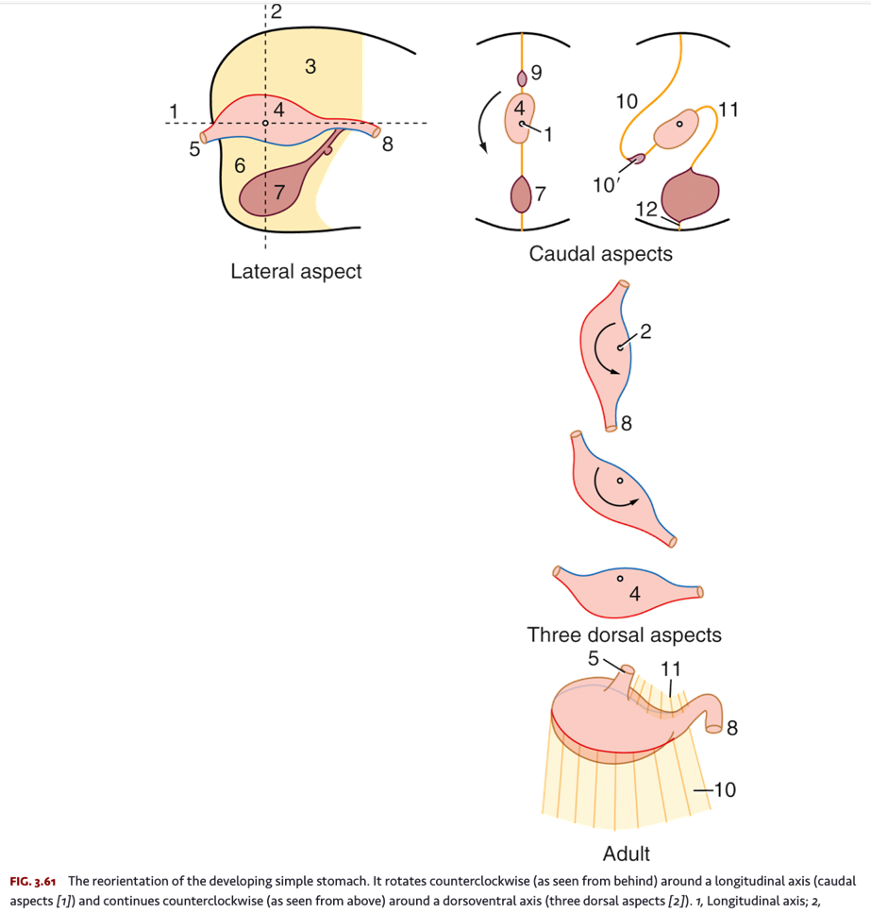
cardia
cardiac glands (mucus) and cardiac sphincter

fundus

body
gastric glands
acid secretion

pylorus
pyloric sphincter
pyloric antrum

stomach
simple columnar epithelium
pH 2 (very acidic)
has gastric pits with glands
neck cells
mucus secretion
chief cells
pepsin secretion
parietal cells
HCl secretion
enteroendocrine cells
gastrin
acid secretion
•Active transport by ATPase is indicated by arrows and diffusion is indicated by dotted arrows.
•Under the action of carbonic anhydrase, carbonic acid is produced from CO2.
•Carbonic acid dissociates into a bicarbonate ion and a proton (H+), which is pumped into the stomach lumen in exchange for K+.
•A high concentration of intracellular K+ is maintained by the Na+, K+ ATPase, while HCO3 – is exchanged for Cl– by an antiport.

jejunum
Longest part, visible once the abdominal cavity is open.
small intestine function
•Completion of digestion
•Nutrient absorption
simple columnar
small intestine epithelium
nutrient absorbtion
villi
lacteal (lymph system)- core of each villi
peyer patches (ilium)

large intestine
•Cecum
•Ascending colon
•Transverse colon
•Descending colon
•Rectum
•Anal canal

large intestine epithelium
simple cuboidal epithelium with brush border
for fluid absorbtion, bacteria and cellulose, immune cells.
taenia coli
bundles- division

haustra

anal canal
removal of ingesta
portocaval anastomosis
prolapses of hemorrhoid
paranal sinus
oily secretion
anorectal junction
change in epithelium from cuboidal to stratified squamous

foregut
•Esophagus-descending duodenum.
midgut
(cranual mesenteric a.)
•Descending duodenum-transverse colon.
hindgut
caudal mesenteric a
•Descending colon to anorectal junction
celiac artery
1.Left gastric artery
2.Splenic artery
3.Hepatic artery
cranial mesenteric artery
1.Jejunal artery
2.Ileocolic
caudal mesenteric artery
1.Left colic artery
2.Cranial rectal artery.
portal vein
1.Cranial mesenteric vein
2.Caudal mesenteric vein
3.Ileocolic vein
receives
1.Splenic vein
2.Gastroduodenal vein
lymphatic centers
Celiac and Hepatic centers
Mesenteric (including jejunal, colic)
Chyli Cistern
Thoracic duct
Iliac lymphatic centers
