Appendicular Skeleton A&P Quiz #4
 Call Kai
 Learn
Learn Practice Test
 Spaced Repetition
 Match
Match Flashcards
 Knowt Play
1/114

There's no tags or description
Looks like no tags are added yet.
Name | Mastery | Learn | Test | Matching | Spaced | Call with Kai | Chat |
|---|
No analytics yet
Send a link to your students to track their progress
115 Terms
What kind of bones does the appendicular skeleton consist of?
Bones of the limbs & supporting elements or girdles
How many bones in the appendicular skeleton
126 bones
Pectoral girdle (4)
Upper limbs (60)
Pelvic girdle (2)
Lower limbs (60)
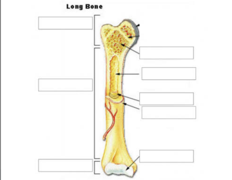
Long Bone Anatomy
**Also remember medullary cavity houses bone marrow → lined by endosteum

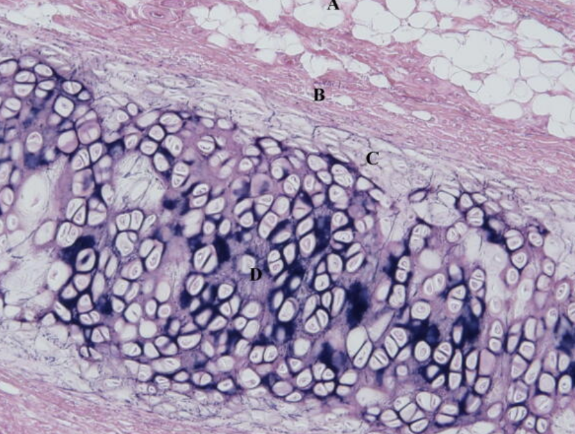
Hyaline Cartilage
Most abundant type of connective tissue in the human body
Contains chondrocytes (which are located in lacunae)
Avascular
Found:
On the ends of bones where they form movable joints, the walls of the nose, larynx, trachea, and bronchi.
The costal cartilage connecting the ribs to the sternum.
**Lets bones glide smoothly past each other, holds open the tubes of the respiratory system

Epiphyseal plate
Hyaline cartilage plate where new bone growth occurs in long bones
It allows children and teenagers to grow taller by adding new bone length
Fibrocartilage
A tough, dense, and shock-absorbing type of cartilage made of thick bundles of collagen fibers
Contains a mix of dense fibrous tissue and chondrocytes (cartilage cells)
Lacks a perichondrium
Avascular
It is the strongest and least flexible type of cartilage in the human body.
Located: Between intervertebral discs, meniscus, pubic symphysis, labrum
Help stabilize, absorb shock, and distribute weight

Elastic Cartilage
A flexible type of connective tissue packed with elastin fibers that allows body structures to bend repeatedly and return to their original shape.
Found in: Ear, auditory tubes, epiglottis, larynx
Contains: Elastic fibers, chondrocytes, perichondrium
Pectoral girdle
The set of bones in the human body that connects the upper limbs to the axial skeleton
Contains these bones: Clavicle, scapula
Contains these joints:
Sternoclavicular joint (only site of attachment to axial skeleton), Acromioclavicular (AC) joint (connects clavicle to scapula)
Glenohumeral joint (ball and socket joint)
Scapulothoracic joint (Where the scapula glides against the back of the rib cage using muscle support)

Name this joint
Sternoclavicular joint
Connects clavicle to manubrium of sternum
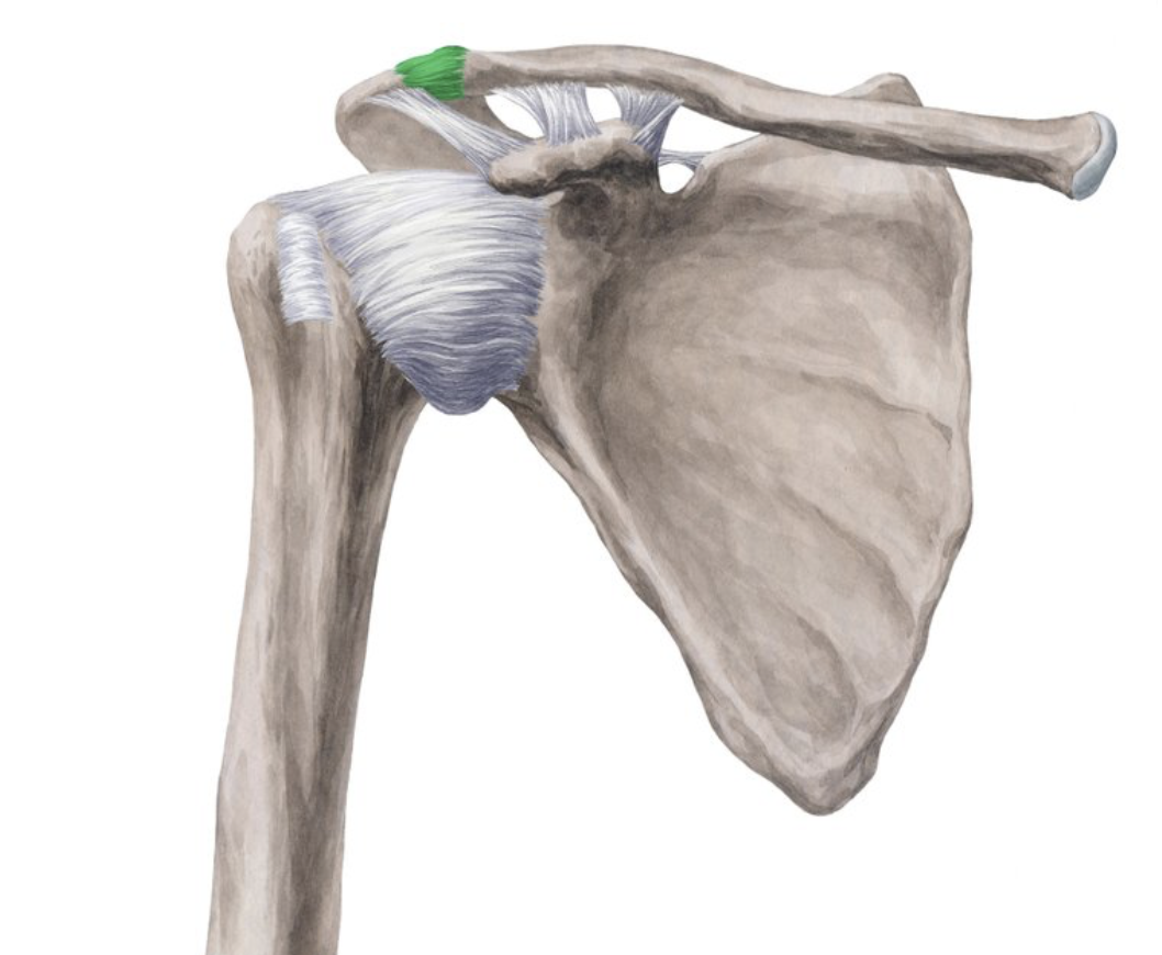
Name this joint
Acromioclavicular (AC) joint

Label these sections of the clavicle
Also note:
smoother side of clavicle = superior, more groves and bumps = inferior side


Name this bone
Scapula

Name this
Coracoid process of scapula

Name this
Acromion

Name this structure
Glenoid Cavity
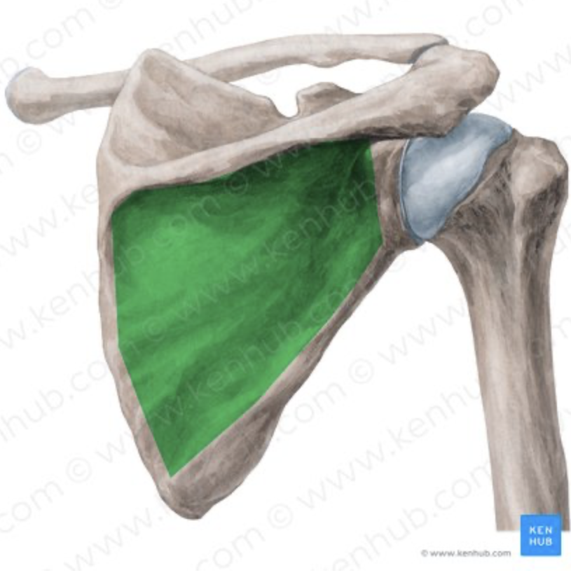
What is this?
Infraspinatus fossa

What is this?
Supraspinatus fossa

Name this bone
Humerus

Name this landmark
Head of humerus

Name this landmark
Anatomical neck of humerus

Name this landmark
Greater tubercle of humerus

Name this landmark
Lesser tubercle of humerus

Name this landmark
Intertubercular groove of humerus
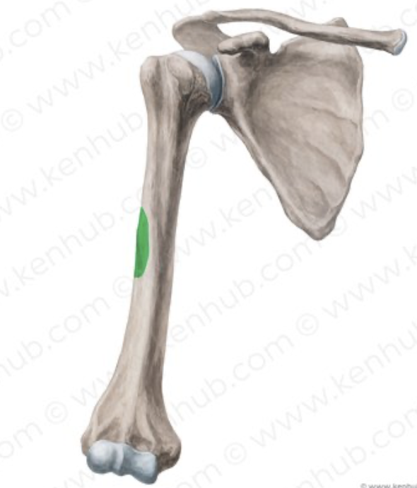
Name this landmark
Deltoid tuberosity of humerus

Name this landmark
Lateral Epicondyle of humerus

Name this landmark
Medial epicondyle of humerus

Name this landmark
Capitulum
**On the lateral side of humerus

Name this landmark
Trochlea of humerus
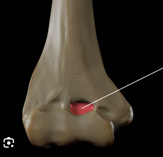
Name this landmark, is it anterior or posterior?
Coronoid Fossa of humerus
Anterior

Name this landmark, is it anterior or posterior?
Olecranon fossa of humerus
Posterior

Is this a left or right humerus?
RIGHT
Tips: Orient so its similar to how it is in body, look for olecranon fossa and make sure its in the posterior position, make sure capitulum is lateral and trochlea is more medial

Name this bone
Ulna
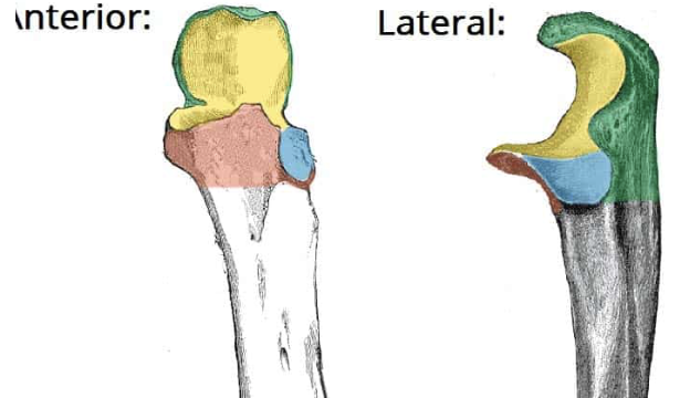
What is the yellow area?
Trochlear notch of ulna

What is this process?
Coronoid Process

What is this process?
Olecranon process

What is this landmark?
Radial Notch
TIP: Remember ulna is medial, thus, to determine if its a right or left ulna the radial notch must be pointing lateral!!
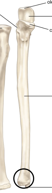
What is this?
Ulnar head

What is this landmark?
Styloid process of the ulna

What bone is this?
Radius
**Remember its the lateral bone of forearm

What is this structure
Radial head

What is this landmark?
Radial neck

What is this landmark?
Radial tuberosity
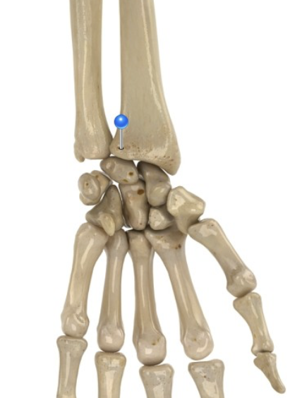
What is this?
Ulnar notch
**Remember at distal end and points medial

What is this?
Styloid process of radius

Is this a right or left radius?
RIGHT
How do we know:
Smooth on front, bumpy on back → this photo shows the smooth distal end of the radius which means the bone is facing anterior, once that is oriented look where the styloid process is pointing. Remember styloid process should always point out towards thumb.
What are the carpals and how many are there?
Tiny little wrist bones
8 bones per hand (16 total on body)


Name the carpals 😔
So long to pinky, here comes the thumb
If you can see pisiform you are looking at the anterior side
If you can’t → posterior side


What are these bones and in what direction do you count them?
Metacarpals (create the palm of your hand)
Number 1 starts from thumb → end with #5 at pinky
How many phalanges do we have?
14 phalanges per hand → 28 total from hands
BUT
Don't forget about feet phalanges
14 per foot → 28 total from feet
THUS WE HAVE 56 TOTAL

What phalanges are these
Proximal phalanges

What phalanges are these
Middle phalanges

What phalanges are these
Distal phalanges
Bony pelvis
2 coxyl bones (that consist of illium, ischium, pubis) & sacrum & coccyx
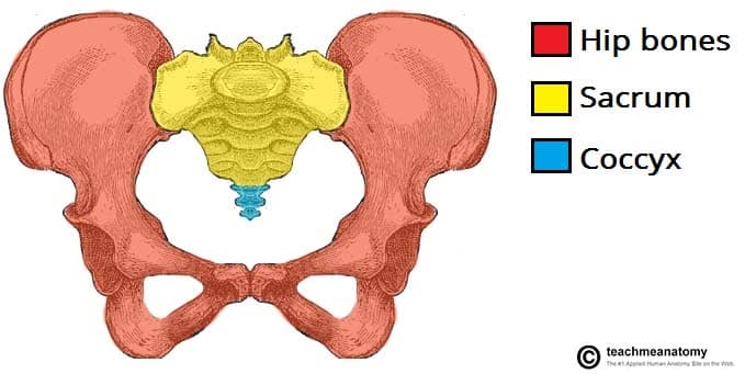
Pelvic girdle
Consisting of the hip (coxal) bones (ilium, ischium, pubis)
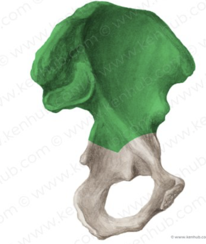
What bone is this?
Ilium

Name this
Iliac fossa

Name this
Iliac crest

What joint is this?
Sacroiliac joint

What is this yellow structure?
Sacral promontory

What bone is this?
Pubis

What bone is this
Ischium

What is the highlighted green structure and what cartilage is it made of?
Pubic symphysis
Made of fibrocartilage and some hyaline cartilage

What is the red circle structure?
(pink box is covered up term)
Pubic arch
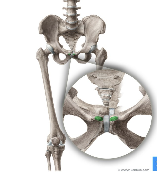
What is this?
Pubic crest
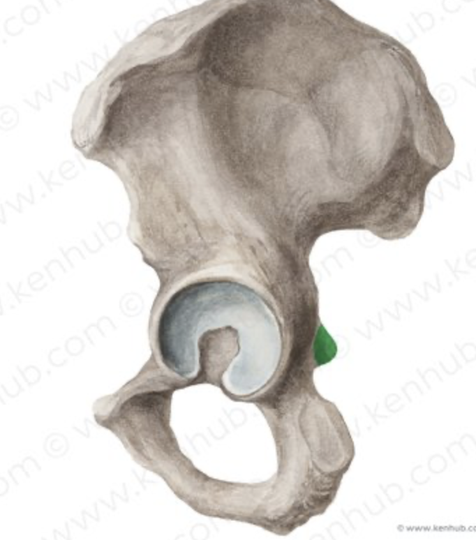
What is the green highlighted structure?
Ischial spine
Pelvic brim vs pelvic inlet
Pelvic brim: The bony boundary/rim around the opening
Pelvic inlet: The actual opening/space enclosed by that boundary
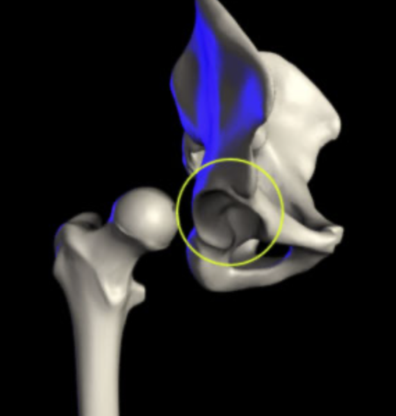
What structure is this?
Acetabulum/ cotyloid cavity

What is this?
Ischial tuberosity
Also called the sit bone
Supports your upper body weight when you sit.

What is this?
Obturator foramen
Male vs female pelvis differences
Female pelvis:
Lighter and thinner
Female inlet is wide, large, and oval or round
The angle under the pubic bones in females is wide and greater than 90 degrees (obtuse)
The female sacrum is short, wide, and curved outward
The female coccyx is more flexible and straighter.
Male pelvis:
Pelvis is thick, heavy, and dense.
Inlet is narrow and heart-shaped
Pubic arch is narrow and less than 90 degrees
Male sacrum is long, narrow, and straighter.
Coccyx is less flexible and curves inward

Name the ligament with the arrow pointing to it
Ligamentum teres
→ Connects the ball (head of the femur) to the socket (acetabulum).

What ligament is this?
Acetabular labrum

What ligament is this
Transverse acetabular ligament

What is the red ligament?
Iliofemoral ligament
**Look for Y shape in the anterior view
Strongest ligament in the human body
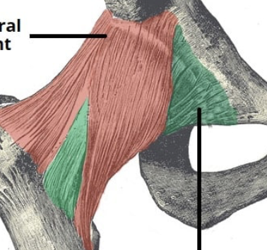
What is the green ligament?
Pubofemoral ligament

What ligament goes here?
Ischiofemoral ligament

What is this?
Fovea capitis

What is this
Femoral head

What is the yellow structure
Femoral neck

What is this?
Greater trochanter

What is this?
Lesser trochanter

What is this? **Acknowledge anterior view
Intertrochanteric Line

What is this? **Acknowledge posterior view
Intertrochanteric crest

What is this? **Landmark on posterior view
Gluteal tuberosity

What is this?
Linea aspera

What are these lines (Area where it splits)
Medial and lateral suprachondral lines

What is this? (posterior view)
Intercondylar notch

What is this
Medial condyle

What is this?
Lateral condyle

What is this?
Lateral epicondyle of femur

What is this?
Medial epicondyle

What is the line with the grey box referring to?
Abductor tubercle of femur
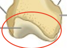
What is this?
Patellar surface of femur

What bone is this?
Patella

What bone is this?
Tibia


What is this landmark?
Tibial tuberosity

What is this?
Medial condyle of tibia

What is this?
Lateral condyle of tibia

What is the pin pointing to?
Medial malleolus

What is this?
Intercondylar eminence