Neuro Exam 2
 Call Kai
 Learn
 Practice Test
 Spaced Repetition
 Match
 Flashcards
 Knowt Play
1/216

Earn XP
Description and Tags
SC tracts, sensory receptors, pain
217 Terms
sensory receptor
specialized nerve cell designed to respond to a specific sensory stimulus (touch, pressure, pain, temperature, light, sound, position in space)
3 Types of Sensory Receptors
exteroceptors
interoceptors
proprioceptors
Exteroceptors
sensory receptors that receive stimuli from the external world
ex: visual, auditory, tactile, olfactory, and gustatory receptors
Interoceptors
sensory receptors that receive information from inside the body (viscera, hollow organs, glands, etc.)
What type of receptor would detect body sensations such as stomach pain, pinched spinal nerves, or processes within the deep layers of skin?
interoceptors
Proprioceptors
sensory receptors located in the muscles, tendons, and joint of the body and in the utricles, saccules, and semicircular canals of the inner ear; detect body position and movement
Developmental Classifications of Sensory Receptors
protopathic
epicritic
Protopathic Sensory Receptors
receptors that identify gross bodily sensations rather than specific regions of sensations
cannot locate precise origin of pain
detects crude touch and dull pain
Epicritic Sensory Receptors
receptors that detect sensation with precision, accuracy, and acuteness
can locate the precise origin of pain
detect discriminative touch, sharp pain, and exact joint position
allows organism to detect imminent danger
What is the difference between protopathic and epicritic sensory receptors?
protopathic detect crude touch and dull pain and are unable to determine the precise origin of pain
epicritic detect discriminative touch, sharp pain, and exact joint positioning and can locate the precise origin of pain
T or F: Protopathic sensory receptors can locate the exact origin of pain
false
T or F: Epicritic sensory receptors can locate the exact origin of pain
true
Do protopathic or epicritic sensory receptors allow the organism to detect imminent danger?
epicritic
A child who does not receive adequate sensory stimulation from the environment, or the sensory information is dulled may show more self-stimulating behaviors.
What sensory system is dominant?
protopathic sensory system
A child who is hypersensitive to sensation from the environment may display tactile defensiveness (e.g. dislike the feeling of wet clothes) and/or gravitational insecurity (e.g. swinging is frightening).
What sensory system is dominant?
epicritic sensory system
Sensory Receptors Classified by Anatomical Position
cutaneous reeptors
proprioceptors
visceral receptors
special sense receptors (visual, auditory, olfactory, gustatory, equilibrium)
Cutaneous Receptors (function, location, type, occupational implications)
function: respond to pain, temp, pressure, vibration, and discriminative touch
location: superficial and deep layers of skin
type: exteroceptors and interoceptors
occupational implications: development of ulcers; burns; difficulty detecting trains, trucks, or earthquakes; distinguishing objects in one’s pocket
Proprioceptors (function, location, type, occupational implications)
function: detect muscle length, tendon joint position, deep muscular and joint pain, tendonitis
location: muscles, tendons, and joints
type: proprioceptors
occupational implications: fall risk, impaired muscle tone affecting AROM in the limbs and trunk
Visceral Receptors (function, location, type, occupational implications)
function: respond to pressure and pain from internal organs
location:
type: interoceptors
occupational implications: at risk for missed identification of serious injury or illness due to impaired pain detection in internal organs
Special Sense Receptor: Visual Receptors (function, types, occupational implications)
function: respond to light information from external environment
type: exteroceptors
occupational implications: difficulty visually identifying all environmental stimuli (e.g. dangerous crossing the street)
Rods and Cones
visual receptors
Special Sense Receptor: Olfactory Receptors (function, location, type, occupational implication)
function: respond to scent and odor stimuli from the ext. environment
location: mucous lining of the nasal cavity
type: exteroceptors
occupational implications: difficulty smelling spoiled food, mold, burning, gas leaks, etc.
What are the olfactory receptors?
hair cells found in the mucous lining of the nasal cavity
Special Sense Receptor: Auditory Receptors (function, location, type, occupational implications)
function: respond to sound stimuli from the external environment
location: cochlea of ear
type: exteroceptors
occupational implications: difficulty participating in social communication; difficulty detecting alert signals (smoke alarm, carbon monoxide detector, horns, etc)
Special Sense Receptor: Gustatory Receptors (function, location, type, occupational implications)
function: respond to taste stimuli from the external environment
location: tongue
type: exteroceptors
occupational implications: difficulty detecting burnt or spoiled food
What are the gustatory receptors?
the taste buds
Special Sense Receptors: Equilibrium (function, location, type, occupational implications)
function: provide info about the position of the head in space in relation to the body
location: semicircular canals, utricles, and saccules of the inner ear
type: proprioceptors
occupational implications: impaired balance; fall risk; positional vertigo
Sensory Receptors Classified by Structural Design
mechanoreceptors
thermoreceptor
chemoreceptors
photoreceptors
Mechanoreceptors
sensory receptors that detect mechanical pressure and/or deformation
detect deep touch, pressure, vibration, proprioception, equilibrium, and audition
several types
Types of Mechanoreceptors and what they detect
Pacinian Corpuscles (rapid vibration)
Meissners Corpuscles (light touch and texture change)
Merkel Discs (sustained pressure)
Ruffini Endings (deep pressure, tension, deep skin layers)
Free Nerve Endings (touch, pressure, stretch)
Baroreceptors (stretch of blood vessles)
Ligamentous Receptors (ligament stretch)
Pacinian Corpuscles
mechanoreceptor that detects rapid vibration
Meissners Corpuscles
mechanoreceptors that detect light touch and texture change
Merkel Discs
mechanoreceptors that detect sustained pressure
Ruffini Endings
mechanoreceptors that detect deep pressure and tension from deep skin layers
Free Nerve Endings
mechanoreceptors that detect touch, pressure, and stretch
Barorecptors
mechanoreceptors that detect the stretch of blood vessels
Ligamentous Receptors
mechanoreceptors that detect ligament stretch and proprioception
Thermoreceptors
sensory receptors that are stimulated by changed in temp
While thermoreceptors are found throughout the body, where are the highest density of cold receptors found?
facial skin, tongue, bladder, and cornea
What are the special pain receptors that detect change in harmful temps called?
nociceptors
Chemoreceptors
sensory receptors that detect changes in chemical stimuli from the environment
2 types: direct and distance
Direct Chemoreceptors
respond to immediate contact
ex: taste buds are stimulated when they make contact with food
Distance Chemoreceptors
repond to gaseous molecules that emerge from a distant odorant
ex: olfactory receptors respond to the gaseous molecules (scent of cookies) given off by the distant odorant (cookies)
Two Categories of Chemoreceptors
direct
distance
Photoreceptors
sensory receptors located in the retina that detect light from the environment
two forms: rods and cones
Rods
visual receptors / photoreceptors that are responsible for night vision and black-and-white vision
located in the retina periphery
Where are the rods located?
retina periphery
Where are the cones located?
fovea (central region of the eye)
Cones
visual receptors / photoreceptors that work in daytime lighting and are responsible for color vision
Large Receptor Fields (function, location, cortical representation)
function: gross discrimination of sensation
location: body regions with less discrimination (legs, abdomen, arms, back)
representation: small

Small Receptor Fields (function, location, cortical representation)
function: fine discrimination of sensation
location: body regions with greatest sensitivity (lips, hands, face, soles of feet)
representation: large

What areas of the body have small receptor fields?
lips, hands, face, soles of feet

What areas of the body have large receptor fields?
legs, abdomen, arms, back

T or F: Both large and small receptor fields are represented on both right and left sides of the brain
true

How does our brain allocate sensory and motor function to the various aspects of the body?
Sensory Homunculus and Motor Homunculus

T or F: Small receptors fields have large cortical representaiton in the brain
true

T or F: Large receptor fields have large cortical representation in the brain
false
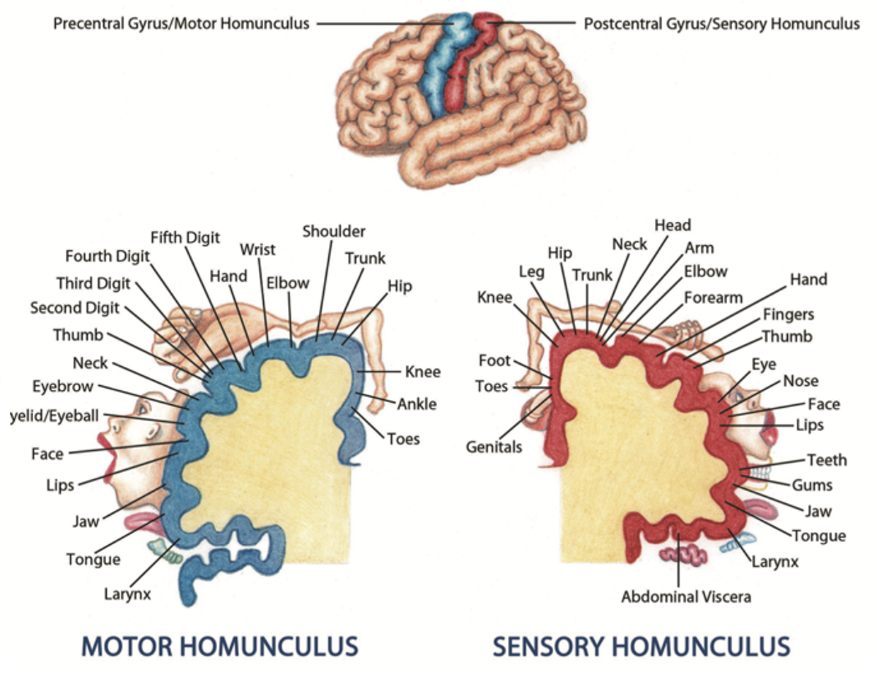
Motor Cortex
variety of areas that control motor movement, including the primary motor cortex which holds the motor homunculus
Association Cortex
area that includes higher order functions that contribute to motor planning, spatial awareness, planning responses, etc.

How do the basal ganglia and cerebellum help mediate movement?
they do not directly communicate with the cerebrum—instead the thalamus acts as a relay station between the basal ganglia/cerebellum and the cerebral cortex

Describe the Organization of Motor Systems
Four Levels:
Motor Cortex: controls motor movement and hold the motor homunculus (motor map of the body)
Association Cortex: includes higher-order functions that contribute to motor planning, spacial awareness, planning responses, etc.
Spinal Cord: receives info from the brainstem
Brainstem: assists with coordination of movement to be sent to the SC

UMN
carry info from the cortex to cranial nerve nuclei in the brainstem or to cell bodies in the ventral horn
UMN are part of the (CNS/PNS)
CNS
UMN Lesions
Below Level of Lesion:
increased muscle tone
hyperreflexia (exaggerated DTRs)
clonus
Babinski Sign
At Level of Lesion:
flaccidity
LMN
carries info from cell bodies in the anterior horn to the skeletal muscles in the periphery
the LMNs are considered part of the (CNS/PNS)
PNS
LMN Lesions
paralysis or paresis of movement
hypotonia
hyporeflexia (decreased) or areflexia (absent)
fibrillations and/or fasciculations
muscle atrophy
Spinal Cord Tracts
bundle of nerve fibers within the spinal cord that carry specific sensory or motor info either up towards the brain (ascending tracts) or down from the brain to the body (descending tracts)
What is termed the “highway” for neural signals to travel along the SC?
SC tracts
Where are spinal cord tracts located?
white matter
How are SC tracts named?
place of origin and destination
Ascending Spinal Nerves
carry sensory information from the periphery (sensory receptor) to the SC
How do ascending spinal nerves enter the SC? What do they do once they enter?
enter the SC through the dorsal root/rootlets (PNS) > enter the dorsal horn and synapse with an interneuron (CNS) > interneuron synapses with an ascending SC tract (CNS)
Ascending Spinal Cord Tracts
carry sensory information up the SC, to the brainstem, thalamus, and cortex
(some carry infor from brainstem to cerebellum)
T or F: While most ascending SC tracts carry sensory info to the brainstem, thalamus, and cortex, some carry info from the brainstem to the cerebellum
true
Dorsal Columns Function
carries conscious info regarding fine touch, vibration, and proprioception to the cortex
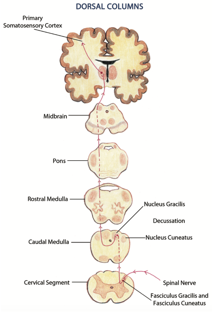
Dorsal Column Decussation
caudal medulla level

Lesions to the dorsal column below the caudal medulla would result in…
ipsilateral loss of fine touch, vibration, and proprioception

Lesions to the dorsal column about the caudal medulla would result in…
contralateral loss of fine touch, vibration, and proprioception

The client has difficulty distinguishing between coins when reaching into his pocket, has developed a umber of pressure sores, comes dangerously close to fragile items in an antique store, and is unable to detect when his car has started.
What tract is likely to be lesioned?
Dorsal Columns
Lateral Spinothalamic Tract Function
carries conscious sensory info to the cortex regarding pain and temp

Lateral Spinothalamic Tract Decussation
SC Level (as soon as the spinal nerve enters the cord and synapses on tract)

Your client often burns himself when showering and is unable to detect the development of pressure sores, a foot that fell off the footplate, or his hand caught in wheelchair spokes.
What tract is likely damaged?
lateral spinothalamic

The Lateral Spinothalamic Tract is a (ascending/descending) tract
ascending
The Dorsal Columns is a (ascending/descending) tract
ascending
Name the Ascending Sensory SC Tracts
Dorsal Columns
Lateral Spinothalamic
Anterior Spinothalamic
Posterior Spinocerebellar
Anterior Spinocerebellar
Cuneocerebellar
Rostral SPinocerebellar
Name the Descending Motor SC Tracts
Lateral Corticospinal Tract
Anterior Corticospinal Tract
Corticobulbar Tract
Rubospinal Tract
Medial Reticulospinal Tract
Lateral Reticulospinal Tract
Vestibulospinal Tract
What SC tracts collect proprioception from the LE?
posterior spinocerebellar and anterior spinocerebellar
What SC tracts collect proprioception info from the UE?
cuneocerebellar and rostral cerebellar
The brain has bilateral representation of unconscious proprioception. What does this mean?
the posterior and anterior spinocerebellar tracts both detect proprioception info from the LE
the cuneocerebllar and rostral cerebellar tracts both detect proprioception info from the UE
If one tract of each set is lost, the other will still send proprioceptive info the cerebellum. An individual will not lose unconscious proprioceptive info from UE or LE unless both tracts in the set are lost
T or F: While most descending SC tracts carry motor info from the cortex, through the internal capsule, thalamus, and brainstem to the SC, some motor SC tracts actually originate in the cerebellum or brainstem
true
When descending SC tracts are ready to exit the cord, they synapse with…
an interneuron in the ventral horn which then synapses on a motor neuron in the ventral horn which then synapses on a descending motor spinal nerve and exits the SC through the ventral root
Pyramidal Tracts
descending motor SC tracts that (1) originate in the cerebral cortex, (2) travel through the pyramids of the medulla oblongata, and (3) control voluntary muscle movement in the face and body
List the Pyramidal Tracts
Lateral Corticospinal
Anterior Corticospinal
Corticobulbar
Extrapyramidal Tracts
descending motor tracts that (1) originate in the brainstem, (2) do not travel through the pyramids, and (3) control involuntary muscle movement (balance, posture, muscle tone)
(Pyramidal/Extrapyramidal) tracts originate in the brainstem
Extrapyramidal
(Pyramidal/Extrapyramidal) tracts originate in the cortex
pyramidal
(Pyramidal/Extrapyramidal) tracts control voluntary muscle movement of the face and body.
pyramidal
(Pyramidal/Extrapyramidal) tracts control involuntary muscle movement (balance, posture, tone)
extrapyramidal
(Pyramidal/Extrapyramidal) tracts include the basal ganglia and cerebellum
extrapyramidal