Cardiac Muscle Cells + Autorhythmicity
 Call Kai
 Learn
 Practice Test
 Spaced Repetition
 Match
 Flashcards
 Knowt Play
1/16

There's no tags or description
Looks like no tags are added yet.
17 Terms
Autorhythmicity
Cardiac muscle sets is own rhythm without a need for input from the nervous system
done with pacemaker cells
Pacemaker Cells
Electrical activity is coordinated by a small, unique population of modified cardiac muscle cells called Pacemaker Cells that rhythmically and spontaneously generate Action Potentials
Action potentials trigger the other type of cardiac muscle cell, known as Contractile Cells, to also have action potentials
Cardiac Muscle Cells
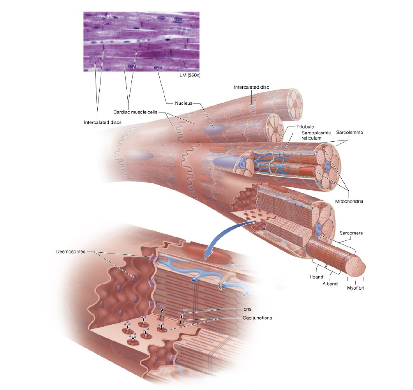
Have Striations, or light and dark bands that show the arrangement of the contractile proteins, transverse tubules and a sarcoplasmic reticulum, which share the same functions as skeletal muscle and generate tension through the sliding filament mechanism
have individual short, branched cells with a single nucleus, abundant Myoglobin, which carries oxygen, and multiple mitochondria
intercalated discs
Intercalated Discs
Structures with desmosomes and gap junctions that link adjacent cells and allow ions to pass from one cell to the next
Functional Syncytium
Gap junctions allow the electrical activity generated by the pacemaker cells to spread rapidly to all cardiac muscle cells which permits the heart to contract as a unit and produce a Heart Beat
cells of the heart are referred to as a Functional Syncytium
Voltage-Gated Sodium Ion Channels
sarcolemma contains sodium ion channels (voltage, calcium, potassium, nonselective)
Open in response to voltage changes across the membrane
found in all cardiac muscle cells except certain pacemaker cells
Calcium Ion Channels
Exhibit voltage-gated opening but time-gated closing, so will close after a certain period regardless of the voltage
present in all cells
Potassium Ion Channels
One or more types per cell
some are ligand-gated while others are voltage-gated
Nonselective Cation Channels
HCN Channel found on some pacemaker cells that is activated by hyperpolarization
allows sodium ions to enter and potassium ions to exit simultaneously

Voltage
Difference in electrical potential between two points
Membrane Potential
Voltage or charge difference that exists across the membranes of all cells, including excitable cells
Resting Membrane Potential
Membrane potential of an excitable cell at rest—averages between −60 and −90 mV
Ion Gradient
Difference in the concentration of ions across a plasma membrane
Current
Flow of ions or electrons with a chemical or electrical gradient
Depolarization
Change in resting membrane potential to a less negative value
occurs when positive charges (usually sodium or calcium ions) rush into the cell
Repolarization
Return of a cell to its negative resting membrane potential
occurs when positive charges (potassium ions) leave the cell
Hyperpolarization
Change in resting membrane potential to a more negative value
occurs when positive charges (potassium ions) exit the cell or when negative charges enter the cell
cause its potential to become more negative than the resting membrane potential
Remember that the concentration of sodium and calcium ions in the ECF is higher than in the cytosol, and the concentration of potassium ions in the ECF is lower than in the cytosol
gradients are maintained by the Na+/K+ pump and a active transport pump for calcium