Lecture 1: T cell activation + signaling
 Call Kai
 Learn
 Practice Test
 Spaced Repetition
 Match
 Flashcards
 Knowt Play
1/51

There's no tags or description
Looks like no tags are added yet.
52 Terms

Review

Review:

Review
****What is the difference between Innate + Adaptive immune response in terms of their antigen specificity?
Innate = no antigen specificity
Adaptive = has antigen specificity = conferred by the Antigen receptors (TCR/BCR)
What are the key cells involved in ADAPTIVE immunity (focus of this module)
Lymphocytes: T + B cells
APC: communicate the infection to T cells (which then coordinate the adaptive response and generate immune memory)
Professional APC = DC, macrophages + B cells
What are the general KEY MOLECULES involved in the Adaptive immune response?
MHC 1/2: expressed by APCs Present peptide to the TCR
TCR (T cell receptor): recognize peptide presented by MHC
Co stimulatory molecules = accessory signals that help activate lymphocytes
*****What are the SIGNALS required for T cell activation?
3 signals
TCR signaling = TCR associating with Ag bound to MHC
Costimulatory interaction + survival signals
Cytokine signaling

********What are the 2 CYTOKINE signals responsible for PROLIFERATION?
IL-2 +IL-2R = proliferation
Other cytokines = for DIFFERENTIATION
****Signal 1: Which cells express MHC 1 + which express 2?
MHC = ALL nucleated cells
MHC2 + CO-STIMULATORY MOLECULES = professional APC only (they also express MHC 1)
Note The key features of APCs
DC + Macrophage vs. B cells
Need to get activated by PAMPs DAMPs +/or Cytokines
B cells do not REQUIRE them to present Ag on their MHC2 as they are always highly expressed. DC + macrophage are upregulated with these signals(AI answer)

Review*****What are the 2 ways of Processing Peptide to present on MHC?
Endogenous vs. exogenous?
Source of antigen?
which one is Cytoplasmic?
Endogenous/ cytoplasmic = MHC 1 → CD8 T cells (killer T)
Endogenous antigens (Virus or cancer) → marked with ubiquitin → CYTOPLASMIC proteasome = processes the Ag into peptide
Exogenous = MHC 2 → CD4 T cells (Helper T)
Exogenous antigens = phagocytosis → endocytic compartments = processes the Ag into peptide

*****Signal 1: What is the synapse?
The interaction between TCR + peptide presented on MHC (antigen is only recognized when associated with MHC)
note that CD3 = signal transduction molecules

****Signal 1: Does TCR have high or low affinity for Ag + MHC?
LOW
*****Signal 1: If TCR has low affinity for Ag + MHC how can the complex stay together long enough to generate a signal?
ACESSORY MOLECULES
Co stim molecules are a type of accessory molecule
Coreceptors
Adhesion Molecules
*****Signal 1: What are some important Accessory molecules involved in the T cell to APC interaction?
What is the Molecule expressed on T cells
What is the molecule expressed on the APC?
What type of accessory molecule are they?
Co-receptor: CD4/8 on T cells → interact with MHC2/1 on the APC
Costimulatory molecule: CD28 on T cells → interact with CD80 or 86 on APC
Adhesion molecule: LFA-1 (Leukocyte Function Antigen 1) an INTEGRIN on T cell → interact with ICAM-1 (Intercellular Adhesion Molecule 1) on APC

*****CD8 + 4 as CORECEPTORS required for Signal 1
Where do they each bind?
CD4 Binds a conserved region in the MHC II beta2 domain
CD8 Binds a conserved region in the MHC I α3 domain
they have a signaling chain in the cytosolic side of the T cell

*****Signal 1: signaling steps - what are the types of proteins involved in TCR + BCR signaling?
5 types
What role do they each play?

*****What is ITAM?
Immuno tyrosine based activation motif
Located on the CD3 (signaling portion) of the TCR
it is phosphorylated to generate down stream signaling

*****What is the Amino acid sequence of ITAM?
Y = tyrosine L = Leucine

What type of enzymes in general Phosphorylate and which dephsophorylate?
Kinases = Phosphorylate
Phosphatases = Dephosphorylate
******How does Tyrosine phosphorylation of a protein modify its function? (aka. what does it do specifically to the protein)
Regulate Interactions between proteins - When a kinase adds a negatively charged phosphate group to a tyrosine residue, it creates a unique binding site
Regulate a proteins enzymatic activity (via Conformational changes)
Regulate a proteins subcellular localization (via intermolecular docking)
*****What are 2 proteins that commonly interact + bind with Phosphorylated tyrosine?
Src Homology 2 (SH2)
Protein tyrosine binding domain (PTB)
*****How does the Phosphorylated Tyrosine bind + interact with SH2? + PTB? How does phosphorylation promote this interaction?
The addition of the NEGATIVELY charged phosphate group promotes interaction with the positively charged ARGININE residue located in the SH2 domain
Additionally there is interaction of the Hydrophobic phenyl group with the hydrophobic pocket of the protein
Hydrophobic interactions = the main driver of interactions with PTB

*******What is the name of the kinase that phosphorylates the ITAM for TCR signaling?
Lck (Lymphocyte specific protein tyrosine kinase)
the TCR and CD3 lack their own intrinsic enzymatic activity thus cannot phosphorylate the ITAM on their own
*****What is Lck
what is the structure
(Lymphocyte specific protein tyrosine kinase)
critical Src family tyrosine kinase (SFK)
Required to initiate T-cell receptor (TCR) signaling.

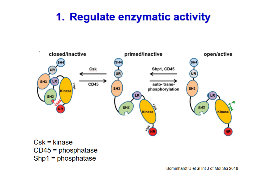
*******What is the Mechanism of Lck’s Kinase activity. What are the steps required for it to perform it’s kinase activity. REGULATION OF ENZYMATIC ACTIVITY(one of the roles of tyrosine phosphorylation)
what are the different enzymes involved in the phosphorylation + dephosphorylation
Step 1: Closed / Inactive State (Intramolecular Inhibition) - In resting T cells, the majority of Lck is kept in a tightly clamped, locked conformation to prevent accidental T-cell activation.
Regulatory Phosphorylation: The C-terminal tyrosine residue, Y505, is phosphorylated (by an enzyme called Csk = kinase).
Intramolecular Clamp:
This phosphorylated Y505 folds backward and binds directly to Lck’s own SH2 domain on the same molecule.
SH3 Domain Locking: Concurrently, the linker region (LR) interacts with Lck's own SH3 domain.
Result: The kinase domain is sterically hindered and buried, rendering it completely inactive.
Step 2: Primed / Inactive State (Dephosphorylation by CD45 = phosphatase)
To prepare for TCR signaling, the regulatory molecular clamp must be released.
CD45 Action: The transmembrane tyrosine phosphatase CD45 removes the phosphate group from the regulatory Y505 residue.
Releasing the Clamp: Without the negative phosphate group on Y505, the SH2 domain loses its binding anchor and releases the C-terminus.
Result: The protein opens up into an extended primed state. It is no longer locked, but it is not yet fully active because its catalytic engine hasn't been switched on.
Step 3: Open / Active State (Auto-Trans-Phosphorylation)
Full activation requires a cluster of primed Lck molecules to interact with TCR
Clustering & Cross-Phosphorylation: When the TCR binds an antigen-MHC complex, primed Lck molecules are brought into close proximity with one another.
Activation Loop Phosphorylation: The kinases undergo auto-trans-phosphorylation, where one Lck = kinase molecule phosphorylates the activation loop tyrosine, Y394, of a neighboring Lck molecule.
Result: Phosphorylation of Y394 stabilizes the catalytic pocket in its = fully open/active state.

*****What are the enzymes needed for TURNING OFF tyrosine kinase signaling of Lck
CD45 = phosphatase
Shp1 = phosphatase —> critical negative regulator (or "off-switch")
dephosphorylates Y394 = deactivates Lck kinase activity
Csk = Kinase
phsophylates the Lck kinase at Y 505 = closes the protein
******now that Lck Activation is covered, what is the mechanism behind Phosphorylation of Tcr (CD3 ITAM) by Lck?
10 steps (involving tyrosine kinases + phosphatases)
TCR binds to MHC+Ag
CD4/8 binds to MHC
association of CD4/8 with MHC brings along Lck kinase in proximity to ITAM on CD3 (LCK is bound on the tail of CD4/8)
Activation of Lck
CD45 dephosphorylates Lck + opening it up
Lck aggregates and auto phosphorylates itself at another tyrosine site = Activates the Kinase activity
Lck phosphorylates ITAMS of CD3 -→ they become P-ITAM
NOTE: As the physical contact zone tightens, the bulky CD45 molecule is mechanically squeezed and excluded from the synapse, preventing it from turning the signal off prematurely.
ZAP70 is attracted to Phosphorylated ITAMS (P-ITAMS). Uses its twin SH2 domains like a key to lock onto the dual-phosphorylated tyrosines.
ZAP70 is phosphorylated by Lck = P-ZAP70 (activated it’s KINASE activity)
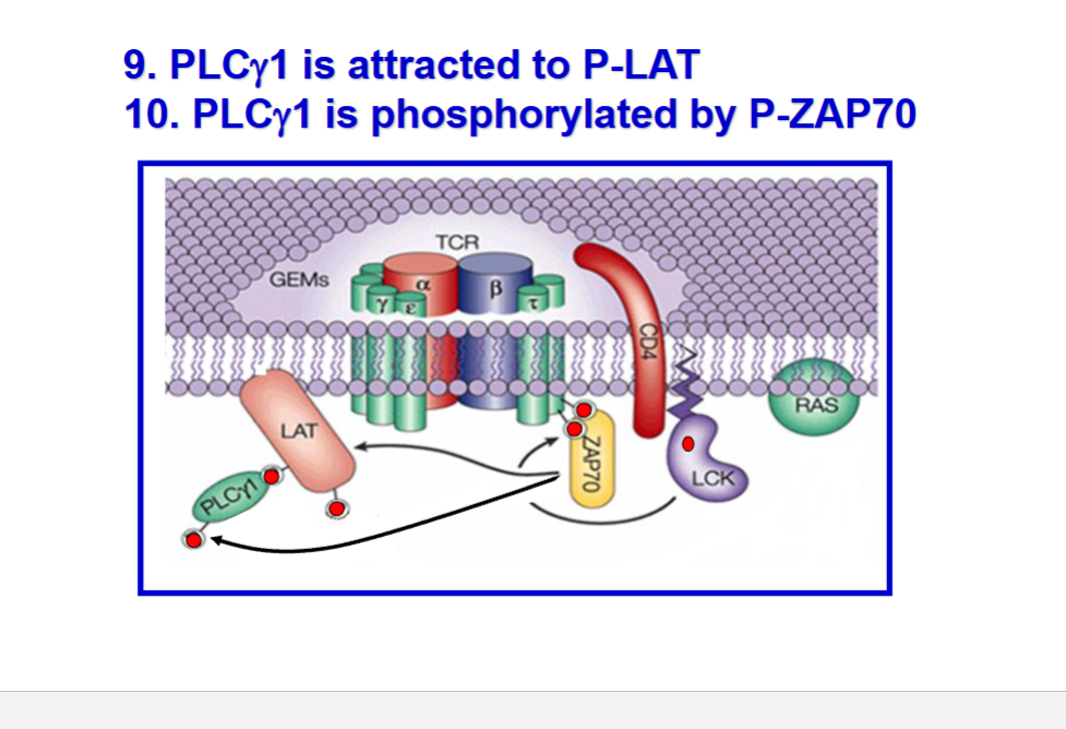
P-ZAP70 = Phosphorylates LAT (Linker protein of Activated T cells ==> P-LAT
P-LAT attracts PLCgamma1
PLCgamma1 is phosphorylated by P-ZAP70 = becomes activated P-PLCgamma1


Flip for more steps


Flip for more steps
*******Continue the TCR signaling pathway from PLCgamma1 being phosphorylated by P-ZAP70 into P-PLCgamma1
What is PLCgamma1? + what does it do?
PLCgamma1 = Phospholipase C-gamma
Phospholipases = an enzyme that breaks down (hydrolyzes) phospholipids into fatty acids and other lipophilic substances.
Step 11. P-PLCgamma1 metabolizes PIP2 (Phosphatidylinositol biphosphate = phospholipid located in the inner leaflet of the plasma membrane)
PIP2 —> DAG (diacylglycerol) + IP3 (Inositol triphosphate)
DAG: The hydrophobic tail portion that remains trapped in the greasy plasma membrane.
IP3: The hydrophilic head group that detaches and diffuses freely into the watery cytoplasm.
*******What does IP3 do?
product after cleavage of PIP2 into IP3 by PLCgamma1
IP3 opens CALCIUM CHANNELS
allows CA2+ into the ER + extracellular fluid
Influx of Ca2+ in the cytoplasm = Calmodulin Activation: This sudden flood of cytoplasmic calcium binds to and activates a sensor protein called calmodulin.
*******What does DAG do?
PKC Activation
Recruiting PKC: After PIP hydrolysis, the DAG left behind in the plasma membrane acts as a docking tag to recruit + Dock PKC (Protein Kinase C) to the cell surface.
Co-Activation: To become fully active, PKC requires two keys: it must bind to DAG at the membrane, AND it must bind to the free Ca2+ ions floating in the cytoplasm.
The Result: Once activated by both DAG and Ca2+
******What is PKC + what does it do?
Protein kinase C
initiates a phosphorylation cascade that ultimately activates the transcription factor NF-\(\kappa \)B.
******What are the 2 other transcription factors other than Nf-kB that is activated by TCR
Which cascade signals associate with each?
AP-1 = Activated by G protein kinase (Ras → MAP kinase cascade)
NFAT= activated by PLC gamma pathway
*****Summarize all the steps from TCR + MHC-Ag binding to Transcription factor activation

ABOVE was all SIGNAL 1 now we move onto SIGNAL 2
*****What is SIGNAL 2?
Co-Stimulation + Survival signals

*****What does Signal 2 do? +What is required for it to act?
Costimulatory molecule - increases T cell response in conjunction with TCR + turns on SURVIVAL GENES
cannot act unless TCR is engaged both signals must be present
Signal 1: The Activation Signal (Circle 1)
The Interaction: The T-cell Receptor (TCR) and its CD4 coreceptor bind to the specific foreign Antigen presented by an MHC class II molecule on the antigen-presenting cell (APC).
The Outcome: This interaction triggers the downstream proximal signaling pathway + turns on activation genes inside the T cell.
The Limitation: As noted at the bottom of the slide, this signal alone is not enough. If a T cell receives Signal 1 without Signal 2, it will enter a state of permanent unresponsiveness (anergy) or undergo apoptosis.
Signal 2: The Co-stimulatory Signal (Circle 2)
The Interaction: The CD28 receptor constitutively expressed on the naive T cell binds to CD80/86 (also known as B7-1 and B7-2) on the surface of the APC.
The Outcome: This interaction delivers separate, crucial metabolic and survival signals into the cell, turning on survival genes (such as Bcl-xL) and growth factor genes (like IL-2).

*********What is required for the Upregulation of Co-stimulatory molecules?
ACTIVATION of APCs
by PAPMs + CYTOKINES

*****What are the 2 key co-stim molecules upregulated on the APCs? What molecules do they bind to on the TCR?
CD80/86 (on APC) binds to CD 28on the Tcell
******Anergy + relation to signal 2?
Anergy = mechanism of self regulation + tolerance decrease response of T cells. Anergic T cells are non-responsive even when co-stimulation becomes available
Signal 1 but NO signal 2 (INACTIVE APC) = anergy
Lack of CD28-CD80/86 interaction turns on anergic genes in T cell
Alternatively the presence of inflammatory signals eg. PRRs + Cytokines = Activate APC = upregulate costim. molecules

******Now that we have covered signal 2 → What is SIGNAL 3?
CYTOKINES
mediation of Proliferation + Differentiation

****What Cytokines are responsible for Proliferation vs. differentiation?
IL-2 = proliferation
Other cytokines = differentiation
***moving on from the 3 signals → DILEMA
HOW are NAIVE CD8+ T cells activated to become CTLs (cytotoxic T lymphocytes)
What is the dilemma + what is the solution?
2 rules that must be followed:
The MHC Class I Rule: Naive CD8+ T cells only recognize antigens processed through the endogenous pathway and presented on MHC Class I. Normally, this means the antigen must come from a virus replicating inside that specific cell.
Naïve T cells can only be activated when there is costimulation by professional APCs = a naive T cell will not activate unless it gets Signal 2 (CD80/86) from a professional Antigen-Presenting Cell (APC), like a dendritic cell.
DILEMA = a naive CD8+ T cell could only be activated if the DC isDIRECTLY INFECTED by that specific virus.
CD8 must be activated by MHC1 + MHC only presents endogenous proteins
SOLUTION = CROSS PRESENTATION = Some DCs are specialized in this process

****What is CROSS presentation again?
Specialized immune process where professional antigen-presenting cells (primarily dendritic cells) take up extracellular (exogenous) antigens and present them on MHC class I molecules to CD8+ cytotoxic T cells
allowing extracellular antigens to be routed into the MHC class I pathway instead,
= Allow DC cells to present Exogenous proteins (norm MHC2 pathway) on MHC 1 to present + activate CD8 naive (no need to be infected)
***What is a basic principle for vaccines and anti-tumor immunity?
Cross presentation

********What is Licensing?
What allows DC (APC) cells to cross present
The extra step needed for CD8+ to get activated

*****What does the LISCENCING?
Done by activated CD4 helper T cells
******What are the 2 ways a DC cell can be LISCENCED?
1. Activated Th1 cells express CD40L ( activated CD4⁺ T cell upregulates CD40 )
CD40L on an activated Th1 cell interacts with CD40 on APC
CD40–CD40L interaction, often combined with helper cytokines + Co-stimulatory molecules like B7 (CD80/CD86) on the DC interacting with CD28 on the T cell also reinforce this cross-talk. = fully "licenses" the DC cell
2. Microbial products
Toll-like receptor activation


Flip for 1 more diagram
