IFS Exam 1
 Call Kai
 Learn
Learn Practice Test
 Spaced Repetition
 Match
Match Flashcards
 Knowt Play
1/36

There's no tags or description
Looks like no tags are added yet.
Name | Mastery | Learn | Test | Matching | Spaced | Call with Kai | Chat |
|---|
No analytics yet
Send a link to your students to track their progress
37 Terms
Stylohyoid (intermediate - suprahyoid)
O: Styloid process of temporal bone
I: Body of hyoid
N: Stylohyoid (preparotid) branch of facial nerve (CN VII)
A: Elevates and retracts hyoid, thus elongating floor of mouth

Mylohyoid (intermediate - suprahyoid)
O: Mylohyoid line of mandible
I: Mylohyoid raphe and body of hyoid
N: Nerve to mylohyoid, a branch of inferior alveolar nerve, from mandibular nerve, CN V3
A: Elevates hyoid, floor of mouth, and tongue during swallowing and speaking

Geniohyoid (intemediate - suprahyoid)
O: Inferior mental spine of mandible
I: Body of hyoid
N: C1 via hypoglossal nerve (CN XII) and nerve to geniohyoid
A: Pulls hyoid anterosuperiorly; shortens floor of mouth; widens pharynx

Digastric (suprahyoid)
O:
Anterior belly: digastric fossa of mandible
Posterior belly: mastoid notch of temporal bone
I: Intermediate tendon to body and greater horn of hyoid
N:
Anterior belly: Nerve to mylohyoid, a branch of inferior alveolar nerve
Posterior belly: digastric branch of facial nerve (CN VII)
A: Digastric: Working with infrahyoid muscles, depresses mandible against resistance; elevates and steadies hyoid during swallowing and speaking
Omohyoid (intermediate - infrahyoid)
O: superior border of scapula near suprascapular notch
I: Inferior border of hyoid
N: C1-C3 by branch of ansa cervicalis
A: Depreses, retracts, and steadies hyoid

sternohyoid (intermediate - infrahyoid)
O: manubrium of sternum and medial end of clavicle
I: Body of hyoid
N: C1-C3 by a branch of ansa cervicalis
A: Depresses hyoid after elevation during swallowing

thyrohyoid (intermediate)
O: Oblique line of thyroid cartilage
I: Inferior border of body and greater horn of hyoid
N: C1 via hypoglossal nerve (CN XII) and nerve to thyrohyoid
A: Depresses hyoid and elevates larynx

sternothyroid (intermediate - infrahyoid)
O: Posteiror surface of manubrium of sternum
I: Oblique line of thyroid cartilage
N: C2 and C3 by a branch of ansa cervicalis
A: Depresses hyoid and larynx

Longus Capitis (prevertebral - deep)
Superior Attachment (I): Basilar part of occipital bone
Inferior Attachment (O): Anterior tubercles of C3-C6 transverse process
Nerve: Anterior rami of C1-C3 spinal nerves
Action: Flex head

Longus colli (prevertebral - deep)
Superior Attachment (I): Anterior tubercle of C1 vertebra (atlas); bodies of C1–C3 and transverse processes of C3–C6 vertebrae
Inferior Attachment (O): Bodies of C5–T3 vertebrae; transverse processes of C3–C5 vertebrae
Nerve: Anterior rami of C2–C6 spinal nerves
Action: Flexes neck with rotation (torsion) to opposite side if acting unilaterally

rectus capitis anterior (prevertebral - deep)
Superior Attachment (I): Base of cranium, just anterior to occipital condyle
Inferior Attachment (O): Anterior surface of lateral mass of atlas (C1 vertebra)
Nerve: Branches from loop between C1 and C2 spinal nerves
Action: Flex head

rectus capitis lateralis (prevertebral - deep)
Superior Attachment (I): Jugular process of occipital bone
Inferior Attachment (O): Transverse process of atlas (C1)
Nerve: Branches from loop between C1 and C2 spinal nerves
Action: Flex head and helps stabilize it

Anterior scalene (deep)
Superior Attachment (O): TP of C3-C6
Inferior Attachment (I): 1st rib
Nerve: Cervical spinal nerves C4-C6
Action: Flexes neck laterally; elevates 1st rib during forced inspiration - laterally flexes ipsilaterally (contraction bends the neck to the same side)
Middle scalene (deep)
Superior Attachment: Posterior tubercles of TP of C5-C7
Inferior Attachment: Superior surface of 1st rib; posterior to groove for subclavian artery
Nerve: Anterior rami of cervical spinal nerves
Action: Flexes neck laterally; elevates 1st rib during forced inspirationa
Posterior scalene (deep)
Superior Attachment: Posterior tubercles of TP of C5-C7
Inferior Attachment: External border of 2nd rib
Nerve: Anterior rami of cervical spinal nerves C7 and C8
Action: Flexes neck laterally; elevates 2nd rib during forced inspirtaion
Platysma - Superficial
Superior/Medial Attachment: Inferior border of mandible, skin, and subcutaneous tissues of lower face
Inferior/Lateral Attachment: Fascia covering superior parts of pectoralis major and deltoid muscles
Nerve: Cervical branch of facial nerve (CN VII)
Action: Draws corners of mouth inferiorly and widens it as in expressions of sadness and fright; draws skin of neck superiorly when teeth are clenched

Sternocleidomastoid - Superficial
Superior/Medial Attachment (I): Lateral surface of mastoid process of temporal bone and lateral half of superior nuchal line
Inferior/Lateral Attachment (O):
Sternal head: anterior surface of manubrium of sternum
Clavicular head: superior surface of medial third of clavicle
Nerve: Spinal accessory nerve (CN XI, motor); C3 and C4 nerves (pain and proprioception)
Action: Unilateral contraction: tilts head to same side (i.e., laterally flexes neck) and rotates it so face is turned superiorly toward opposite side
Bilateral contraction: (1) extends neck at atlanto-occipital joints, (2) flexes cervical vertebrae so that chin approaches manubrium, or (3) extends superior cervical vertebrae while flexing inferior vertebrae, so chin is thrust forward with head kept level
With cervical vertebrae fixed, may elevate manubrium and medial ends of clavicles, assisting pump-handle action of deep respiration
Action Unilaterally:
Laterally flex the neck to same side
Rotate the neck to opposite side
Action Together Bilaterally:
Flex the neck
Assists in elevation of rib cage during inhalation

Trapezius - Superficial
Proximal Attachment (O): Medial third of superior nuchal line; external occipital protuberance; nuchal ligament; spinous processes of C7–T12 vertebrae
Distal Attachment (I): Lateral third of clavicle; acromion and spine of scapula
Nerve: Spinal accessory nerve (CN XI) (motor fibers) and C3, C4 spinal nerves (pain and proprioceptive fibers)
Action: Descending part elevates; descending and ascending parts act together to rotate glenoid cavity superiorly
Action: middle part (or all parts together) retracts scapula
Action: ascending part depresses; descending and ascending parts act together to rotate glenoid cavity superiorly
**Cervical Extension - upper
Upper trapezius — unilateral contraction
Ipsilateral lateral flexion → bends the neck toward the contracting side
Contralateral rotation → turns the head away from the contracting side
Example: Right upper trapezius contracts
→ right lateral flexion
→ left rotation

Latissimus Dorsi
(Superficial Layer - Extrinsic Back Muscles - axio-appendicular)
Proximal Attachment (O): Spinous processes of inferior 6 thoracic vertebrae, thoracolumbar fascia, iliac crest, and inferior 3 or 4 ribs
Distal Attachment (I): Floor of intertubercular sulcus of humerus
Nerve: Thoracodorsal Nerve (C6. C7. C8)
Action: Extends, adducts, and medially rotates humerus; raises body toward arms during climbing
Same-side shoulder extension, adduction, medial rotaiton - Right lat contracts —> right arm ext, add, IR

Rhomboid Major
(Superficial Layer - Extrinsic Back Muscles - axio-appendicular)
Proximal Attachment (O): SP of T2-T5 vertebrae
Distal Attachment (I): medial border of scapula from level of spine to inferior angle
Nerve: Dorsal scapular nerve (C4, C5)
Action: Retract scapula and rotate its glenoid cavity inferiorly; fix scapula to thoracic wall
Right rhomboids act on the right scapula

Rhomboid Minor
(Superficial Layer - Extrinsic Back Muscles - axio-appendicular)
Proximal Attachment (O): nuchal ligament; spinous processes of C7 and T1 vertebrae
Distal Attachment (I): smooth triangular area at medial end of scapular spine
Nerve: Dorsal scapular nerve (C4, C5)
Action: Retract scapula and rotate its glenoid cavity inferiorly; fix scapula to thoracic wall

Levator Scapulae
(Extrinsic back muscle - superficial layer)
Proximal Attachment (O): Posterior tubercles of transverse processes of C1–C4 vertebrae
Distal Attachment (I): Medial border of scapula superior to root of scapular spine
Nerve: Dorsal scapular (C4, C5) and cervical (C3, C4) nerves
Action: Elevates scapula and rotates its glenoid cavity inferiorly by rotating scapula (**scapula downward rotation)
Extends neck (with trapezius)
Laterally flexes neck to the same side
Right lateral flexion of neck —> ipsilateral; right scapular elevation —> ipsilateral

Serratus Posterior Superior
(Intermediate Layer - Extrinsic Back Muscles - thoracic wal)
Superior Attachment (O): Nuchal ligament, SP of C7-T3 vertebrae
Inferior Attachment (I): Superior borders of 2nd-4th ribs
Nerve: 2nd-5th intercostal nerves
Action: Proprioception (elevate ribs)

Serratus Posterior Inferior
(Intermediate Layer - Extrinsic Back Muscles - thoracic wal)
Superior Attachment (O): SP of T11-L2 vertebrae
Inferior Attachment (I): Inferior borders of 8th-12th ribs near their angles
Nerve: Anterior rami to T9–T12 thoracic spinal nerves
Action: Proprioception (depress ribs)

Splenius Cervicis
(Superficial Layer of Intrinsic Back Muscles)
Proximal Attach (O): Nuchal ligament and SP of C7-T6
Distal Attach (I): Tubercles of transverse processes of C1-C3 or C4 vertebrae
Nerve: Posterior rami of spinal nerves
Action:
Unilaterally: laterally flexes the neck and rotates the head to side of active musces
Bilaterally: extend the head and neck
Right splenius cervicis contracts —> right lateral flexion + right rotation

Splenius Capitis
(Superficial Layer of Intrinsic Back Muscles)
Proximal Attach (O): Nuchal ligament and SP of C7-T6
Distal Attach (I): Fibers run superolaterally to mastoid process of temporal bone and lateral 1/3rd of superior nuchal line of occipital bone
Nerve: Posterior rami of spinal nerves
Action:
Unilaterally: laterally flexes the neck and rotates the head to side of active musces
Bilaterally: extend the head and neck


Iliocostalis
(Intermediate Layer of Intrinsic Back Muscles)
Proximal Attachment (O): Arises by a broad tendon from the posterior part of the iliac crest, posterior surface of the sacrum, sacro-iliac ligaments, sacral and inferior lumbar spinous processes, and supraspinous ligament
Distal Attachment (I): lumborum, thoracis, cervicis; fibers run superiorly to angles of lower ribs and cervical transverse processes.
Nerve: Posterior rami of spinal nerves
Action:
Bilaterally: Extend vertebral column and head; as back is flexed, control movement via eccentric contraction
Unilaterally: Laterally felxes vertebral column
**rotation
Laterally flexes ipsilaterally (not really a rotator, but ipsilateral)
Longissimus
(Intermediate Layer of Intrinsic Back Muscles)
Proximal Attachment (O): Arises by a broad tendon from the posterior part of the iliac crest, posterior surface of the sacrum, sacro-iliac ligaments, sacral and inferior lumbar spinous processes, and supraspinous ligament
Distal Attachment: thoracis, cervicis, capitis; fibers run superiorly to ribs between tubercles and angles to transverse processes in thoracic and cervical regions and to mastoid process of temporal bone
Nerve: Posterior rami of spinal nerves
Action:
Bilaterally: Extend vertebral column and head; as back is flexed, control movement via eccentric contraction
Unilaterally: Laterally felxes vertebral column
**Rotation

Spinalis
(Intermediate Layer of Intrinsic Back Muscles)
Proximal Attachment (O): Arises by a broad tendon from the posterior part of the iliac crest, posterior surface of the sacrum, sacro-iliac ligaments, sacral and inferior lumbar spinous processes, and supraspinous ligament
Distal Attachment (I): thoracis, cervicis, capitis; fibers run superiorly to spinous processes in the upper thoracic region and to cranium.
Nerve: Posterior rami of spinal nerves
Action:
Bilaterally: Extend vertebral column and head; as back is flexed, control movement via eccentric contraction
Unilaterally: Laterally felxes vertebral column

Semispinalis
(Major Deep Layer of Intrinsic Back Muscles)
Inferior Attachment (O): Arises from TP of C4-T12 vertebrae
Superior Attachment (I): thoracis, cervicis, capitis; fibers run superomedially to occipital bone and spinous processes in thoracic and cervical regions, spanning 4–6 segments.
Nerve: Posterior rami of spinal nerves
Actions: Extends head and thoracic and cervical regions of vertebral column and rotates them contralaterally
Contralateral rotation

Rotatores
(Major Deep Layer of Intrinsic Back Muscles)
Inferior Attachment (O): Arises from TP of vertebrae; best developed in thorcic region
Superior Attachment (I): Fibers pass superomedially to attach to junciton of lamina and TP or SP of vertebra immediately (brevis) or 2 segments (longus) superior to vertebra of attachment.
Nerve: Posterior rami of spinal nerves
Action: Stabilizes vertebrae and assist with local extension and rotatory movements of vertebral column; may function as organs of proprioception
Contralateral rotation

Multifidus
(Deep layers of intrinsic back muscles)
Inferior Attachment (O): arises from posterior sacrum, posterior superior iliac spine of the ilium, aponeurosis of erector spinae, sacro-iliac ligaments, mammillary processes of lumbar vertebrae, transverse processes of T1–T3, articular processes of C4–C7
Superior Attachment (I): thickest in lumbar region; fibers pass obliquely superomedially to entire length of spinous processes, located 2–4 segments superior to proximal attachment.
Nerve: Posterior Rami of spinal nerves
Action: stabilizes vertebrae during local movements of vertebral column
**Rotation and lateral bending
(Extension for all transversospinalis)
Contralateral rotation, ipsilateral lateral flexion

Transversospinalis group (semispinalis, multifidus, rotatores)
(Major Deep Layer of Intrinsic Back Muscles)
Inferior Attachment (O): Transverse process
Superior Attachment (I): SP of more superior vertebrae
Nerve: Posterior rami of spinal nerves
Action: Extension

Interspinales
(Minor Deep Layer of Intrinsic Back Muscles)
Inferior Attachment (O): Superior surfaces of spinous processes of cervical and lumbar vertebrae
Superior Attachment (I): Inferior surfaces of spinous processes of vertebra superior to vertebra of proximal attachment
Nerves: Posterior rami of spinal nerves
Action: Aid in extension and rotation of vertebral column

Intertransversarii
(Minor Deep Layer of Intrinsic Back Muscles)
Inferior Attachment (O): TP of cervical and lumbar vertebrae
Superior Attachment (I): TP of adjacent vertebrae
Nerve: Posterior and anterior rami of spinal nerves
Action: Aid in lateral flexion of vertebral column; acting bilaterally, stabilize vertebral column
Right lateral flexion - ipsilateral

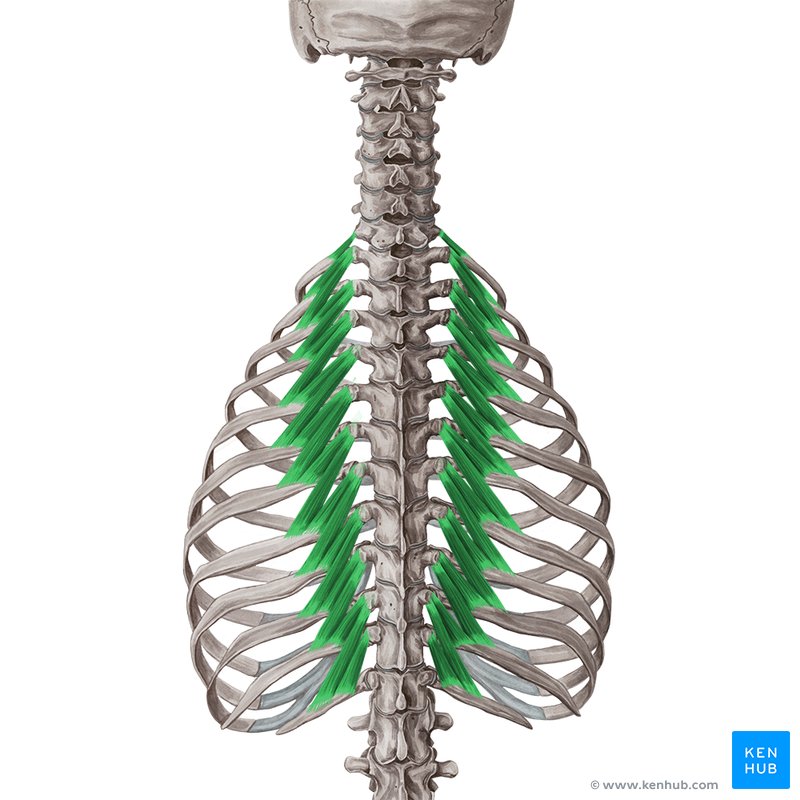
Levator costarum
(Minor Deep Layer of Intrinsic Back Muscles)
Superior Attachment (O): Tips of transverse processes of C7 and T1–T11 vertebrae
Inferior Attachment (I): Pass inferolaterally and insert on rib between tubercle and angle
Nerve: Posterior rami of C8-T11 spinal nerves
Action: Elevate ribs, assisting respiration; assist with lateral flexion of vertebral column
lateral felxion —> ipsilateral
Quadratus Lumborum
(Muscle of Posteiror Abdominal Wall)
Inferior Attachment (O): Iliolumbar ligament and internal lip of iliac crest
Superior Attachment (I): Medial half of inferior border of 12th ribs and tips of lumbar transverse processes
Nerve: Anterior branches of T12 and L1–L4 nerves
Action: Extends and laterally flexes vertebral column; fixes 12th rib during inspiration
Lateral flexion —> ipsilateral
