Lecture 3 - Cell Cycle Mitosis and Meiosis
 Call Kai
 Learn
Learn Practice Test
 Spaced Repetition
 Match
Match Flashcards
 Knowt Play
1/97

There's no tags or description
Looks like no tags are added yet.
Name | Mastery | Learn | Test | Matching | Spaced | Call with Kai | Chat |
|---|
No analytics yet
Send a link to your students to track their progress
98 Terms

Binary fission
Type of cell division by which prokaryotes (bacteria and archaea) reproduce; a form of asexual reproduction

Process of binary fission
Chromosome replicates starting at the origin of replication → the two copies of the origin move toward opposite ends as the cell elongates → a division septum forms → the cell separates into two daughter cells
Meiosis (life cycle definition)
Process where a diploid cell gives rise to haploid cells (gametes)
sexual life cycle
Fertilization
Process where two haploid cells (gametes) fuse to form a diploid zygote
restoring the diploid state; gametes usually come from different individuals
Three main categories of sexual life cycles
1) Diploid-dominant
2) Haploid-dominant
3) Alternation of generations
Diploid-dominant life cycle
The multicellular diploid stage is the most obvious life stage
the only haploid cells are the gametes; found in humans and most animals
Haploid-dominant life cycle
The multicellular (or sometimes unicellular) haploid stage is the most obvious life stage;
the single-celled zygote is the only diploid cell; found in fungi and some algae
Alternation of generations
Both the haploid and diploid stages are multicellular (dominant to different degrees in different species)
found in plants and some algae
Gamete
A reproductive cell of an animal or plant (e.g., sperm or egg);
haploid

Germ cells
Special diploid cells made in the gonads early in animal embryo development;
they divide by mitosis to make more germ cells, and some undergo meiosis to make haploid gametes

Gonads
Organs that produce gametes: testes and ovaries

Zygote
A diploid cell resulting from the fusion of two haploid gametes; a fertilized ovum

Human sex cells
Egg and sperm cells
contain a single set of chromosomes and are haploid

Organisms with a haploid-dominant life cycle
Most fungi and some protists (unicellular eukaryotes)
the "body" (mature, ecologically important form) is haploid
Ex: Black bread mold

Hyphae
Multicellular, thread-like haploid structures of fungi
in bread mold sexual reproduction, hyphae from two compatible individuals (+ and − mating types) grow toward each other

Zygosporangium
Structure formed where two compatible hyphae meet;
contains multiple haploid nuclei from the two parents

Zygospore
Diploid structure formed after nuclear fusion
can stay dormant for long periods, then under the right conditions its diploid nuclei undergo meiosis to make haploid spores

Spores (bread mold)
Single cells containing haploid nuclei released after meiosis of the zygospore
they grow by mitosis into new hyphae

Phases in alternation of generations
A haploid sexual phase (gametophyte) and a diploid asexual phase (sporophyte)

Gametophyte
The haploid multicellular plant (or alga)
makes gametes using specialized cells

Why is meiosis not directly involved in making gametes in gametophytes?
Because the gametophyte is already haploid, so it makes gametes by mitosis
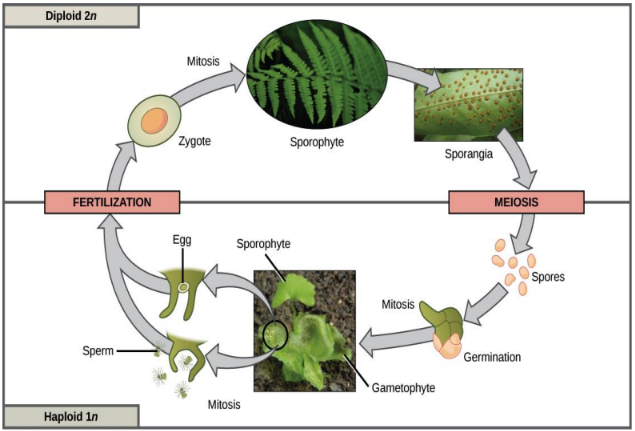
Sporophyte
Diploid multicellular plant formed from the zygote by many rounds of mitosis
specialized cells undergo meiosis to produce haploid spores
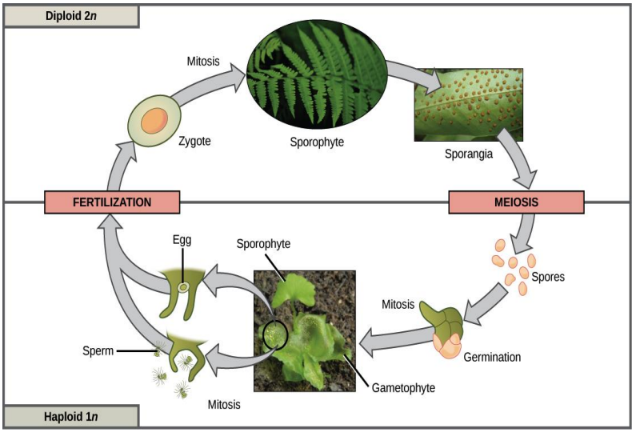
Alternation of generations cycle order
Haploid gametes fertilize → diploid zygote → mitosis → sporophyte → meiosis (in sporangia) → haploid spores → germination/mitosis → gametophyte → makes egg and sperm

Sporophyte vs gametophyte in seed plants
In seed plants (e.g., magnolia trees, daisies), the sporophyte is much larger
what we consider the "plant" is almost entirely sporophyte tissue

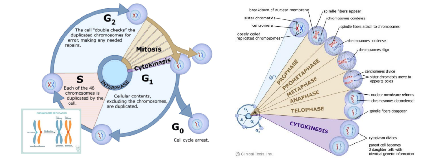
Cell cycle
Four-stage process:
cell increases in size (G1), copies its DNA (S), prepares to divide (G2), and divides (M)

Interphase
The part of the cell cycle made of G1, S, and G2 (everything except M phase)

G1 phase
Gap 1: cell grows
metabolic changes prepare the cell for division
cellular contents (excluding chromosomes) are duplicated
many cells check that nutrient levels are high enough to complete division

Restriction point
Point in G1 after which the cell is committed to division and moves into S phase

S phase
Synthesis phase: chromosomes are duplicated (all 46 in humans)
each chromosome now consists of two sister chromatids

G2 phase
Gap 2: metabolic changes assemble cytoplasmic materials needed for mitosis and cytokinesis
the cell "double checks" duplicated chromosomes for errors and makes repairs

M phase
Nuclear division (mitosis or meiosis) followed by cytoplasmic division (cytokinesis)
G0
Cell cycle arrest
a resting state where the cell exits the cycle

Order of mitosis stages
Prophase → Prometaphase → Metaphase → Anaphase → Telophase (then Cytokinesis)

Prophase (mitosis)
Chromosomes condense and become visible
spindle fibers emerge from the centrosomes
nuclear envelope breaks down
centrosomes move toward opposite poles

Prometaphase
Chromosomes continue to condense
kinetochores appear at the centromeres
mitotic spindle microtubules attach to kinetochores
Metaphase (mitosis)
Chromosomes line up at the metaphase plate
each sister chromatid is attached to a spindle fiber from opposite poles
Anaphase (mitosis)
Centromeres split in two
sister chromatids (now called chromosomes) are pulled toward opposite poles
certain spindle fibers elongate the cell
Telophase (mitosis)
Chromosomes arrive at opposite poles and decondense
nuclear envelope reforms around each set
mitotic spindle breaks down
spindle fibers keep pushing poles apart
Cytokinesis in animal cells
A cleavage furrow separates the daughter cells
Cytokinesis in plant cells
A cell plate (precursor to a new cell wall) separates the daughter cells
Three components required for eukaryotic chromosome maintenance during cell division
Centromeres, telomeres, and origins of replication

Centromere (definition)
Specialized DNA sequence of a chromosome that links a pair of sister chromatids (a dyad)
spindle fibers attach to it via the kinetochore during mitosis
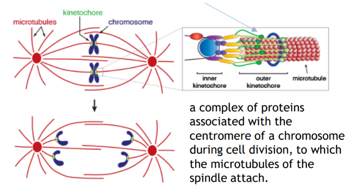
Kinetochore
Complex of proteins associated with the centromere during cell division, to which spindle microtubules attach (has inner and outer kinetochore)

Two major chromosomal events of S phase
1) DNA replication copies each chromosome completely
2) sister chromatid cohesion is established by ring-shaped cohesin molecules

Cohesin
Ring-shaped protein hypothesized to encircle the two copies of replicated DNA, holding sister chromatids together (sister chromatid cohesion)

Chromatid
Each copy of a duplicated chromosome
the two chromatids of a pair are sister chromatids

Three major events of M phase (mitosis)
1) The two kinetochores of each sister chromatid pair attach to opposite spindle poles
2) cohesion is eliminated by destroying the cohesin ring
3) sister chromatids are segregated to opposite poles

How many centromeres does a normal chromosome have?
One
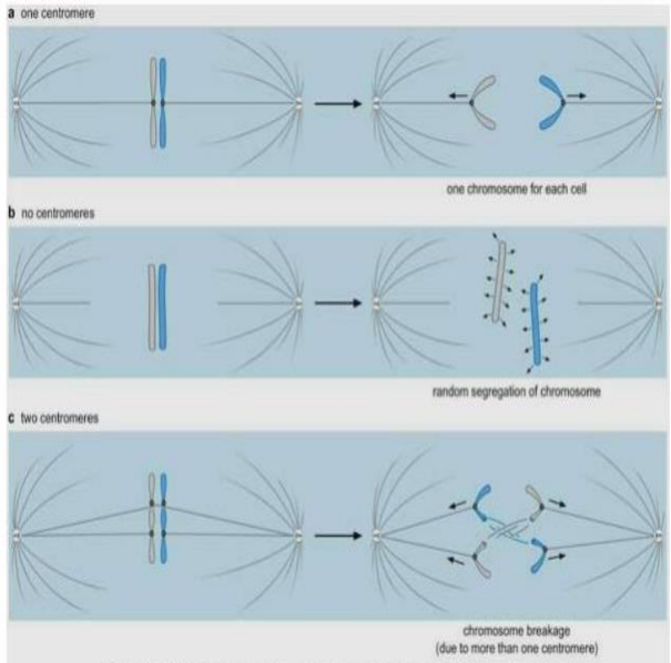
What happens to centromeres after chromosome replication?
Each copy of the centromere directs formation of a kinetochore
the two kinetochores bind opposite spindle poles and are pulled to opposite sides of the cell
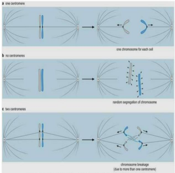
Chromosome with no centromere
Cannot attach to the spindle, so it is randomly distributed to daughter cells (one daughter may get two copies and the other none)

Chromosome with two or more centromeres
Frequently broken during segregation

Centromere size in different organisms
Varies dramatically:
S. cerevisiae ~125 bp (CDE I-III)
S. pombe 40-100 kb
D. melanogaster ~400 kb
human 240 kb to several Mb

Meiosis (overview)
Sex cell division: one germ cell → four gametes
two divisions, each with prophase, metaphase, anaphase, telophase

What precedes meiosis?
Interphase, in which DNA is replicated so chromosomes consist of two sister chromatids

Interkinesis
Optional growth phase between meiosis I and meiosis II; NO DNA replication occurs

Final outcome of meiosis
Four haploid daughter cells, which may all be genetically distinct if crossing over occurs in prophase I
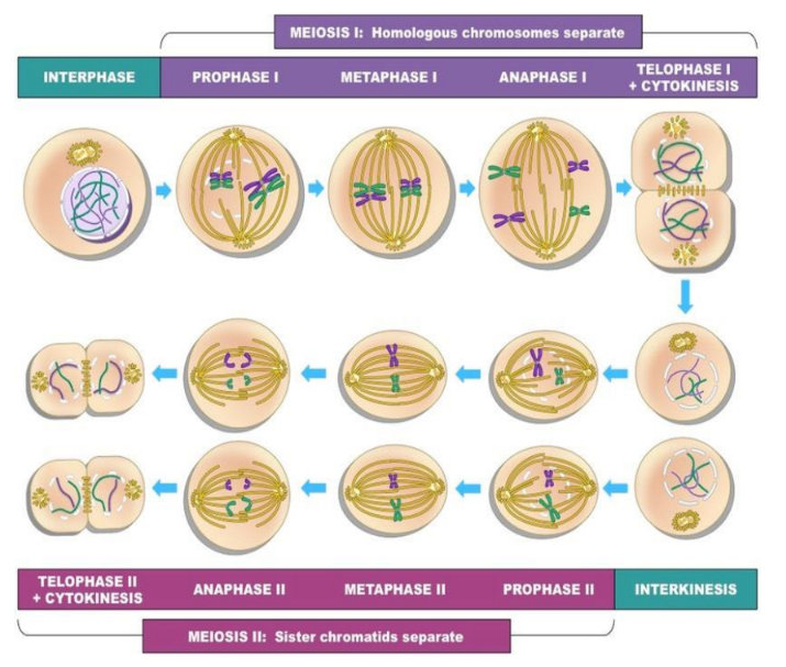
What separates in meiosis I?
Homologous chromosomes

What separates in meiosis II?
Sister chromatids
Reduction division
Meiosis I: diploid → haploid, in which homologous chromosomes are separated, reducing the chromosome number by half

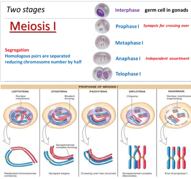
Prophase I
Chromosomes condense, nuclear membrane dissolves, homologous chromosomes pair to form bivalents (synapsis), and crossing over (recombination) occurs
Bivalent (tetrad)
A pair of synapsed homologous chromosomes
contains a total of 4 chromatids
Metaphase I
Spindle fibers from opposing centrosomes connect to bivalents (at centromeres) and align them along the middle of the cell
Anaphase I
Spindle fibers contract and split the bivalent
homologous chromosomes move to opposite poles
independent assortment occurs

Telophase I
Chromosomes decondense, nuclear membrane may reform, cell divides (cytokinesis) to form two haploid daughter cells

Linkage
Co-inheritance of two genes (loci) on the same chromosome

How does distance affect linkage?
The closer two genes are, the more likely they show linkage, because crossovers occur by chance and more DNA between genes means a greater chance of a crossover

Linkage group
Group of genes mapped (using crossovers) to show their relative order on a chromosome or part of a chromosome
Map unit (mu)
Unit of distance between genes, based on recombination frequency
1 mu = 1% recombination between 2 genes

Parental vs recombinant chromosomes
Parental: allele combinations same as the parents (e.g., AB, ab) from meioses with no crossover between genes
Recombinant: new combinations (e.g., Ab, aB) from a crossover between the genes
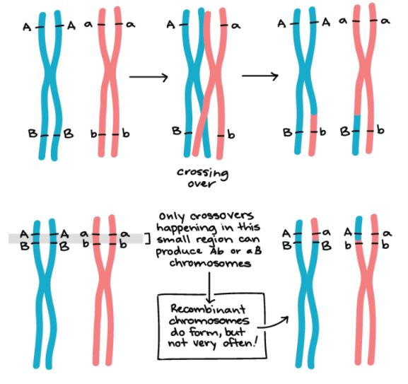
Why does recombination frequency reflect gene distance?
Crossovers in meiosis I happen at random positions, so a short distance is a small "target" for crossovers (few events) while far-apart genes recombine more often

Use of recombination frequency
Estimating the relative distance between two genes on a chromosome (their degree of linkage)
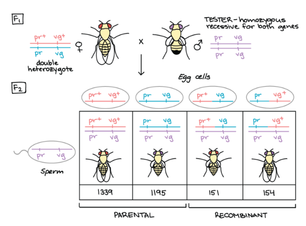
How do you know two genes are linked in a test cross?
The four classes of offspring are not produced in equal numbers
parental types are over-represented and recombinant types are under-represented

Recombination frequency (RF) formula
RF = (Recombinant offspring ÷ Total offspring) × 100%

Meiosis II (overview)
Second division
separates sister chromatids (which may not be identical due to crossing over in prophase I), producing four haploid gametes
Prophase II
Chromosomes condense, nuclear membrane dissolves, centrosomes move to opposite poles (perpendicular to before)


Metaphase II
Spindle fibers from opposing centrosomes attach to chromosomes at the centromere and align them along the cell equator

Anaphase II
Spindle fibers contract and separate sister chromatids; chromatids (now called chromosomes) move to opposite poles

Telophase II
Chromosomes decondense, nuclear membrane reforms, cells divide (cytokinesis) to form four haploid daughter cells
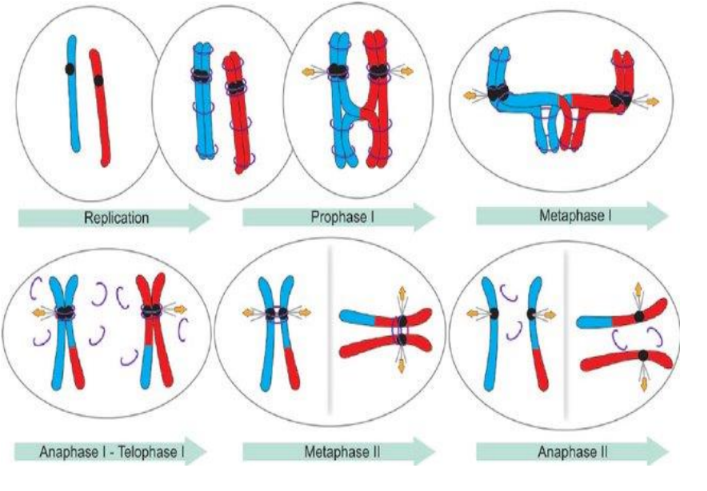
Chromosome behavior in meiosis
Two rounds of chromosome segregation follow a single replication
sister chromatids are held together by cohesion

How many crossovers per pair of homologues?
At least one crossover (CO) per pair of homologues is always observed

What forms the bivalent at metaphase I?
Crossovers + sister chromatid cohesion physically link the recombined homologous chromosomes

Cohesion at anaphase I
Released everywhere except at the centromeres
homologs segregate while sister chromatids stay together

Cohesion at metaphase II / anaphase II
Centromeric cohesion is destroyed, allowing sister chromatids to segregate to opposite poles starting at anaphase II
Mitosis: duplications and divisions
Chromosomes duplicate once; cell divides once
Meiosis: duplications and divisions
Chromosomes duplicate once; cell divides twice
Outcome of mitosis
Two diploid daughter cells that are genetically identical
Outcome of meiosis
Four haploid daughter cells that are NOT genetically identical
they contain different combinations of homologues due to crossover
DNA synthesis: mitosis vs meiosis
Occurs in S phase of interphase in both
Synapsis and crossover: mitosis vs meiosis
Meiosis: during prophase I; Mitosis: do not occur
Lining up at the metaphase plate: mitosis vs meiosis
Homologous pairs: metaphase I only (not in mitosis)
sister chromatids: metaphase II in meiosis and metaphase in mitosis
Trait
A specific characteristic of an individual; determined by both genes and environment
Genotype
The genetic composition of a trait
Phenotype
The visible or otherwise observable, measurable trait produced by a genotype
Homozygous
Genotype with two identical alleles in a gene pair
Heterozygous
Genotype with two different alleles in a gene pair
Dominant allele
Allele whose trait appears in both homozygous and heterozygous individuals
Recessive allele
Allele whose trait appears only in homozygous individuals
Codominant allele
Allele whose trait reflects the combination of two different alleles (both show)