oct 20th Lipids: Ketone Bodies and Cholesterol Synthesis
 Call Kai
 Learn
 Practice Test
 Spaced Repetition
 Match
 Flashcards
 Knowt Play
1/16

There's no tags or description
Looks like no tags are added yet.
17 Terms
How does the liver fuel the body as fasting continues?

During fasting, the liver has two important roles: maintaining blood glucose for tissues that need it and providing alternative fuels. Ketone bodies made from fatty acids are exported for use by other tissues, while fatty-acid oxidation also supplies ATP for the liver's own energy needs.
The brain normally depends heavily on glucose and uses little circulating fatty acid for energy. Ketone bodies readily cross the blood–brain barrier, so they can serve as an alternative fuel for the brain. As fasting continues, the brain gradually increases its use of ketone bodies, although it still requires some glucose.
This adaptation helps preserve muscle protein. Without an alternative fuel, more glucose would need to be produced from amino acids obtained partly through muscle protein breakdown. By reducing the brain's glucose requirement, ketone bodies reduce the need for these amino acids.
How does the brain's use of ketone bodies during prolonged fasting help preserve muscle protein?
Ketone bodies replace part of the brain's glucose requirement, reducing the need to break down muscle protein to supply amino acids for gluconeogenesis.
Why does the liver during fasting make ketone bodies?

During fasting, insulin levels fall, increasing the release of fatty acids from adipose tissue and their delivery to the liver. At the same time, malonyl-CoA levels decrease, relieving inhibition of CPT-I. This allows more longchain fatty-acyl groups to enter mitochondria for β-oxidation.
β-Oxidation produces acetyl-CoA, NADH, and FADH₂ equivalents. Normally, acetyl-CoA enters the TCA cycle by combining with oxaloacetate to form citrate. During fasting, oxaloacetate is also needed for gluconeogenesis, which helps the liver maintain blood glucose. This reduces the availability of oxaloacetate for the TCA cycle. In addition, increased NADH production during fatty-acid oxidation slows several reactions of the TCA cycle.
As a result, acetyl-CoA may be produced faster than the TCA cycle can oxidize it. The liver converts some of this acetyl-CoA into ketone bodies.
Why does the fasting liver convert acetyl-CoA into ketone bodies instead of oxidizing it all in the TCA cycle?
β-Oxidation produces acetyl-CoA faster than the TCA cycle can oxidize it. Oxaloacetate is diverted to gluconeogenesis, and the increased NADH slows several TCA-cycle reactions. The liver converts some of this acetyl-CoA into ketone bodies.
What are the ketone bodies?

Ketone bodies are small, water-soluble molecules produced mainly by the liver from acetyl-CoA. Because they are water-soluble, they circulate without requiring albumin for transport, unlike fatty acids.
Ketone bodies are normally present in blood at low concentrations. Their concentrations increase during fasting, prolonged exercise, and carbohydrate restriction.
Although β-hydroxybutyrate (also called 3-hydroxybutyrate) is chemically a hydroxy acid rather than a ketone, it is classified as a ketone body because it can be converted to and from acetoacetate.
β-Hydroxybutyrate has no ketone group. Why is it classified as a ketone body?
It is interconverted with acetoacetate, so it functions as part of the same ketone-body pool.
How are ketone bodies made?

Ketone-body synthesis, called ketogenesis, occurs in liver mitochondria. Two acetyl-CoA molecules combine to form acetoacetyl-CoA. Mitochondrial HMG-CoA synthase adds a third acetyl-CoA to form HMGCoA, which is then cleaved (HMG-CoA lyase) to produce acetoacetate and acetyl-CoA.
Acetoacetate can be converted to β-hydroxybutyrate using NADH. Increased fatty-acid oxidation produces more mitochondrial NADH, favoring β-hydroxybutyrate formation. As a result, β-hydroxybutyrate can become the predominant circulating ketone body.
A small fraction of acetoacetate spontaneously loses CO₂ to form acetone, which is volatile and exhaled.
Ketogenesis and cholesterol synthesis use separate mitochondrial and cytosolic pools of HMG-CoA, respectively. Ketogenesis does not use HMG-CoA reductase, so statins inhibit cholesterol synthesis without blocking ketogenesis.
How does acetone form, and why is it not a significant fuel?
Acetoacetate spontaneously loses CO₂ to form acetone. Acetone is a minor, volatile product that is mainly exhaled rather than used for energy
Some tissues can use ketone bodies, and some cannot

Ketone bodies enter cells and provide energy through mitochondrial oxidation.
In tissues that use ketone bodies, β-hydroxybutyrate is converted back to acetoacetate, producing NADH. An enzyme called SCOT (succinyl-CoA:3-ketoacid CoA-transferase) enables the conversion of acetoacetate to acetoacetyl-CoA, which is then split into two acetyl-CoA molecules for the TCA cycle.
Although the liver produces ketone bodies, it lacks appreciable SCOT activity and therefore cannot use them for energy. Red blood cells cannot use ketone bodies because they lack mitochondria and depend on glucose for ATP production.
Why can the liver make ketone bodies but not use them?
Hepatocytes lack appreciable SCOT activity, which is needed to convert acetoacetate into acetoacetyl-CoA for subsequent oxidation. The liver therefore exports ketone bodies to other tissues
Why can ketone levels be too low or too high?

During fasting, the liver depends on fatty-acid β-oxidation to supply acetyl-CoA for ketone-body synthesis.
Therefore, in a fasting patient with low blood glucose, a low ketone level does not mean the body has enough fuel; it can signal that fatty-acid oxidation has failed.
In MCAD deficiency, the liver cannot oxidize fatty acids efficiently during fasting. This limits both the ATP needed to support gluconeogenesis and the acetyl-CoA needed for ketogenesis. The resulting combination of low blood glucose and an inadequate rise in ketone bodies is called hypoketotic hypoglycemia.
In severe insulin deficiency, the opposite problem occurs: excessive fatty-acid release supplies the liver with more substrate for ketogenesis. The resulting excessive ketone-acid production can cause diabetic ketoacidosis, as explained on the following slides.
A child with MCAD deficiency and an adult with untreated type 1 diabetes are both fasting. Predict their ketone-body levels and explain the difference.
The child with MCAD deficiency has an inadequate rise in ketone bodies because β-oxidation is impaired. The adult with severe insulin deficiency has elevated ketone bodies because excessive fatty-acid delivery increases hepatic ketogenesis.
When do ketone bodies cause acidosis?

During fasting, the liver increases ketone-body production to provide fuel for other tissues. This rise in circulating ketone bodies is called ketosis. Ketosis is a normal response to fasting and can also occur during carbohydrate restriction or prolonged exercise.
Acetoacetate and β-hydroxybutyrate are the anions of organic acids. Normally, the body uses and eliminates ketone bodies sufficiently to prevent a major disturbance in blood pH. However, when ketone-acid production greatly exceeds utilization and elimination, the excess acid consumes bicarbonate, the buffer that helps maintain blood pH. As bicarbonate falls, blood pH decreases, producing metabolic acidosis.
Insulin helps prevent excessive ketone production by limiting fatty-acid release from adipose tissue. During fasting, insulin levels decrease but remain sufficient to restrain excessive lipolysis. In severe insulin deficiency, this restraint is lost, allowing much more fatty acid to reach the liver. Excessive ketone-acid production can then lead to diabetic ketoacidosis (DKA).
Why can ketone-body levels increase during an overnight fast without causing acidosis, whereas excessive ketone production in DKA causes acidosis?
During normal fasting, remaining insulin limits excessive fatty-acid release, and ketone production stays controlled. In DKA, severe insulin deficiency permits excessive ketone-acid production, which consumes bicarbonate and lowers blood pH.
How does insulin deficiency lead to diabetic ketoacidosis?

In diabetic ketoacidosis (DKA), severe insulin deficiency allows excessive fatty-acid release from adipose tissue, and a high glucagon-to-insulin ratio promotes ketone-body production in the liver. The liver converts these fatty acids into ketone bodies. When ketone-acid production exceeds utilization and elimination, bicarbonate is consumed, causing metabolic acidosis. Blood β-hydroxybutyrate is typically elevated.
Insulin deficiency also reduces glucose uptake by insulin-sensitive tissues and increases glucose production by the liver, causing hyperglycemia. Excess glucose enters the urine and draws water with it, contributing to dehydration. Deep, rapid breathing helps compensate for metabolic acidosis by removing CO₂, while exhaled acetone may produce fruity breath.
Treatment: Insulin suppresses fatty-acid release and excessive ketone production. Fluids and electrolyte management address associated losses. Blood glucose may decrease before ketoacidosis resolves, so insulin treatment must continue until ketoacidosis resolves.
Why must insulin treatment continue even after blood glucose begins to fall?
Falling blood glucose does not mean that excessive ketone production and metabolic acidosis have resolved. Insulin is still needed to suppress lipolysis and ketogenesis until ketoacidosis resolves. As blood glucose falls, dextrose may be added to IV fluids so insulin can continue until ketoacidosis resolves. Potassium must also be monitored and corrected as needed during treatment.
Why can ketoacidosis occur without marked hyperglycemia?
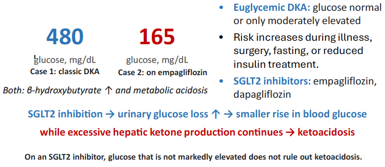
SGLT2 inhibitors increase urinary glucose loss, so blood glucose may remain only moderately elevated even when insulin action is insufficient to restrain excessive ketone production. Illness, surgery, fasting, and reduced insulin treatment can increase this risk. Rarely, patients taking these drugs develop euglycemic diabetic ketoacidosis (EDKA).
As in classic DKA, excessive ketone-acid accumulation causes metabolic acidosis, but without marked hyperglycemia.
Why can a patient taking empagliflozin develop ketoacidosis even when blood glucose is only moderately elevated?
Empagliflozin increases urinary glucose loss, limiting the rise in blood glucose. However, excessive hepatic ketone production can continue, causing metabolic acidosis despite the absence of marked hyperglycemia.
A patient has been fasting for two days. The liver produces ketone bodies to supply energy to other tissues, but cannot use them for its own ATP production. Why?
A. Hepatocytes lack mitochondria
B. Hepatocytes lack appreciable SCOT activity
C. Ketone bodies cannot cross the hepatocyte membrane
D. Hepatocytes lack mitochondrial HMG-CoA synthase
B
During an overnight fast, blood ketone levels increase, but blood pH remains approximately normal. What best explains why ketoacidosis does not develop?
A. The kidneys eliminate all ketone bodies produced during fasting.
B. Glucagon suppresses fatty acid release from adipose tissue.
C. Remaining insulin limits excessive lipolysis and ketone production.
D. Liver glycogen breakdown prevents ketone-acid formation.
C
A patient taking empagliflozin develops metabolic acidosis after surgery and poor intake. Glucose is 165 mg/dL; β-hydroxybutyrate is elevated. Which mechanism best explains why ketoacidosis can occur without marked hyperglycemia?
A. Poor food intake and a reduced insulin effect increase hepatic ketone production, while empagliflozin increases urinary glucose loss and limits the rise in blood glucose.
B. Empagliflozin inhibits HMG-CoA reductase, diverting HMG-CoA to ketogenesis
C. Empagliflozin increases glucose uptake into muscle and adipose tissue, lowering blood glucose while excessive ketone production continues
D. Empagliflozin blocks ketone-body use by the brain
A
What does the body use cholesterol for?

Cholesterol has several essential functions, so the body must maintain an adequate supply. The liver synthesizes cholesterol and helps regulate its distribution and elimination.
Because cholesterol is poorly soluble in water, it travels in blood within lipoprotein particles. Low-density lipoprotein (LDL) carries cholesterol to tissues, where it can be taken up by cells, including liver cells, through LDL receptors.
Elevated LDL cholesterol increases the risk of atherosclerosis. This is why drugs that reduce cholesterol synthesis or increase LDL removal from the blood are clinically important.
Why does the body need cholesterol even though elevated blood LDL cholesterol increases cardiovascular risk?
Cholesterol is essential for cell membranes and for producing steroid hormones, bile acids, and vitamin D. The clinical concern is excessive circulating LDL cholesterol, not the presence of cholesterol itself.
How is cholesterol made from acetyl-CoA?

Cholesterol synthesis occurs in the cytosol and at the endoplasmic reticulum. It requires acetyl-CoA, ATP, and NADPH.
As discussed in fatty-acid synthesis, mitochondrial acetyl-CoA cannot cross the inner mitochondrial membrane directly. Instead, its carbon is transported to the cytosol as citrate. ATP-citrate lyase then produces cytosolic acetyl-CoA, which supplies carbon for cholesterol synthesis.
Three acetyl-CoA molecules contribute to the formation of HMG-CoA. HMG-CoA reductase converts HMGCoA to mevalonate, using NADPH. This is the major regulated step in cholesterol synthesis and the target of statins. Mevalonate then undergoes several reactions to form squalene, which is converted to cholesterol.
Bempedoic acid inhibits ATP-citrate lyase, reducing the supply of cytosolic acetyl-CoA for cholesterol synthesis.
Why don't statins block ketogenesis?

Although HMG-CoA is an intermediate in both pathways, ketogenesis and cholesterol synthesis occur in different cellular compartments and use different enzymes.
During ketogenesis, mitochondrial HMG-CoA is cleaved to produce acetoacetate and acetyl-CoA. During cholesterol synthesis, HMG-CoA reductase converts cytosolic HMG-CoA to mevalonate.
Why does inhibiting HMG-CoA reductase not block ketogenesis?
Ketogenesis uses a separate mitochondrial pool of HMG-CoA and does not require HMG-CoA reductase.
How does cell cholesterol control synthesis and LDL uptake?

SREBP-2 is a transcription factor that helps liver cells respond to their cholesterol supply. When cellular cholesterol decreases, SREBP-2 increases the expression of genes involved in cholesterol synthesis and LDL uptake. This helps replenish cholesterol inside the cell. When cellular cholesterol is sufficient, SREBP-2 activity decreases, reducing production of both.
LDL receptors on the hepatocyte surface bind LDL particles and bring them into the cell, removing LDL from the bloodstream.
During statin therapy, SREBP-2 can increase HMG-CoA reductase production even though the statin inhibits the enzyme's activity. These effects are not contradictory: SREBP-2 increases how much enzyme is made, while the statin inhibits its function.
Why does a decrease in cholesterol inside hepatocytes increase LDL removal from the blood?
Lower cellular cholesterol activates SREBP-2, increasing LDL-receptor production. More LDL receptors allow the liver to remove more LDL particles from the bloodstream.

decrease
decrease
increase
decrease