a&p 1 - nervous system
 Call Kai
 Learn
 Practice Test
 Spaced Repetition
 Match
 Flashcards
 Knowt Play
1/83

There's no tags or description
Looks like no tags are added yet.
84 Terms
CNS
integrating, processing, and coordination
higher cognitive functions
brain and spinal cord
PNS
connects CNS to rest of body
delivers sensory info to CNS
carries motor commands to the body
cranial nerves, spinal nerves, and ganglia
sensory and motor divisions
nervous system functions
sensation - monitor external (room temp) and internal (core temp) changes
integration - process sensory input and determine response
response - by activating effector organs
sensory nervous system - in PNS
also called afferent system
Transmits information from receptors to the CNS
divides into somatic and visceral subcategories
somatic sensory
division of sensory nervous system in PNS
sensation from skn, muscles, bones, and joints
receptors for:
touch, pressure, vibration
pain, temperature
proprioception
special senses - hearing, vision, taste
visceral sensory
division of sensory nervous system in PNS
sensation from organs
mainly stretch, temperature, and pain
stomach stretching when you eat
somatic motor
a motor division in PNS
contraction of skeletal muscles
voluntary movement
somatic reflexes - involuntary contractions of skeletal muscle
visceral motor / autonomic nervous system
a motor division in PNS
regulates smooth and cardiac muscles, also glands
involuntary
parasympathetic and sympathetic division
what makes up nervous tissue?
neurons and glia
glia
support cells in nervous tissue
neurons roles
excitability - respond to stimuli
conductivity - produce and transmit electrical signals
secretion - release neurotransmitters
neuron features
cells in nervous tissue that have high metabolic rates (need constant oxygen and glucose) and live for a long time that do not go through mitosis
parts:
soma (body)
dendrites (receive signal)
axon (transmit signal)
axon terminal (communicate with target cell)
types of neurons
sensory (afferent)
interneurons
motor (efferent)
sensory (afferent) neurons in PNS
neurons that conduct signals from receptors and send to CNS
interneurons in CNS
neurons that integrate incoming signals and mediate responses
motor (efferent) neurons in PNS
conduct signals from CNS to effector
brain telling muscle to do something
neuron categories
multipolar -one axon and multiple dendrites extending from the cell body
pseudounipolar -a single axon that splits into two branches, bypassing the cell body
bipolar - cell body in middle with a long dendrite and then axon and axon terminals at ends
multipolar neurons examples
motor neurons and interneurons

pseudounipolar neuron example
sensory neurons

bipolar neuron example
special sensory neurons

dendrites
part of neuron that provide surface area for receiving signals from other neurons and conveys incoming signals towards the cell body
astrocytes - type of glial cells of the CNS
provides structural support
regulate extracellular environment
part of blood-brain barrier - projections from them help mediate what can leave blood and go into CNS
divide to replace damaged tissue since the neurons do not divide
formation of scar tissue
types of glia in the CNS
astrocytes
oligodendrocytes
microglia
ependymal cells
oligodendrocytes - glial cells of the CNS
wrap around axons to form myelin sheath
microglia - glial cells of the CNS
phagocytotic defense cells
ependymal cells - glial cells of the CNS
form and help circulate CSF
ciliated cells that go around the central canal
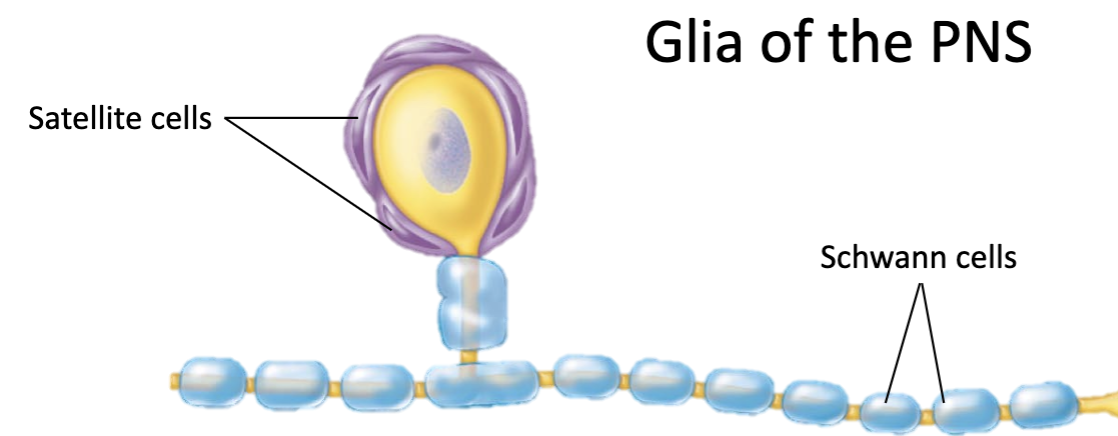
glial cells of the PNS
satellite cells
provide structural support
regulate extracellular environment
schwann cells
form myelin sheath - myelinate the CNS
help repair damaged axons
anatomy of a nerve
axon - surrounded by myelin sheath
fascicle - group of axons
nerve - group of fascicles with blood vessels
myelin sheath
electrical insulation around axons
plasma mebrane of schwann cells or oligodendrocytes
facilitates signal transmission
segmented
what are the segmented parts of a myelin sheath?
unmyelinated nodes or myelinated internodes
myelination in the PNS
begins prenatally
schwann cells spiral around a single axon
myelination in the CNS
oligodendrocyte processes wrap around multiple axons
begins postnatally
need enough lipids during childhood for these to be made
grey matter
somas, dendrites, and unmyelinated axons
white matter
myelinated axons
synapses
point of communication between neuron and adjacent cell (postsynaptic cell)
electrical
chemical
electrical role of synapses
gap junction joins adjacent cells and allows for rapid signal transmission
chemical role of synapses
neurotransmitters released from synaptic vesicles and are received by receptors on postsynaptic cell membrane
NTs
chemical signals that alter postsynaptic cell physiology
ACh, NE, GABA, serotonin
excitatory like ACh and NE often
GABA often inhibitory
can vary based on receptor type
membrane potential
differences between intra and extracellular charge caused by unequal electrolyte distribution between ICF and ECF
K+ is higher in ICF
Na+ is higher in ECF
resting membrane potential
what is the resting membrane potential at rest?
more polar → more negative internally
ligand-gated ion channels
ligand = chemical messenger
only open when specific ligand is bound (normally closed)
receptors on protein channel
chemical messengers must bind with their receptor for the channel to open
where are the receptors for a ligand-gated ion channel?
on a protein channel
Immediately after an action potential has reached its depolarization peak, what opens?
voltage-gated potassium channels
what are the kinds of ion channels?
voltage, mechanically, and ligand gated
voltage gated ion channel
channels that open or close in response to membrane potential changes
stimulated by action potentials
more voltage outside cell, but it increases (-70 to -50) when gates are open
local anesthetics
work by blocking voltage-gated Na+ channels to prevent depolarization
blocks afferent neurons from carrying sensory info to the brain
work non selectively and can impact efferent motor neurons leading to numbness and muscle weakness
examples include novocaine and lidocaine
mechanically gated ion channels
open in response to mechanical stimulation
pressure and stretch
close when stimulus is removed
important for things like sense of touch
sequence of membrane potential changes
resting potential
depolarization
repolarization
hyperpolarization
what happens when ions move in the electrochemical gradient?
it alters the membrane potential
depolarization
increase in internal cell charge
repolarization
return to RMP
hyperpolarization
decrease in internal charge below RMP - linked with refractory period by making it longer
local potentials
small and local changes in membrane potentials
response varies with stimulus strength
length of stimulus, number/type of ion channels that open
reversible - neurons turn to RMP if stimulus is too small/short
effects get weaker with distance
where do local potentials take place?
at dendrites or somas but they do not reach axons

signal integration
summation - combined effect of multiple local potentials trigger action potential
temporal summation
spatial summation
temporal summation
frequent stimulation of the same location
multiple APs from a single presynaptic cell

spatial summation
simultaneous stimulation of many locations
APs from multiple presynaptic cells

action potentials
all or none
irreversible
maintained along length of axon
self propogating
all or none - APs
proceeds only if threshold is reached
irreversibility of APs
continues to completion once threshold is reached
APs across distance
they are maintained along the length of an axon to allow signals to be sent over long distances
self propagating - trait of APs
meaning that each one triggers the next
AP generation
NTs open ligand-gated Na+ channels
local depolarization of cell membrane → local potential
summation opens voltage-gated Na+ channels
full AP triggered if depolarization reaches axon
rapid depolarization of axon
Na+ channels close, K+ channels open
rapid depolarization of axon in AP generation
regional depolarization triggers adjacent Na+ channels - positive feedback loop
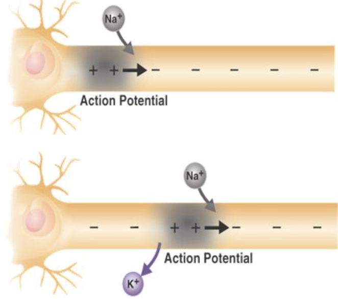
during AP generation Na+ channels close and K+ channels open at one point, what does this lead to?
the outflow of K+ repolarizes the cell
what happens at the axon terminal during the end of the AP?
voltage gated Ca2+ channels open
what happens that opens voltage gated Ca2+ channels?
depolarization of axon terminals
what happens after Ca2+ flows in at a chemical synapse (axon terminal)?
it triggers exocytosis of neurotransmitters which then bind to receptors on postsynaptic cell
finally is signal cessation
signal cessation at axon terminal during AP
presynaptic cells stop NT release
NT cleared away from postsynaptic receptors
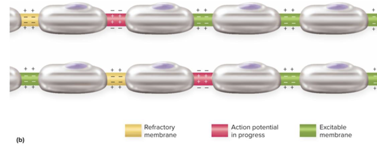
refractory period
resistance to new stimulation
either absolute or relative
local effect - other parts of neuron can be stimulated
maintains unidirectional movement of AP toward axon terminals
absolute refractory period
time in which no stimulus can trigger another AP, comes before other period - red here

relative refractory period
time in which only a very strong stimulus can trigger another AP and this period lasts until restoration of RMP - comes after other period - yellow here

Na+/K+ antiport
antiport is a membrane protein
exchanges K+ ions for Na+ ions
K+ moves into cell and Na+ moves out
important for maintaining electrochemical gradients
but, requires ATP
continuous signal conduction
unmyelinated fibers
voltage-gated channels along entire length of axon
opening of Na+ channels causes local depolarization → excites adjacent Na+ channels
initiates new AP distally
wave of successive APs continues down length of axon
ensures unidirectional spread of the AP

what kind of channels are in continuous signal conduction?
voltage gated channels which are along the entire length of axon
what are the two types of signal conduction?
continuous and saltatory
saltatory signal conduction
myelinated fibers
myelin insulates axons - APs only generate at nodes
signal seems to “jump” from node to node
faster than the other kind of conduction
is saltatory signal conduction myelinated or unmyelinated?
myelinated - segmented myelin sheath causes signals to have to “jump” from node to node - APs only need to be generated at unmyelinated segments
is saltatory or continuous signal conduction faster?
saltatory
signal conduction speed
myelination increases speed of conduction
faster in larger fibers
small unmyelinated fibers < small and myelinated < large and myelinated
multiple sclerosis
autoimmune destruction of oligodendrocytes → demyelination of CNS neurons
interrupts saltatory conduction
common symptoms: muscle weakness, numbness, visual impairments, speech deficits
no cure but anti-inflammatories and pt can help
nerve regeneration
only possible in PNS + if soma is intact
fiber distal to injury degenerates because macrophages clear away debris
axon stump sprouts multiple growth processes
schwann cells form regeneration tube
single growth process continues to growh
schwann cells release gH
regeneration tube guides growth
synaptic contact re-establishes
re-myelination over time
can take up to 2 years
axon stump
forms earliest formation of newly growing axon, called growth processes
what do schwann cells do in nerve regeneration?
it forms the regeneration tube and dominant growth process continues to grow because this cell releases growth factors and the tube guides it toward the target cell