PCB3713C - Final
 Call Kai
 Learn
Learn Practice Test
 Spaced Repetition
 Match
Match Flashcards
 Knowt Play
1/106

There's no tags or description
Looks like no tags are added yet.
Name | Mastery | Learn | Test | Matching | Spaced | Call with Kai | Chat |
|---|
No analytics yet
Send a link to your students to track their progress
107 Terms
Circulatory System Function
Transport system of nutrients, wastes, and others
Driving Pressure
Pressure difference of two points in a vessel causing flow of blood
Transmural Pressure
Pressure difference between the inside and outside of the vessel
Cardiac Output
Volume of blood pumped by the heart per unit of time. 5L/min
Hagen-Poiseuille Equation
Equation that equates flow to the change of volume, fluid viscosity, and time
Turbulent Flow
Noisy flow in a tube due to viscosity and driving pressure. Calculated using Reynolds number (Re)
Capillary Exchange
Movement of a solute through the capillary due to concentration and permeability
Fick’s Law
The rate of diffusion is proportional to how steep the concentration gradient is
Transcapillary hydrostatic pressure difference
Difference in hydrostatic pressure driving water out the capillary
Oncotic pressure difference
Difference in oncotic pressure in the capillary that attracts water in the capillary
Net Filtration Pressure (NFP)
Driving force across a capillary by hydrostatic and oncotic pressures
Lymphatics
System that collects fluid filtered by capillaries that are not reabsorbed
Sinoatrial Node (SA Node)
A pacemaker cell at the top of the heart that regulates hearbeat
Atrioventricular Node (AV Node)
Cell that delays and relays the electrical signal form the atria to the ventricles
Na+ Current
Inward current responsible for rapid depolarization of the heart
Ca2+ Current
Inward current responsible for slow depolarization of the heart
K+ Current
Outward current responsible for repolarization of the heart
Abnormal Cardiac Rhythms
Arrythmias of the heart that result in issues with conduction or automaticity
Atria
Blood receiving chambers of the heart
Ventricles
Large blood pumping chambers of the heart
Cardiac Valves
Passively opens to allow blood to flow one way based on pressure difference
Ventricular Filling
AV valves open to allow blood to fill
Isovolumetric Contraction
Heart contracts to increase pressure. Valves are still closed.
Ventricular Ejection
Semilunar valves open to eject blood out the heart
Isovolumetric Relaxation
Valves close to decrease pressure in the heart
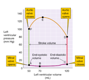
Pressure-volume loop
Diagram depicting the pressure and volume in the LV at certain times of the heart
Mean Arterial Pressure (MAP)
Pressure exerted on the arteries that is universal along the entire body
Neural MAP Regulation
Use of neural pathways for short-term control of MAP
Baroreceptors
Stretch-sensitive mechanoreceptors in the blood vessel walls that increase firing rate based on MAP
Chemoreceptors
PO2 and PCO2 receptors that cause a change in heart rate and vasoconstriction
Low-Pressure Baroreceptors
Stretch-sensitive mechanoreceptors that detect changes in blood volume and causes changes in cardiac output
Integrated Cardiovascular Homeostasis
Vasoconstriction and other cardiovascular adjustments due to physical and mental changes
Hemorrhage Compensation
Loss of blood causing the body to vasoconstrict and start quick RBC production to return to normal MAP
Nephron
Function filtration unit of the kidney
Bowman’s Capsule
A nephron structure of a nephron that surrounds the glomerulus and collects fluid filtered from blood
Glomerular Filtration Barrier
Fenestrations in the glomerular membrane that helps solutes permeate the membrane
Glomerular Filtration Rate (GFR)
Volume of fluid filtered into Bowman’s capsule per unit of time
Ultrafiltration
Process of separating solutes by forcing a solution through a semipermeable membrane
Starling Forces
Fluid forces (Hydrostatic forces and oncotic force) that dictate the filtration of fluid
Renal Blood Flow (RBF)
Blood flow through the renal system. Dictated by constriction of arterioles
Peritubular capillaries
Renal tubules that reabsorb fluid and solutes in the interstitial space
Myogenic Constriction
An increase in pressure causes Ca2+ release that leads to the smooth muscle to contract and resist pressure
Renin-Angiotensin-Aldosterone Axis (RAAS)
A hormonal system activated by low blood pressure or low sodium that increases blood volume and blood pressure through vasoconstriction (angiotensin II) and sodium/water retention (aldosterone
Kidney Na+ Reabsorption
Reabsorption of Na through the proximal tubule and loop of Henle
Transcellular Reabsorption
Ion reabsorption through facilitated diffusion across the apical membrane and an pump out the cell. Remember the charges.
Apical Cotransporters
Usage of a downhill electrochemical gradient to uptake another solute uphill in the same direction
Apical Exchangers
Usage of a downhill electrochemical gradient to uptake another solute uphill in the opposite direction
Aquaporin (AQP)
Channel that allows for water to more readily permeate a membrane
Urea Transport
Passive movement of urea through the nephron to aid in water reabsorption
Glucose Reabsorption
Reclaim of filtered glucose in the Proximal Tubule
Potassium Kidney Transport
Highly regulated potassium is filtered and only essential amount of it is reabsorbed
Water diuresis
High water volume causing kidneys to produce a large volume of dilute urine
Antidiuresis
Low water volume causing kidneys to produce small volume of concentrated urine
Kidney Water Transport
Use of salts to passively move water across membranes
Interstitial Osmolality
Increase in osmolality on the inner side of the medulla due to high Na, Cl, and urea concentrations
Urea Recycling
Reabsorption of urea due to the reabsorption of water causing urea concentration to increase
Bicarbonate
Blood pH buffer used to neutralize acids
Urine Acid Secretion
Excess H+ ions are excreted into urine
Bicarbonate Reabsorption
Secretion of H+ into urine consumes bicarbonate, which causes a new bicarbonate to appear in blood
Respiratory Acidosis
Production of bicarbonate to compensate for increased H+ secretion
ECF Volume
Amount of fluid in the extracellular matrix and primarily controlled by adjusting the total-body NaCl content
ECF Osmolality
Amount of salts in liquid in the extracellular matrix, regulated by adjusting the total-body water content
Hormones
Signaling molecules released by an endocrine tissue into the extracellular fluid
Carrier Proteins
Allows for hydrophobic hormones such as steroid and thyroid hormones to move in the blood
Hypothalamic-pituitary axis
Hypothalamus control over the production of pituitary hormones responsible for growth, stress, reproduction, and metabolism
Tropic Hormones
Gland stimulating hormone to produce further hormones
Trophic Hormones
Growth promoting hormones
Anterior Pituitary
Produces and releases tropic hormones responsible for stimulation of other glands
Posterior Pituitary
Releases AVP to control water balance, and oxytocin
Peptide Hormones
Hormones responsible for signal-transduction systems
Amine Hormones
Small, amino acid–derived hormones that typically act rapidly via second messengers (catecholamines) or alter metabolic rate (thyroid hormones)
Steroid Hormones
Fat-soluble hormones that change gene expression for slow, long-term effects (e.g., cortisol, aldosterone, sex hormones)
Thyroid hormones
Lipid-soluble amine hormones that boost metabolism and support growth and development
Somatotrophs
Growth hormone secreting and producing glands
Thyroid Receptors
Nuclear receptors that alter gene transcription by diffused thyroid hormone
Hypothalamic-pituitary-thyroid axis
Synthesis and secretion of thyroid hormones through a cascade of TRH, TSH, and thyroid hormones
Hypothalamic-pituitary-adrenocortical axis
CRH → ACTH → Adrenal Cortex; the body's main stress hormone pathway
Renin–angiotensin–aldosterone (RAA) axis
Renin → angiotensin I → angiotensin II → aldosterone; increases blood pressure and blood volume.
Islets of Langerhans
Endocrine glands of the pancreas
Pancreatic A Cells
Cells that secrete glucagon
Pancreatic B cells
Cells that secrete proinsulin and insulin
Pancreatic D Cells
Cells that secrete somatostatin
Glucagon
Increases blood glucose by stimulating glycogenolysis and gluconeogenesis
Parathyroid Hormone (PTH)
Increases blood calcium via bone resorption, kidney Ca²⁺ retention, and vitamin D activation
Intestine Wall Anatomy
Structured with a mucosal epithelial layer with layers of smooth muscle outside, and then complete with connective tissue
Digestion
Conversion of food into absorbable products
Absorption
Intake of food and nutrients into the body
Enteric Nervous System (ENS)
A specialized division of the autonomic nervous system embedded in the GI tract that can control digestion independently, regulating motility, secretion, and blood flow
Brain-Gut Axis
Bidirectional system of ENS, GI Hormones, and Immune System components that control GI function
Stomach
A muscular organ that stores, mixes, and digests food using acid and enzymes (HCl, pepsin), protected by mucus, and regulates emptying into the small intestine
Parietal Cell Acid Secretion
CO2 from the basolateral membrane catalyze to form bicarbonate and H+. Results in active transport of HCl across the apical membrane
Pepsinogens
Chief-cell enzyme precursor activated by acid into pepsin for protein digestion. Regulated by ACh
Gastric Acid Neutralization
The use of bicarbonate to neutralize stomach acid prior to the duodenum
Pancreas
Secretes juices that neutralize gastric acidity and secretes digestive enzymes (proteases, amylases, lipases, and nucleases)
Villi
Intestinal microprojections that aid in nutrient and electrolyte absorption. Increases surface area for absorption
Crypts
Base of villi that aid in electrolyte and water secretion
Intestinal Apical Na Absorption
Facilitated transport downhill using the electrochemical gradient
Intestinal Basolateral Na Absorption
Active transport into the ECF using Na-K pumps
Bile
Fluid produced by the liver that aids in lipid digestion and allows excretion of liver byproducts
Sex Determining Region Y (SRY Gene)
A gene on the Y chromosome responsible for determining sex