PNB exam 3
 Call Kai
 Learn
 Practice Test
 Spaced Repetition
 Match
 Flashcards
 Knowt Play
1/132

There's no tags or description
Looks like no tags are added yet.
133 Terms
nervous system consists of 2 major parts what are they
what is the first part made out of
what is the second part made out of
CNS/central nervous system
-brain and spinal cord
PNS
all other nervous tissue (spinal and cranial nerves ganglia)
extra: 97 percent of nervous tissue is in the brain and 3 percent is in the spinal cord

what groups of neuronal cells exist in the Central nervous System
the central nervous system has what kind of matter
what are in the bundles of axons for the central nervous system
white matters consists of what of the cell body in CNS
neural cell bodies
nuclei(neurons) /grey matter
part of the white matter-called tracts or fibers
axons
extra: neurons=grey matter
in the peripheral nervous system/PNS what are the groups of neuronal cell bodies
bundles of axons in PNS are
ganglia
nerves coming off the spinal cord or cranial nerves coming off of cranium
extra: dorasal root ganglia, ont he side of the spinal chord, consists of the cell bodies of the sensory neurons
white matter in bottom left, the rest is grey matter
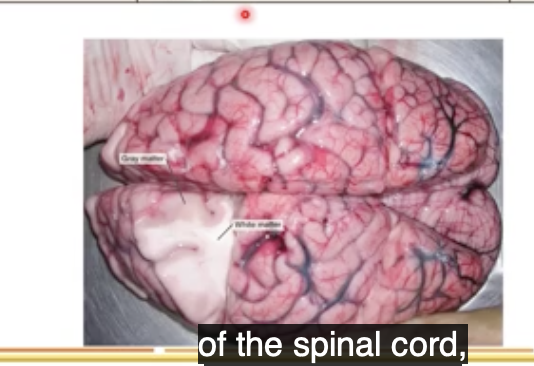
major cell types in CNS pt 1
name all 5 of them
what does the first one do
what 4 things make up glial cells
what are astrocytes , what do they support, what do they create, waht two other things do they do ,
neurons, astrocytes, microglia, oligodendrocytes, ependymal cells
neurons-receiving, processing, integrating and transmitting signals between cells
astrocytes, microglia, oligodendrocytes, ependymal cells
nerve cells, blood brain barrier, ion homeostasis, synaptic formation
extra: Astrocytes=glial cells

what are microglia
they have blank functions, they are also activated in blank or blank, and they recast lot’s of blank factors in the enviro
what is the main function of onigodendoctyes
what is the function of ependymal cells: these are cells that blank blank, and are improtant for blank
macrophages of the brain
protective, inflammation or injury, toxin
production of the myelin sheath around axons
line ventricles, CSF(cerebrospinal fluid)
what is the protection that is most outwards of the brain called
under skin what comes next that is good protection of the brain
the brain has how many meninges , also what are they also called
dura matter: what is it closest to, describe it’s thini/thickness
what is the layer after dura matter called, describe it’s structure
what layer comes after this one/is also close to grey matter of the brain, is it thick or thin
skin
cranial bones(flat bones)
3-(connective tissue)
the bone, thickest membrane
arachnid-more mesh like
Pia matter-thin matter
extra: 7 mm thick
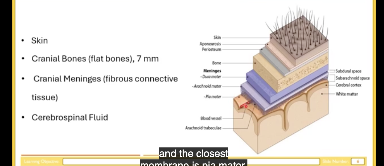
the subarachnoid space is the space between what two structures
what does it contain what does this thin provide
what does this thing also do
space between arachnoid and Pia matter
cerebrospinal fluid
additional protection of the brain from crashing toward the cranial bone
decrease weight of the brain
extra: (brain is inside of the skull, swimming in CSF, )
extra: if not in cerebral spinal fluid, is much bigger weight
brain Meningie:
match these
dura matter, arachnoid, and Pia matter
with thin fibrous and filamentous men, thick fibrous, and thin fibrous
where do you find most blood vessels (of the 3 meninges)
brain Meningie
dura: mater: thick fibrous
arachnoid: thin fibrous and filamentous mesh)
pia mater: thin fibrous
in arachnid and Pia though
extra:
pia=only microscopically seen, sometimes can’t distringioush arachnoid and Pia mater
big mesh of blood supply and blood vessels in dura ,

diseases which can be caused by infection of meninges
what is bacterial meningitis caused by
what is viral meningitis caused by , is it more or less serious than bacterial meningitis
what can also cause meningitis
bacterial infection, can cause stroke, hearing loss, permanent brain damage, early treatment of this involves antibiotics, which are important to be admitted as soon as possible can cross blood brain barrier and decrease the symptoms of the disease
enteroviruses, less serious
final infections
left and right side of brain
top of the brain-protection of skin, then bone
what is dipole
dipole has 3 layers, what are the 2 layers made out of, what is the one layer made out of
what is the thickest membrane out of the 3 meningitis
where does CSF circulate, where is it drained
bone/scalp obones=flat bones
2 compact bone, 1 spongy bone
dura mater-thickest membrane
subarachnoid space, superior sagittal sinus

cerebrospinal fluid is produced by what two things
what is the main function of the cerebrospinal fluid
out of the two, which one produces most of the CSF
what are ependymal cells contained in/
the choroid plexus (here in red)+ eppendymal cells
to support the brain
ependymal cells(they make the choroid plexus)
(here in purple)
the choroid plexus (in the ventricles)
extra: and average brain is between 1400-1600 grams, suspended in cerebral spinal fluid but when it’s suspended in cerebral spinal fluid, it’s 60 grams on the body


what are capillaries lined with
what wraps around endothelial cells, what layer is then outside of the one wrapped around them
out of CSF and plasma which one is more diluted/has more water
what are the ONLY two things that CSF has more than plasma of
what is the one thing that CSF and plasma have equal parts of
why is it good that CSF has less protein concentration than plasma /is a good thing
endothelial cells,pericytes, then ependymal cells
in CSF
CSF
water, sodium and chloride-for excitability of cells
osmolarity
bc it decreases the ability of infections and proliferation of microorganisms in cerebrospinal fluid

another 20 percent of CSF is not produced by choroid plexus, it is produced by blank blank blank
what is it
CSF flows through the subarachnoid space of the brain AND blank blank
when it is produced in the choroid plexus, it is released from the blank blank to the blank blank, then the blank blank to the blank blank. it passes through the blank blank of the blank blank then it will be reabsorbed in the blank blank sinus
superior sagital spines-have specific blank blank which allows it do do what
brain interstitial system
fluid filled space between neurons and capillaries
spinal cord
lateral ventricle, third ventricle, cerebral aqueduct, 4th ventricle,, subarachnoid space, spinal cord superior sagittal
arachnoid vili, reabsorb CSF and drain them in the dural venous sinuses

cerebrospinal fluid= is a blank source of different diseases by lumbar puncture in the blank spinal chord
the lumbar spinal chord does not contain what , what does it contain -what is it called
majority of CSF is produced where, some is also where
diagnostic, lumbar
spinal chord, spinal nerves called-a caudal equina
in the choroid plexus of the lateral ventricle, 4th ventricle
extra:
take sample of CSF, analyze for cell count, glucose level, protein level, can detect some other features like oligoclonal bands-useful for diagnostic purposes of multiple sclerosis,
can detect other diseases like meningitis neurosyphilis and encephalitis-IS improtant
why is the blood brain barrier improtant
what is one of the cells that form blood brain barrier/BBB
astrocytes have special blank, wehre, what are they called
what do they do
capillaries have blank cells, what kind of junctions , what does this prevent
what is the second cell that is a key part of the blood brain barrier
what 3 things makes up the neuromuscular unit
-protection against pathogen
astrocytes
feets on end of processes called astrocyte foot processes
firmly attached to capillaries present in the brain
capillaries have endothelial cells, tight junctions, prevent paracellular transport
pericytes
pericytes, capillary endothelium and astrocytes
extra: tight junctions=v tightly bond between each other, entry of diff toxins across cells
blank blank =v improtant part of neruomusclar unit due to complex blank blank, which prevent/control blank blank (keep toxins out of body)
in order to pass the BBB, you have to be blank, blank molecules or have specific blank to pass through
BBB enables a low blank enviro to prevent blank like microorganisms
endothelial cells, tight junction, molecular traffic,
small, lipophilic, transporters
low protein, proliferation
for brain to function we need what molecule
which molecule passes freely across BBB
what is one of the metabolic needs of the brain for energy source:
there is a high concentration of glucose in the plasma and brain will consume that glucose and abt how much of the bodies glucose is consumed by the brain
oxygen
oxygen
glucose,
half/a lot!
extra: brain receives around 15 percent of the blood pumped by the heart at res
what moves glucose from the plasma into the brain interstitial fluid and enable energy
what is progressive hypoglycemia
membrane transporters
low concentration gf glucose in the blood
extra: causes-confusion,unconsciousness and death)
blood supply of brain
main blood supply of brain = is what
from aorta, to the front of brain anterior, we have blank blank blank
which branch into external and internal carotid arteries
which ones passes into the brain and branch to several blood vessels in the brain
the internal carotid arteries give rise to the blank and blank arteries
aorta
common carotid arteries, carotid artery
and internal carotid artery
anterior, cerebral

vertebral arteries supply what part of the Brian
what does it supply to that part of the brain
what is the circle of wills
what do they prevent the loss of
if there is occlusion of one part of the blood supply, what happens
back of brain (they’re posterior)
oxygen
circular arrangement of the anterior and posterior blood vessels
functions
alternative root supply can provide oxygen to that effected brain
extra: they supply the brain /brain stem and some cerebellum and some of the posterior structures of the bran and occipital lobe of the Brian

blood =returned by venous system, by the …
internal jugular vein exits through the blank blank of the blank bone
to return the blood to the arterial system
main artery=jugular vain
jugular foramen , occipital

cerebrum:
fifth lobe deep buried below frontal, temporal and parietal lobe is called what
what is the cerebrum important for
what is diencephalon important for
the brainstem consists of what three things
what is the brainstem important for
cerebellum is important for what
cerebrum:
insulin
higher brain functions
homeostatic controls
midbrain pons and medulla,
autonomic reflex center-most functions for survival=contorlled by Brains stem
cerebellum=coordination of movement
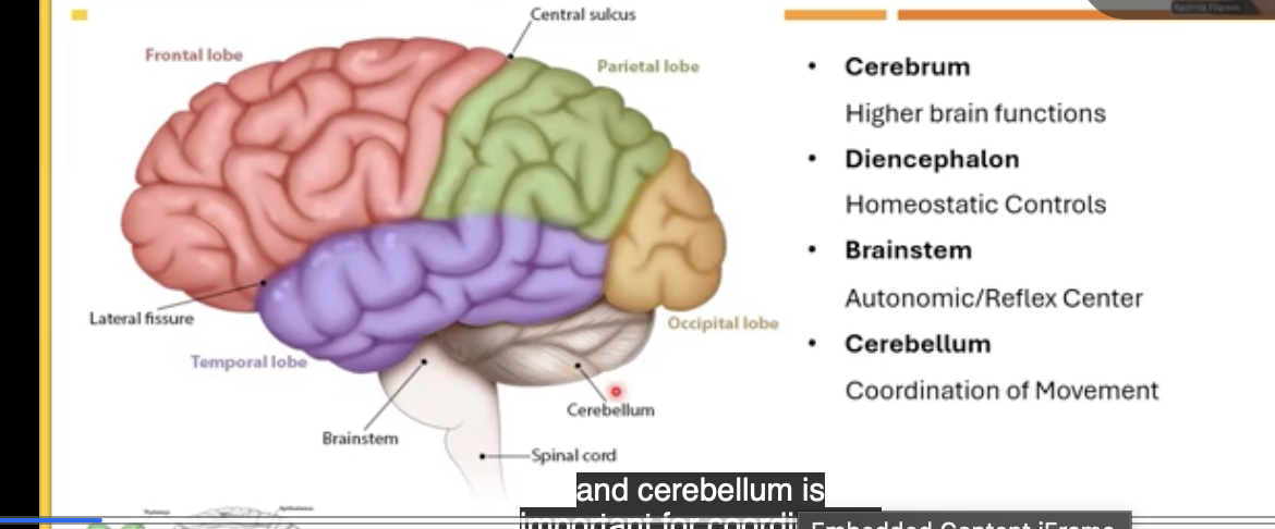

main landmarks of cerebellum
posterior view=left
longitudinal fissure divides what
transverse fissure divides what two structures
lateral fissure divides the lobes such as
the central sulcus divides what two lobes
the motor cortex is in front of what structure
behind the central sulcus you have what cortex
left and right hemispheres
divides cerebrum and cerebellum
frontal, partietal/temporal lobe
frontal from parietal love
the central sulcus
the somatosensory cortex

5 lobes of cerebrum
there is contralateral control: left cerebrum controls what side of the body and right cerebrum controls what side of the body
frontal cortex: below the frontal bone in the skull in the left hemisphere: it is a place where there is a blank blank
what main 4 things is it improtant for
5 lobes of cerebrum
right side of the body , the left side of the body
motor cortex (in precentral gyrus)
improtant for speech, memory, personality, and emotion


2nd lobe
parietal cortex/lobe
it is the blank cortex
it’s positioned where in comparison to the central sulcus/central gyrus
the occipital lobe has what cortex, it is important for blank processing and storing blank blank
somatosensory
posteriorly
occipital cortex- has visual cortex
visual , visual memories
the temporal lobe is important for what 4 things
the fifth lobe/insulin (a separate lobe) when you cut parts of frontal, parietal, and temporal loves, there is insula (island)
what is insula improtant for control of(4 things)
it is also improtant for processing info of the sensation of waht two things
hearing, speech, language, and smell
blood pressure, heart rate, digestion, vital autonomic nervous system
smell and taste


cortex in cerebrum is organized in horizontal layers
left side=sagital view of cortex: diff complexity of cortex I diff regions of cerebrum
left=front, right=back in blue
what three structures make up the neocortex
how many layers do those areas have of the cell
what is the pyriform cortex important for sensation of , how many layers does it have , what is it called
parietal cortex and visual cortex, motor cortex
6
smell, 3, paleocortex

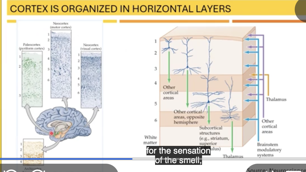
looking ont he RIGHT
the first layer: compare the levels of cells it has
the second and third layer are improtant for blank between blanks and blank areas
layer 4 does what, which one sends lot’s of info to this layer
layers 5 and 6 are responsible for what
waht are the 3 subcortical structure
sparse with cell
communication, hemispheres, cortical
receives info form other cortical areas ,thalamus
sending info from cortex to other subcortical structure like
striatum brainstem, and thalamus

3 major planes of sectioning: name them
frontal/coronal divides brain into what regions
transvere/horiontla divides brain into what two regions
lateral/sagital plane divides brain into what regions
coronal plane, horizontal plan, sagittal view
front/anterior and back posterior (occipital is back in coronal)
upper/superior and lower inferior
left and right regions

grey matter is what
grey matter has a blank appearance with cell bodies around the blank
dark/grey matter/basal ganglia nuclei have what level of cell bodies, it is where
cell bodies of the neurons
darke, cerebrum
a lot of cell bodies, deep inside hemisphere, (not in the top)
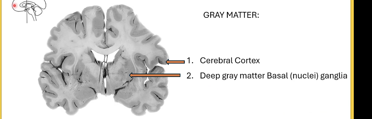
white matter
white matter consists of what , coming out of what
how many major white matter bundles and fibers do we have coming from the cerebral cortex and cerebrum and out to sub cortical regions OR fibers that connect different sides of the hemisphere
what are the names of the 3 fibers
what do the CF do
PF: are bundles of fibers close to blank blank. they are fibers that connect blank to other regions like blank regions
AF: define it
axonal fibers coming out of the cell body of the neurons
commissural fibers, projection fibers , and association fibers
connect the left and right hemispheres
basal ganglia, cerebrum, subcortical
connections within cerebral hemisphere (adjacent gyro too)
subcortical regions liek: Brain stem, spinal cord, the end

Geri: ridges or bumps
the brain has some or lots of them
the brain has few or many folds
what is the advantage of folds, what does it increase
what does an increase of this also cause
this also contributes to more blank control of the sensory motor system and more blank synaptic blank
has lots of ridges/gyri
convoluted, has lot’s of folds
surface area of the cortex, convoluted
more connections,
specific , neuronal , connections
extra: gyrus=singular , gyri=plural, grooves+sulci or sulcus singular-gives Brian cauliflower look
human cortex=highly convoluted to other animals
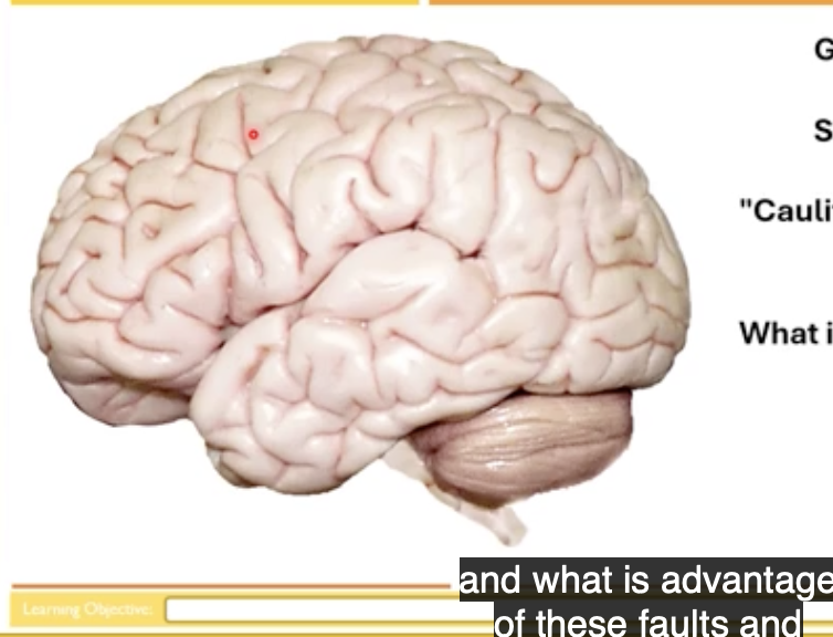
what is the EQ/encephalization cquotient used to compare
the actual size of the brain to the expected brain size of that specific body weight
extra:
ex: human have the highest EQ of any animals, Brian is much larger than expected for mammal of our size
if compare diff neuronal density in diff regions, find conclusions cool
cerebellum makes up what percent of the brain mass, but consists of what percent of the brain neurons, this area is also highly blank, and increase in surface area
cerebral cortex makes up what percentage of brain mass, but has what percentage of brain regions,
what structure has the highest number of neurons/hghest percentage of neurons
what structure of the brain has highest brain mass
ten percent , 80 percent, convoluted
80 percent , 19 percent
cerebellum
cerebral cortex

with maturation, we have Increased blank in human brain
we are born with certain number of neurons, does it change /increase as we age
as we age, we gain more blank between neurons
connectivity
no
connectivity

frontal lobe of cerebrum/cortex is important for what 3 things
frontal love also contains what cortex, what’s it important for
this cortex plays an important role in what two things
hypothesis generation
planning/organizing strategy
personality
motor cortex imprint for motor control
interpersonali social skills, higher level of intelligence

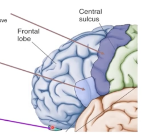
frontal lobe is part of the lateral view
(motor cortex is in front of central sulcus in blank gyrus)
by stimulating this region, your body will move in that particular part, if region is controlling hand, muscles of which hand will move
left cortex has what , what is it important for, injury of it causes a disease known as what, it’s improtant for what
in the top frontal lobe in the inferior part, you have what structure, what is it important for
prefrontal
the hand on the opposite side of the brain that’s being stimulated
Broca’s area, speech control, aphasia + speech
olfactory bulb, odor perception
extra: olfactory buble is not that prominent for humans
but sheep depending on smell for surviival, there’s is larger

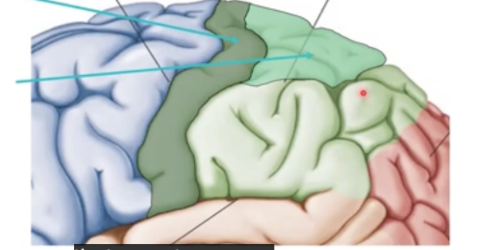
parietal in green
central slices, parietal lobe is improtant for what two things
it’s also important for what two things
what is the dark green structure called
the frontal lobe contains what cortex
in the parietal lobe (posterior part) we have what cortex
what is it improtant for processing
sensation and sensory integration
spacial awareness and perception
primary somatosensory cortex
the motor cortex
somatosensory association cortex, for processing sensory information
extra:
if you receive stimulation on certain body parts, will have or can have high activity of neurons in the somatosensory cortex in opposite side


somatotopic organization of our body
there are cartoons called motor and sensory homunculus
in the middle homunculus looks like small body with huge hands, huge lips, huge hands but small trunks and small legs
the cortex area for processing sensation and controlling movement is equal or not equal across body parts?
what 3 body parts get large areas?
what does contralateral representation mean
not equal
hands, face, and lips
the right cortex receives insinuation from the left side of the body and the left motor cortex controls movement on the right side


temporal lobe is divided by central sulcus from frontal and parietal lobe
temporal lobe contains what cortex , what is it improtant for
the left hemisphere of the temporal lobe-superior temporal gyrus has what area,
what is it important for
what happens if you have damage here, you will be able to do what, but language won’t have what
the temporal lobe also have blank formation and blank retrieval in blank
the hippocampus area is apart of what cortex, and it’s where
auditory cortex, hearing
Wernickes area
language comprehension, speak (no motor impairment), appropriate meaning
memory memory retrieval, hippocampus
limbic cortex, deep in the emporal lobe


image shows location of primary cortex
and association cortex
image shows location of primary cortex
and association cortex
have wernickes area in that area-language comprehension in the left hemisphere

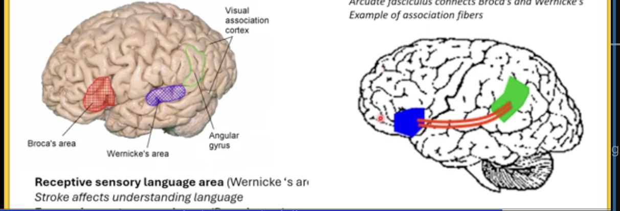
location of braces and wernickes area
brocas area also is the blank blank speech area
what happens if you damage brooks area , what does it affect
having a stroke or damage to wernickes area effects what 2 things
these tw areas are blank via arcuate fascicles
association fibers are connected in….
expressive motor speech area
- expressing language, face will have trouble expressing
understanding language and producing meaningful language
connected, (example of association fibers,)
diff areas of 1 side , both left temporal

occipital lobe does what
what cortex is present here -what does it do, through what
processing, integration, interpretation of vision and visual stimuli
secondary /association visual cortex: processing, interpreting info, the primary visual cortex
primary visual cortex : will achieve info coming from the…
all the interpretation of visual info goes through what
eyes and visual pathways and then form two dimensional sketch of the subject
the secondary or visual association area
insula: fifth lobe
has different and how many separations f anterior and posterior
what is the fifth lobe involved in
what is it’s functions 5 tings
it’s a complex function-it receives what
blank and two different ones
consciousness, role in emotions, interoceptions of receptive info from internal organs, regulation of the body homeostasis,
functions: compassion, empathy processing of sensation taste, smell, awareness of hunger, pain, fatigue,
info coming from diff parts of the brain
connect superior, posterior, inferior, and anterior to how it’s translated when talking about the brain
translate this to when you’re talking abt the brainstem and cerebellum
ex: posterior cortex is superior to corpus collosum

repeat for brainstorm and cerebellum
brainstem and cerebellum: diff terminology
rostra, dorsal, ventral, caudal
cerebellum is dorsal to the brainstem
midstram is rostral to the pons
pons is rostral to medulla

the basal nuclei is considered what
they are groups of blank blank deep inside the blank blank
what are the 3 major parts of basal nuclei
what 4 things ar ether important for
the main parts function in what
what two things does the basal nuclei work with
deep grey matter/grey matter
cell bodies, cerebral hemispheres
caudate nucleus, putamen, globes pallidus
motor control, mood, some control of emotions, semi automatic motions and sub conscious control
modulating(regulating) motor movements
pre central gyrus and cerebellum
what are the 3 major regions of diencephalon
which one is called the pineal gland
which is the biggest part
epithalamus, thalamus, and hypothalamus
epithalamus(pineal gland) -posterior/superiorr
thalamus(biggest part) and superior
hyptohalamus-infreior/anterior
extra:
sagittal view of brain

what is the epithalamus important for
secretion of melatonin (hormone that helps regulate day night cycles, circadian rhythm)
jet lag-melatonin helps to overcome that problem
extra: eptihalamus: -highlighted in red (sagiral view of brain)-in middle of cerebrum
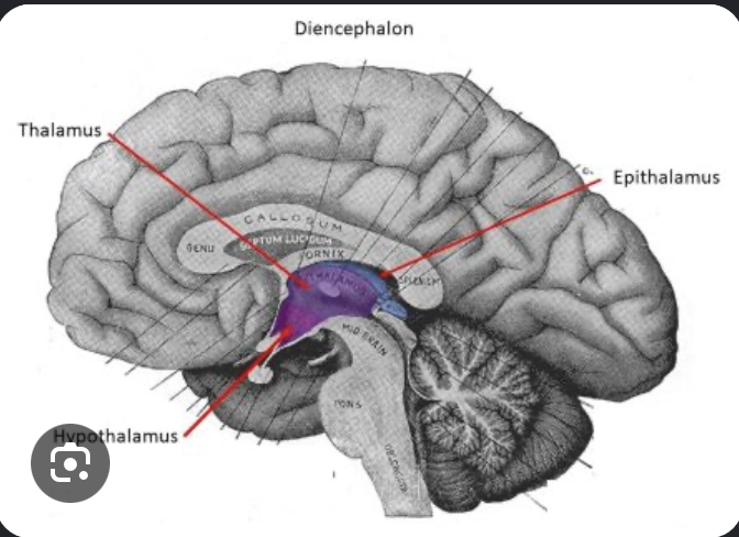
the central part of diencephalon-biggest Parth is known as what
it has a lot of what , why?
v complex structure, many diff types of blank , important for processing of blank and blank info and higher blank as well
between left and right thalamus what do we have
thalamus
routes- so the sensory and motor signals can get to the rest of the brain neurons, sensory, motor, functions between left and right
third ventricle
Extra
thalamus is also called the /switchboard or relay station


another region of the diencephalon is called the hypothalamus
it’s inferior to the thalamus and anterior to the epithalamus
hypothalamus has complex functions, it regulates many blank mechanisms , it’s the master control of the blank blank blank
also the master control of what system, connected to what
what 4 things does the hypothalamus also do
another region of diencephalon is hypothalamus
homeostatic, autonomic nerves system(regulation of sympathetic and parasympathetic nervous system)
endocrine system, pituitary gland (both anterior or posterior)
regulation of body temp, control of emotional behavior, food and water intake, regulation of sleep wake circadian rhythms
hypothalamus has has neurons that regulate the control of the endocrine system through anterior pituitary gland

the brainstem is a biridrectonal passageway for what two things extending to cerebrum and spinal chord
what are the 3major parts of the brainstem
what is highly enlarged in humans
brainstem, important for survival has blank centers, blank center, blank center, and what sensors
sensory and motor tract,
midbrain, pons, medulla /medulla obligata
pons
autonomic, reflex, cardiac, respiratory
extra: midbrain: the most rostral(mesencephalon)

midbrain/mesencephalon two bumps on the posterior side of the midbrain are known as what
Sc is important for what
IC is improtant for what
on the ventral side, there are cerebral peduncles which are…
the superior and inferior colliculi are called what, why
superior and inferior colliculi
improtant for visual reflexes
auditory reflexes
motor pathways that contain fibers coming from the motor cortex and going from motor cortex to spinal chord -descending motor information
corpora qudrigemina-4 of them, two on each sides
extra: SC=superior colliculi
dorsal/posteriro view of the midbrain
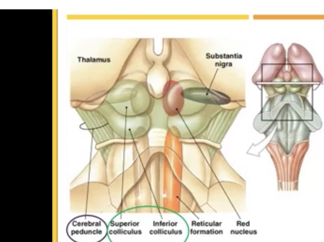
pons: are blank centers, they have blank blank blank, they also contain what -what is this important for
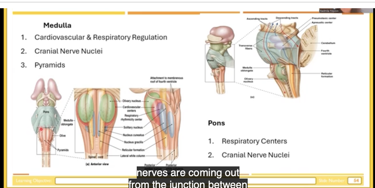
the medulla is improtant for the regulation of what two things , what does the medusa have a lot of 2 things
there are cranial nerves coming out of the junction between blank and blank /also where a majority of the cranial nerves are
respiratory, cranial nerve nuclei, trigeminal nucleus-sensation and motor control of the face
cardiovascular and respiratory, cranial nuclei+ pyramids
pons and medulla

medulla is the formation of pyramids -pyrimidal system: system that contains axonal fibers that contain motor information and motor control
they make the two bumps pry maids in medulla
cerebellum
small brain
found on dorsal part of the brain and brainstem
improtant for adjusting the postural muscles of the body , correcting errors during movement, complex motor learning (like riding a bike)
automating and optimizing behavior, enabling synchronicity and timing of movements (movement are in order and synchronous),
limbic system-emotional brain -localized in many regions of the Brian
strongly related tot he behavior, memory, autonomic nervous system, regulation of hormones, eating habits, processing of smell, other sensory information

major parts of limbic system part 1
amygdala-basal ganglia, anterior part of basal ganglia on inferior and rostral part of basal ganglia is amygdala, process information and helps recognize potnetial threats
hypothalamus-via the pituitary gland to maintain body balance
thalamus: relay station
major parts of the limbic system part 2
cingulate gyrus: above corpus collosum, improtant for behavior responses for perception of pain, regulate emotional processes
basal ganglia: assist with reward processing, habit formation, and movement control
major parts of the limbic system
olfactory bulb: processes smell information and links scents directly to emotional memories and reactions
maxillary body and hippocampus: improtant for formation of longterm memory and short term

spinal cord and brain=CNS
grey matter=outside, white matter is inside
spinal cord it’s opposite; whit matter is around and grey matter is inside
greys matter=in forms of Butterly
spinal chord is in vertebral canal , it is protected by vertebra

spinal chord is connected with periphery through spinal nerves
dorsal root, ventral root which makes spinal cord which come into the spinal cord and connect spinal cord to the periphery
spinal cord through periphery send info to the muscles through the motor roots
spinal cord has 31 pairs of the spinal nervous, come out through the adjacent vertebra (intervertebral space) which has exits of the spinal nerves

sens sensory information from he spinal nerve to the central nervous system
butterfly shape of the grey matter,
top=dorsal,(posterior) bottom-ventral(anterior)
grey matter inside that have diff type of neurons
butterfly shape can be classified as dorsal horn vs ventral horn
dorsal horn-have sensory neurons mostly and some interneurons
ventral horns: motor neurons

in certain regions of spinal cord:sympathetic and parasympathetic nervous system
dorsal root ganglion on the side -the buttons on each side of the spinal cord
root ganglions of the cell bodies of the sensory neurons which send info through the spinal nerve to the spinal cord
ventral root: sends info from the ventral horn to the periphery

besides grey matter we have white matter
white matter consists of the ascending and descending pathway
ascending pathways are the sensory pathways -sensation of touch, proprioception, vibration, pain and temp
descending pathways: the ones in blue on the bottom, go from the brain to the periphery through the ventral root

NEW CHAPTER
the nervous system division consists of sensory receptors, neurons, and the parts of the brain that receive and assign meaning to information
both general sensory and specialized sensory system depend on what
interaction of stimuli with special proteins or receptors on the surface
sensation is due to receptors blank diff stimuli into blank blank that are conducted/transmitted into blank
what is perception
when does perception occur? when sensory info comes to the …..
it requires selective activation to regions of the… blank blank, which does what ….
transducing, nerve impulses, CNS (midbrain/mesencephalon)
assigning meaning to that information within the brain
conscious level in the brain
cerebral cortex, assigns meaning to the stimulus
sensory system contains what major thing
they are blank blank send that info to the brain and parts of the central nervous system (spinal cord and brain) receive an assigned meaning to that info
peripheral nervous system (receptors)
afferent fibers
sensation can be classified as what two things
what are the 4 things that fall in the general sense category
it is sensed by diff recetprpos that are where
what are the 4 factors that fall into special senses
they are processed consciously or unconsciously
general/somatic sense or special senses
touch, pressure, temperature, and pain
inside the skin/muscles
vision, olfaction, hearing, taste/gustation
consciously
extra:
have other stimulus in our bodies processed subosnciously-blood pressure, glucose, osmolarity of body fluids, lung inflation, pHdiff kinds of sensory receptors that are sensoring that info and subosnsciously processing
can classify receptors by the location, 3 of them: what are the 3 of them
what 3 things fall under exteroreceptors
what part of the environment do they sense
what are the two examples of Interoreceptors
what do they sense external or internal enviro of body
interoceptors/proprioreceptors- have which receptors
these receptors are inside the what 3 things
they give us info abt what
exteroceptors, interoceptors, and interoceptors/proprioreceptors
smell, taste, touch
the external enviro
interoreceptors
ex baroreceptors -(blood pressure) and chemoreceptors (o2, Co2, blood level)
internal body receptors/sense inside of the body
-have both receptors
muscles, muscle spindles, or Golgi tendon organs
body position in space and each body part relative to each other
proprioreceptors-example-if you close your eyes and raise your hand without looking at hand you can approach with the other hand that hand
when loosing proprioception: which gives awareness of the body parts and controls what
;;; and position of the muscles to. have what..
without proprioreceptors you can’t have what
the level of stretch of the muscles
coordinated movement without looking at your leg
other classification of receptors is based on type of stimulus or modality
what are the 5 ones that fall into this category
tell me what each of them does
thermoreceptors, mechanoreceptors, nociceptors, photoreceptors, and chemoreceptors
thermoreceptors:; sense changes in temperature
chemoreceptors: respond to specific chemical, ex glucose
mechanoreceptors-detect fluctuations in pressure, gravity, change in volume or shape
nociceptors: receptors that detect pain
photoreceptors: detect light
extra:
thermoreceptors: -can be cooler or warmer temp
same receptors sensing cold temp can be activated by some other stimulus
menthol in mint mint activates the same receptors that detect cold temperatures
each receptor is most sensitive/has a lower threshold to blank type of stimulus
nociceptor will only respond to what stimulus
regardless of how a sensory neuron is stimulated, how many sensory modalities will be recieved
your brain does not care how a nerve is triggered, it only cares about what
one
burning sensation/painful stimulus
only 1!
which nerve sends the message
classification of the receptor by the structure
free nerve endings -ex receptors for what 3 things
these are blank, have receptors that can be activated by blank, where is the stimulus sent
then what happens from here?
encapsulated receptors: what do we have here that’s concentrated? around what
when receive esitmulus, it is sent through where
is sending info faster or slower compared to unmyelinated here , then wehre is the signal sent?
what are two examples of encapsulated
pain, temperature, and smell
to the unmyelnated axon to the cell body,
from cell body to another part of axon that synapses inside of the central nervous system
capsules/concentrated layers of connectied tissue around the enclosed nerve endings
through myelinated axon, info is faster, to central nervous system
pressure and touch

rods and cones-these are specialized receptors where?
give an example here
hair cells; specialized hair cells which has what and what, what does the second thing detect
we have a cell body here, and a blank cell synapse onto what and info is sent along
give two examples for hair cells
in the visual system, sight
small cilia, and protrusion, vibration, have cell body, specialized cell synapse on myelinated axon
hearing and equilibrium

receptor potnetial refers to what
with increase of stimulus intensity, what two things increase as well
what is receptor potnetial
info is sent to the brain in the form of what
increasing stimulus intensity will produce increase of graded potential but it’s not…
graded potential in sensory neuron
response and receptor potential
stimulus intensity(ex how warm the room is or how firm a chair is)
excitatory post synaptic potential
inear increase
amplitude of receptor potnetial correlates to what
the relationship between stimulus intensity and graded potential is blank
receptor potential is graded but long distance transmission requires blank blank
true or false action potential is all or none response, you will have a bigger amplitude of action potential due to more stimulus
stimulus intensity
logarithmic
action potentials
false!you won’t have bigger amplitude of action potential due to more stimulus
how brain sense stronger stimulus
neuron has receptors on it’s membrane Called dendrites, when a stimulus excites them, what do they generate
there is generation of action potential that we can record by electrode where?
while stimulus intensity and receptor potential have a direct relationship, what other potential has a direct relationship with stimulus/increased response and higher frequency with higher stimulus
a stronger stimulus activates more blank including the ones that need more stimulation to fire and together, they make the neuron send action potentials blank blank
receptor potnetial
in the nodes of ranvier on axon hillock
action potential
receptors, more often
extra: stronger stimulus results in 2 things:
higher frequency of action potentials form individual sensory receptors and activates receptors with lower AND higher threshold recruitment
one characteristic of sensory receptor is they have different sensor adaptation
wha tis a rapidly adapting receptor/phasic receptors: (mention what happens with prolonged stimulus)
what are two examples of this
what is slowly adapting receptor/tonic receptors (mention prolonged stimulus)
give two examples
with prolonged stimulus, some of the receptors+ action potential will not respond
ex: meissner’s corpuscles (light touch) receptors, + pacinian (vibration pressure)
with prolonged response, some receptors keep responding for as long as stimulus lasts, receptor potnetial stay s on the whole time, action potentials fire from the start, they become less frequent but never stop
merkel disks (B, tecture, fine touch) like for reading braille
Ruffini (E, grasping objects)
proprioreceptors(sense of position and movement of joints)

why do you need two diff kinds of receptors
bc for some senses you need longer duration of the response (slowly adapting)
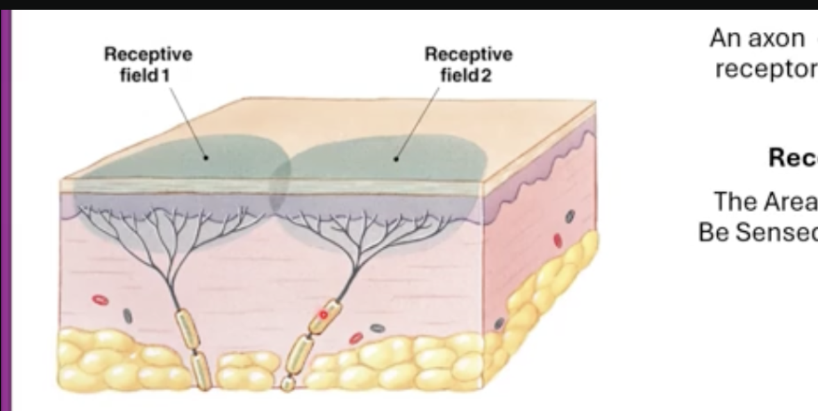
each receptor in image=piece of skin, neuron is myelinated axon, and dendrites and somewhere down is the cell body of neuron
axon is branching, has many branched dendrites,
what is receptive field:
what is a sensory unit
the surface of skin that dendrites from one single neuron occupy
Her definition of receptive field- area/region that can be sensed by sensory unit
an axon of the neuron and all receptors on it’s dendrites

receptive fields can be bigger or smaller, depending on size of receptive field, they are able or not able to sense what
what is two point discrimination
receptor fields are evenly or unevenly distributed throughout the body
two point discrimination on smaller or bigger distances:
my definition: the ability of a person to tell that they’re skin is being touched in two diff areas
hers: the ability of a person to discriminate when their skin is being touched at two separate points
if you point the same distance as in the skin, you will sense this as one stimulus not two
unevenly distributed
extra: ex: small receptive fields in the finger tips, if you apply two stimulation with forceps and very small distance between those stimulus will be sent as two separate stipulations
two point discrimination: in forearm, you apply the forceps but you need to apply in much bigger distance to sense two points of stimulation
if you point the same distance as in the skin, you will sense this as one stimulus not two
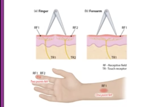
in diff parts of the body you will have more dense receptors, what’s one example of an area that has more dense receptors , is there a high or low distance needed to sense two point discrimination with dense receptors
in fingers and thumbs and low distance
in the belly, back, thigh, calf, you need the two stimuli in much bigger distances to be sensed as two diff stimuli
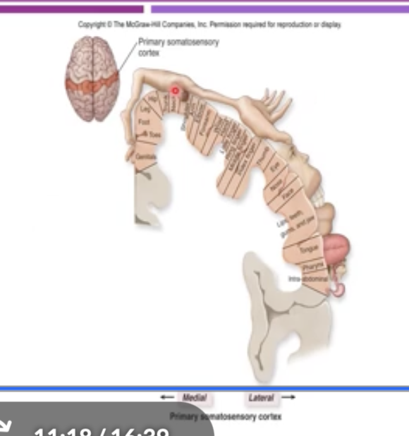
image recpresents sensory hommunculus is representation of the display differences in receptor differences in the body
top: superior part of the brain/view
red slip is the somatosensory cortex, if you lay down somatosensory on the side , see body parts represented in cortical area
what two areas have a big representation of surface area/higher /bigger density of receptors
even though these two areas have a bigger density of receptors, they have a blank receptor field
they also occupy a blank surface area in what cortex
face and hands
smaller
bigger, somatosensory cortex


one coding for stimulation-location, fact that receptive fields are overlapped, by applying stimulus on this position f the skin
you can stimulus either one neuron or all 3 neurons depending where you apply stimulus
if you can record stimulus/ only stimulate only B, that means the stimulus is applied to the receptive field or the overlapping receptive field?
if you stimulate the area between those two /shared by two sensory units you will have recording/can record response in which neurons
why is overlapping of receptive field good in case of injury
receptive field of that neuron
both of the neurons
if those neurons get injured there won’t be complete loss of sensation intuit area bc the other neuron will be able to send info form that area to the brain

in the coding there is lateral inhibition
what does lateral inhibition do, what does it sharpen, why?
example is in visual cortex/system
example: when. center cone is activated, horizontal cells (a third cell type) inhibit the neighboring cones around it, which makes the center cone’s response sharper
when an active receptor suppresses it’s neighbors, sharpening the contrast between them so the Brian can tell exactly where the stimulus is
(lateral inhibition in her words: in the pattern of action potentials received by the CNS allowing a finer resolution of stimulus location)
in the case of tactile info and or pain info, if you’re pinning the skull and inducing painful info, that is appleid in the receptive field which is sensed by three diff neurons,
which neuron will send that info to the brain
what is the name of that neuron
meaning that it does what
which will stimulate the secondary neuron to blank blank blank , then only info from that neuron will be sent to blank neurons to blank neuron/ tertiary neuron to the blank.
inhibition oft the lateral neurons will enhance what ? via lateral inhibition
onlythe neuron closest to stimulation
the primary sensory neuron
secretes the highest amt of the neurotransmitter
inhibit adjacent neurons, secondary, third/tertiary, brain
perception of the stimulus B
perception=sensory awareness

what is presynaptic inhibition via axo anoxic synapse
green neuron is receiving action potential and then releasing the the inhibitory neuron transmitter (GABA) in this cases GABA does what
and it’s needed in this yellow neuron to produce neurotransmitter which will be released and then synapse on the post synaptic membrane of the dendrite
in this case, regulate the amt of neuron that is actually part of the mechanism of the what (3 words)
when one axon synapses onto another axon and adjusts the amt of neurotransmitter produced/ released from it’s dendrites
inactivates calcium channels
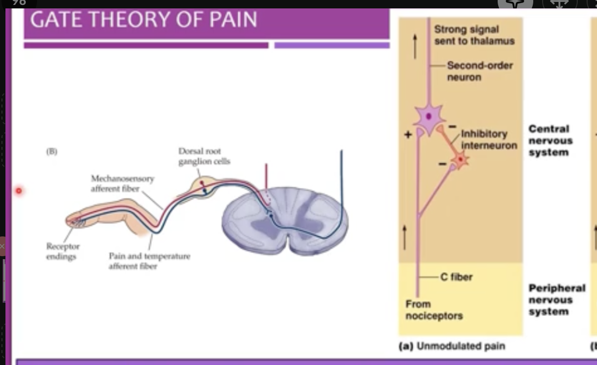
gate theory of pain


ascending pathways are what
cross section of spinal cord
grey matter and white matter is where in this picture
what are the two sensory neurons present in the spinal cord
sensory info comes into the spinal cord then is sent to the blank through where
in blue are the ascending pathway
descending pathways such as corticospinal tract for example descend from blank to blank blank and out to the blank
neuronal tracts that carry sensory info from the body/periphery up through the spinal cord to the brain
grey matter=inside, white matter is outside in the spinal cord
dorsal and ventral root
brain, the dorsal root
brainstem, spinal cord, muscles

two major sensory pathways
one if for blank blank and blank and the other is for what 4 things
they both have system of how many neurons
what is the first order neuron : they carry signals from the..
in the first order neuron what synapses with what
what is decussation
what two things occur in the second order neuron
what is third order neurons
touch proprioception and vibration other pain temperature, crude touch and itch
3 neurons
peripheral tissue to CNS-
their axons/cell body of axon synapse to spinal cord/Brian stem
second order neurons: synapsing in the thalamus + and then signal crosses over to the other side (decussation)
in thalamus from the thalamus
-cell body in thalamus(filters/filtering ) then synapses in the somatosensory cortex-(cortex in post central gyrus)
extra: they start from receptors which are sending their axons…

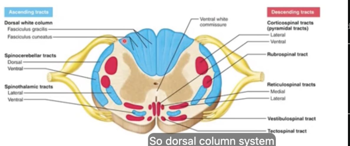
medial lemniscus/dorsal column system
it is medial lemniscus/dorsal column bc it is present int he dorsal part of the blank blank
blue=posterior, light born=anterior
dorsal column system is in dorsal part of the spinal cord, while it’s called dorsal column/medial meniscus system
called medial lemniscus system bc it’s medially oriented int he brain stem/toward the blank
spinal cord
thalamus

dorsal column=system that process info abt what two things
what is the first order neuron here: blank tissue to what, where does it terminate? in the blank
in dorsal column system: first synapse is in the blank does not synapse in the spinal cord, just pass thorough in the same side of blank blank. It synapses in the blank and then blank
second order neuron: terminates where? in the blank , what also occurs in the thalamus
third order neuron is called what: this structure is part of what
inside the encephalon we have the blank
it is a relay station for all blank but blank, what does it also do? (regarding information) then that info goes where
finally we will have blank of fine touch and proprioception
fine touch and proprioception vibration
peripheral tissue—>spinal cord→ medulla
medulla, spinal cord, medulla, crossover
thalamus (contralateral side/oposite side of origin) side of the thalamus
epithalamus, , encephalon,
thalamus: senses, smell
-filtering information, to the somatosensory cortex, perception

first order neuron:
peripheral tissue through spinal cord, terminates in medulla
what is the route for first order neurons in the lower body called
what is the route for first order neurons in the upper body called
recall:whatoccurs in the 2nd and 3rd order neuron
then synapsing occurs in what cortex, leading to what
fasiculus gracilis
fusiculus cunneatus
second order neuron: crosses over (decussation) terminates in the thalamus
cell body in thalamus (filtering)
somatosensory cortex and will have perception of fine touch proprioception

second system=anterolateral system-why? bc it is laterally coming to the lateral side, it is synapsing on the blank in spinal cord
crossing over and then it is positioned in the blank blank of the spinal cord and it stays on that side until it synapses in the blank
what is the first order neuron for anterolateral system blank tissues processes to the blank blank blank , blank blank blank will do what 3 things
from the thalamus, the info Is sent to the blank blank
depending on where this info is coming from blank or blank will end up toward either what two places?
interneurons
anterolateral side, thalamus
peripheral tissue—>processs to the posterior grey horn—>in posterior grey horn,
synapse, cross over(part of secondary neuron) , terminates in thalamus,
cerebral cortex, depending where this info is coming form, cervical or lumbar will e the midline(lumbar) or toward the middle part, cervical region(upper body)

lateral inhibition:
for sensation of pain, there is smth called gate theory of pain what does it mean
in drawing, can see some receptors on the fingertips and those receptors endings will send info of the pain and temp in blue but can also sense info abt fine touch
ex: touch proprioception of vibration
pain/temperature and touch proprioception receptors in the fingertips travel in blank route and then same blank blank
what happens once they get tot eh spinal cord
pain/temperature fibers synapse in what
touch/proprioception fibers ascend where
certain neurons can modulate/adjust the activity of diff neurons
similar, spinal nerve
they split.
in the dorsal horn
ascend straight to the brain

Unmodulated pain: define it
a painful stimulus activates blank, which send the signal along what
blank blank synapses onto a neuron in where blank blank, which sends that signal to what
right next to C fiber, sits an blank blank, the C fiber blanks this blank so it’s switched blank
when the inhibitory neuron is off,
when only pain fibers are active, the pain signal goes through at full strength
nociceptors(paint receptors) slow unmyelinated C fibers
C fiber, the spinal cord, the thalamus
inhibitory interneuron . inhibits, interneuron, off
, nothing blocks the pain pathway so the gate is open and the pain is felt in full

with modulated pain such as rubbing the painful spot, it activates blank receptors which send signals along which fibers, up what pathway
these fibers also send a branch tot he iblank blank which is now blank
the interneuron does what , why?
the gate is blank blank so you feel blank pain=gate theory of pain
mechanoreceptors , fast, myelinated fibers , up the dorsal column/medial lemniscus pathway
inhibitory interneuron, activated (unlike in unmodulated pain where it was shut off)
inhibits the pain pathway neuron, so only a weak pain signal reaches the thalamus
partly closed, less,

what is gate theory of pain:
both sensory pathways: dorsal column medial lemniscus and anterolateral. cross to the blank side to a stroke in one hemisphere affects which side of the body
what is ipsilateral
what is contralateral
damages at different points along the pathway (periphery to the cortex) causes diff symptoms
If the damage is in the cortex, you lose sensation on what side
that activation mechanoreceptors can modulate sensation of pain, example of lateral inhibition
opposite, affects the opposite side of the body
the same side
the opposite side
opposite side
ex:
stroke affecting the right hemisphere will affect left side of the body-contralateral effect
3 major layers of the eye
first layer is called what -what does it consist of
what is the second layer called-it consists of a blank supply , also consists of the blank, has blank blank and blank
the blank blank: holds the blank in place
what does blank do?
the pupil is another structure that does what
the third layer is called what , where is it located it is a blank blank blank
outer fibrous layer- white sclera and transparent clear conreum
vascular layer-vascular, choroid, iliary body, iris(iris is the color part of our eye
ciliary body/muscle -lens
Lens -bends the light that comes through the pupil
p- opens in the iris
retina-on the back of the eye , deep nervous layer
