7: Respiratorisk system
 Call Kai
 Learn
Learn Practice Test
 Spaced Repetition
 Match
Match Flashcards
 Knowt Play
1/157

There's no tags or description
Looks like no tags are added yet.
Name | Mastery | Learn | Test | Matching | Spaced | Call with Kai | Chat |
|---|
No analytics yet
Send a link to your students to track their progress
158 Terms
What are the two functional divisions of the respiratory system?
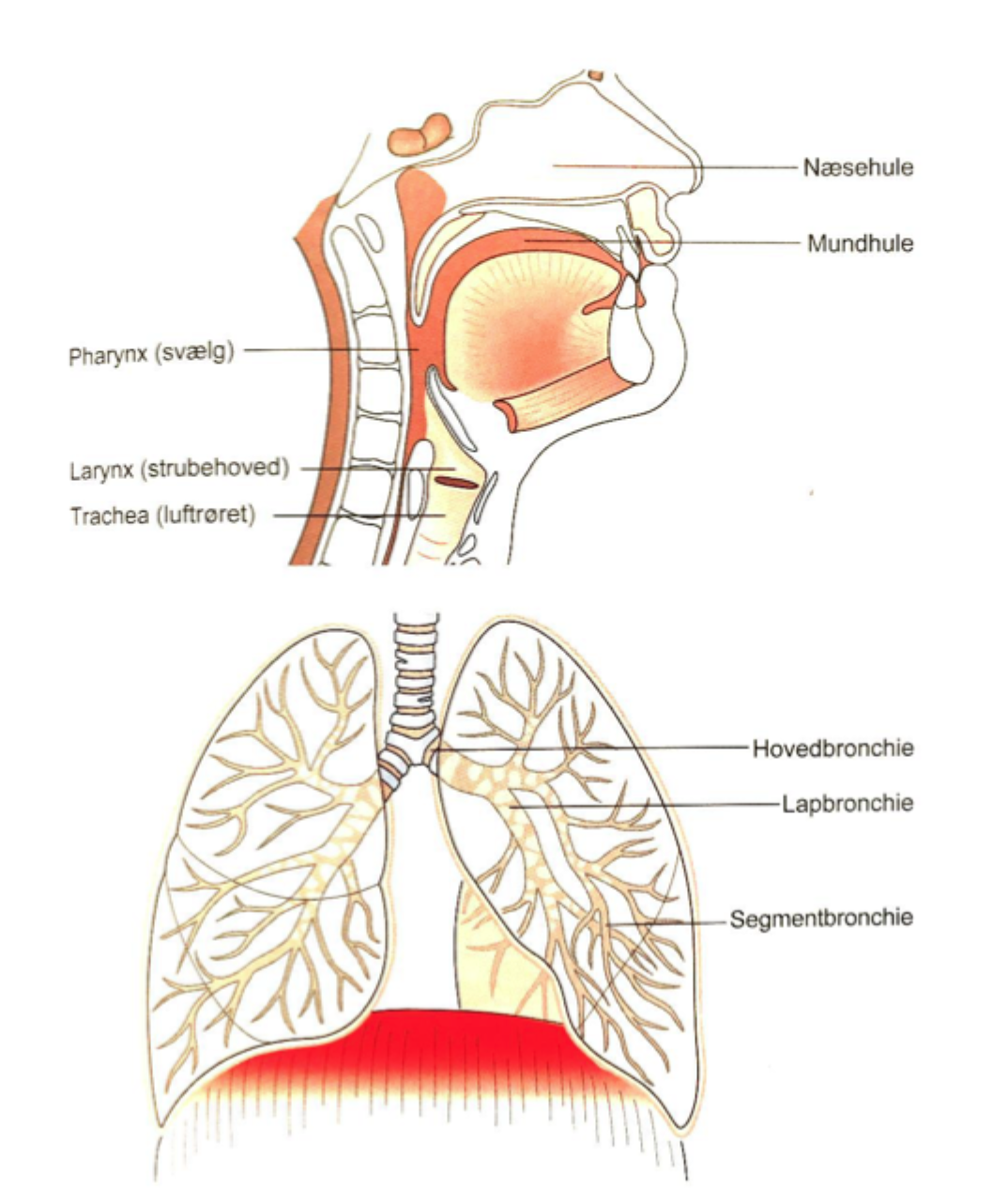
Conducting portion and respiratory portion
What is the main function of the conducting portion?
To conduct, warm, humidify, and filter air. It does not normally perform gas exchange.

Which structures belong to the conducting portion?
Nasal cavities, pharynx, larynx, trachea, bronchi, bronchioles, and terminal bronchioles.

Which structures belong to the respiratory portion?
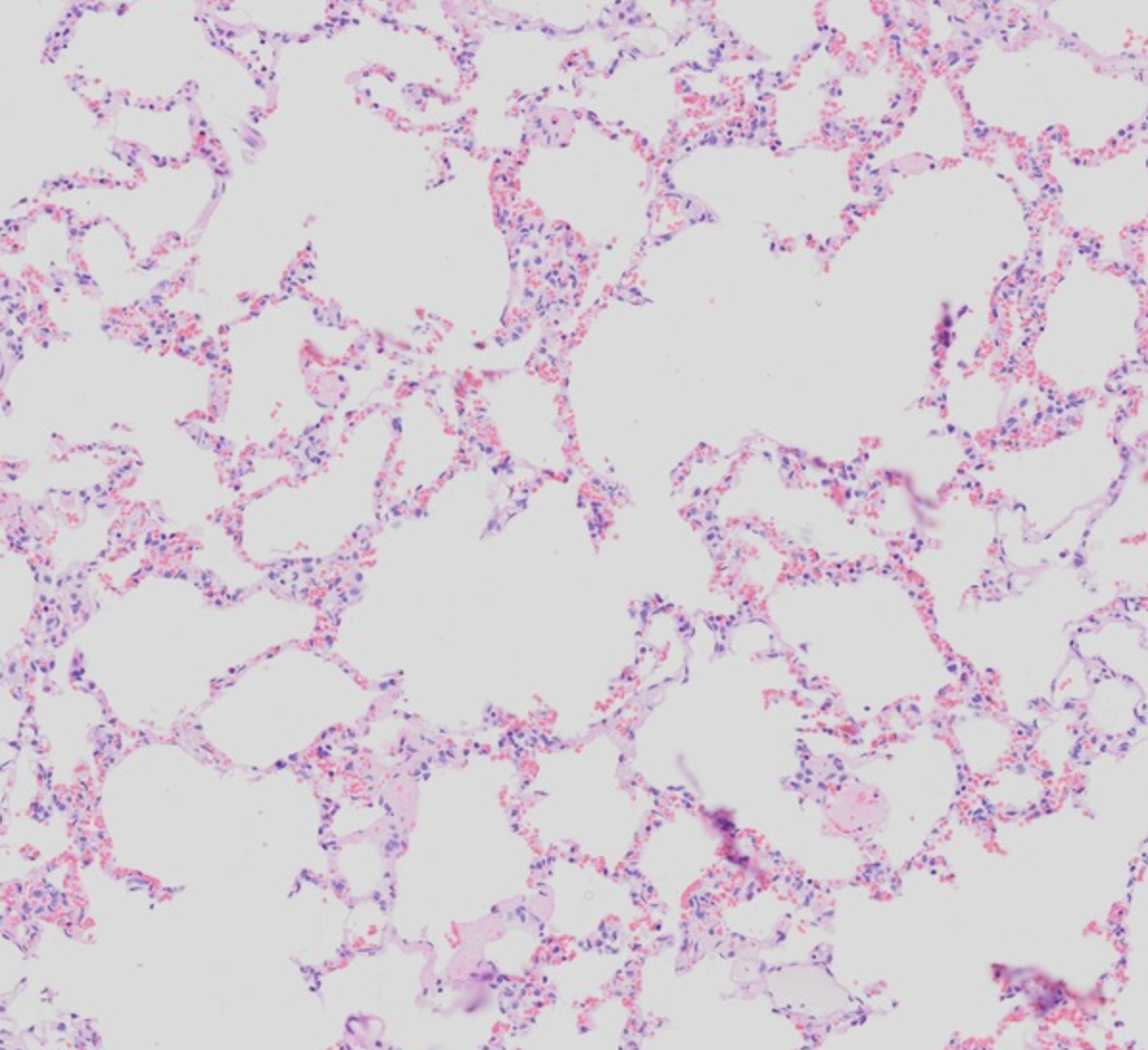
Respiratory bronchioles, alveolar ducts, alveolar sacs, and alveoli.

Where does gas exchange begin?
In the respiratory bronchioles, because alveoli first appear in their walls.
Where does most gas exchange occur?
In the alveoli, across the thin alveolar-capillary barrier.
What is the airflow pathway from nose to alveoli?
Nasal cavity → pharynx → larynx → trachea → bronchi → bronchioles → bronchioles → alveolar ducts → alveoli.
(What is the difference between a terminal and a respiratory bronchiole?)
A terminal bronchiole is the final conducting airway; a respiratory bronchiole has alveoli in its wall and participates in gas exchange.
Why are upper-airway infections often less serious than lower-airway infections?
Upper-airway infections usually do not directly impair alveolar gas exchange. Lower-airway or alveolar infection can reduce ventilation and oxygen uptake.
Why can obstruction in the lower airways be dangerous?
It can reduce airflow to alveoli, impair gas exchange, cause hypoxaemia, and increase the work of breathing.
Which structures are commonly considered upper airways?
Nose, nasal cavity, paranasal sinuses, pharynx, and often the larynx.
Which structures are commonly considered lower airways?
Trachea, bronchi, bronchioles, and lungs.
What is the function of the nasal cavity?
It filters, warms, and humidifies inspired air. It also contributes to smell and resonance of the voice.
What is the function of the pharynx in respiration?
It is a shared passageway that conducts air from the nasal/oral cavity to the larynx.
What is the main role of the epiglottis (strubelåg)?
It helps protect the airway during swallowing by directing food and fluid away from the laryngeal inlet.
What is the main role of the larynx?
It maintains an open airway, protects the lower airways during swallowing, and produces soun
What is the main role of the trachea?
To conduct air to the main bronchi while remaining open and flexible.
Why does the trachea lie anterior to the oesophagus?
The posterior tracheal wall is flexible, allowing the oesophagus to expand during swallowing.
What is ventilation?
The movement of air into and out of the lungs.
What is the main muscle of quiet inspiration?
The diaphragm
What happens when the diaphragm contracts?
It moves downward, increasing thoracic volume and lowering intrathoracic pressure, so air enters the lungs.
(Which muscles assist quiet inspiration?)
The external intercostal muscles, which elevate the ribs
(Is quiet expiration normally active or passive?)
Mostly passive, due to elastic recoil of the lungs and chest wall.
(Which muscles are recruited during forced expiration?)
Internal intercostal muscles and abdominal muscles.
What is the key physical principle driving airflow?
Air moves down a pressure gradient: from higher pressure to lower pressure.
What is the difference between ventilation and gas exchange?
Ventilation is movement of air; gas exchange is diffusion of O₂ and CO₂ between alveoli and blood.
What is the typical epithelium of the trachea and large bronchi?
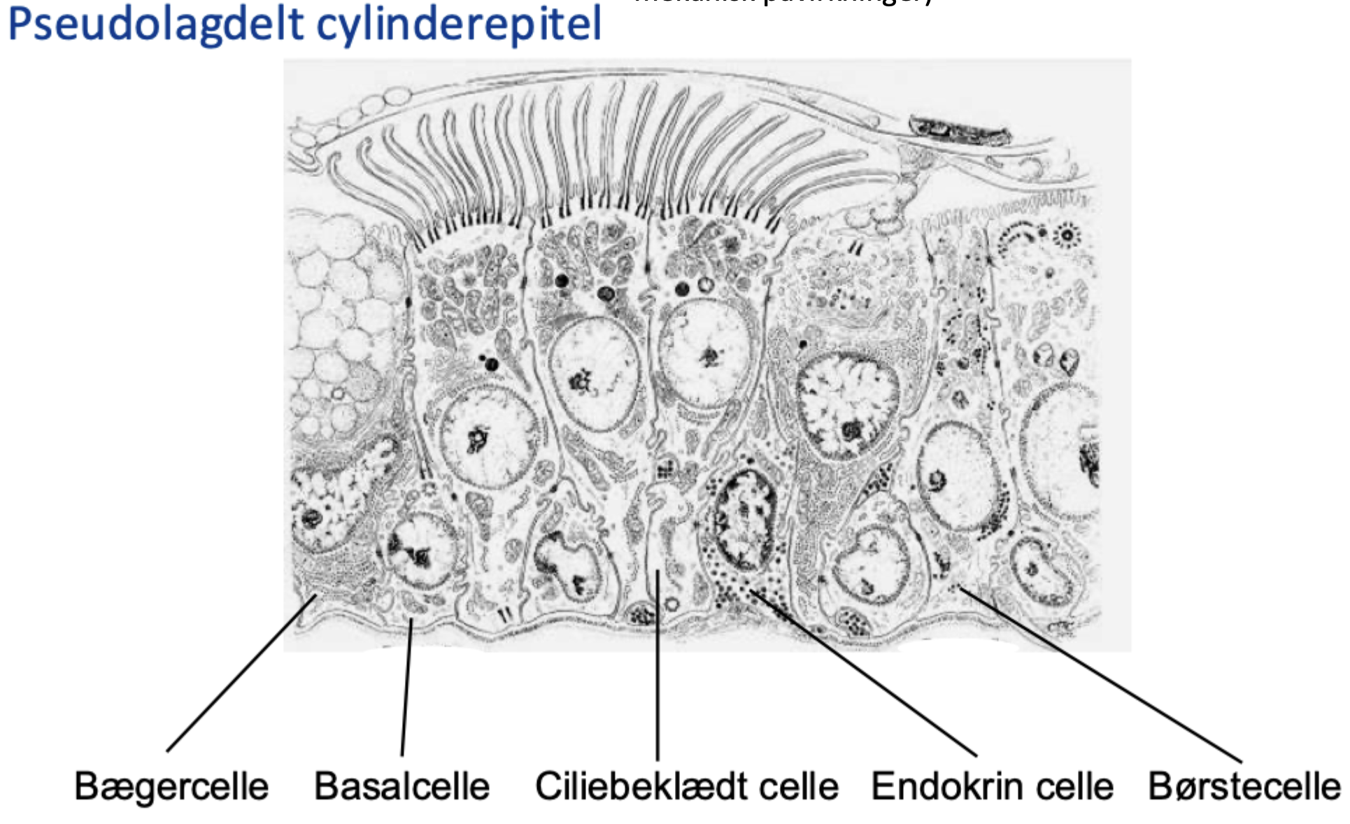
Pseudostratified ciliated columnar epithelium with goblet cells

Why is it called pseudostratified epithelium?
It looks multilayered because nuclei lie at different heights, but all cells contact the basal lamina.
Do all cells in pseudostratified epithelium reach the lumen?
No. All contact the basal lamina, but not all reach the free/apical surface

What is the main function of respiratory epithelium?
Mucus production, particle trapping, mucociliary clearance, epithelial defence, and conditioning of inspired air.
What is the mucociliary escalator?
Cilia move mucus and trapped particles upward toward the pharynx, where they can be swallowed or expelled.
Why is the mucociliary escalator clinically important?
It clears inhaled particles and microbes. Defects increase infection risk and mucus retention.
What happens if ciliary beating is impaired?
Mucus accumulates, airway clearance falls, and the risk of infection and obstruction rises.
How does smoking damage airway defence?
It can impair ciliary function, increase mucus production, promote inflammation, and reduce mucociliary clearance.
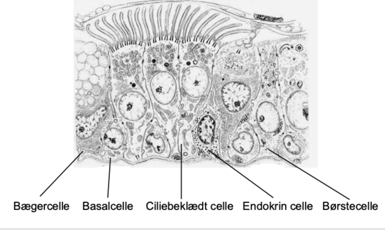
What are the cells of pseudostratified ciliated columnar epithelium:
ciliated cells, goblet cells, basal cells, brush cells, neuroendocrine cells and regulative Lymphocytes and other immune cells

What are ciliated cells?
Columnar epithelial cells with motile cilia that move mucus toward the pharynx

What are goblet cells?
Secretory epithelial cells that release mucins, which contribute to mucus.

What is mucus made of?
Mainly water, mucins, ions, proteins, antimicrobial molecules, and trapped material.
What is the function of goblet-cell mucus?
To trap particles, allergens, and microorganisms before cilia transport them out of the airway.

Why do goblet cells often look pale on routine histology?
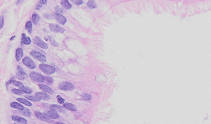
Their mucin content is poorly stained by H&E but stains strongly with PAS.
What does PAS staining highlight in goblet cells?
Carbohydrate-rich molecules, especially mucins.

What are basal cells?
Small cells near the basal lamina that act as progenitor/stem cells for airway epithelial repair.

Why are basal cells important after airway injury?
They can proliferate and differentiate into replacement epithelial cells.

What are brush cells?
Rare airway epithelial cells with apical microvilli; they are generally considered sensory/chemosensory cells
Do not confuse brush cells with which cell type?
Club cells. Club cells are mainly found in bronchioles and are secretory, detoxifying, and progenitor-like.

What are neuroendocrine cells in the airway?
Rare cells that release signalling molecules and participate in local regulation, development, and sensory responses.
Can immune cells be present within airway epithelium?
Yes. Lymphocytes and other immune cells can patrol or migrate through the epithelium.
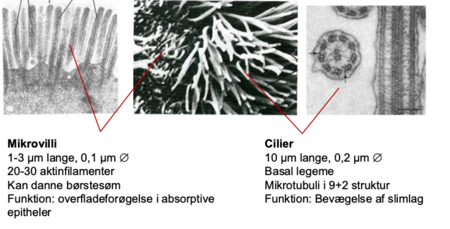
What is the main function of microvilli?
To increase apical surface area for absorption or exchange

What is the main function of motile cilia in the airways?
To move the mucus layer and trapped particles toward the pharynx.

Approximate length of microvilli?
About 1-3 μm.

Approximate length of cilia?
About 10 μm.

What cytoskeletal protein supports microvilli?
Actin filaments.

What is the classic internal arrangement of motile cilia?
9 + 2 microtubule arrangement: nine peripheral doublets surrounding two central microtubules.

What powers ciliary movement?
ATP-dependent dynein activity causes microtubule sliding and ciliary bending.

What are basal bodies?
Structures at the base of cilia that anchor them; they are related to centrioles

Which surface specialisation is motile: microvilli or cilia?
Cilia are motile; microvilli are generally non-motile

What is a brush border?
A dense layer of microvilli that increases surface area, especially in absorptive epithelia.
Why must mucus have the correct water content?
It must be hydrated enough for cilia to move it, but not so watery that it cannot trap particles effectively.
What happens if mucus becomes too thick or dehydrated?
Cilia cannot move it efficiently, so mucus stagnates and can obstruct airways.
What is the periciliary layer?
The watery layer surrounding cilia that allows them to beat effectively beneath the mucus layer
What can happen if the periciliary layer is too shallow?
Cilia become compressed by mucus and cannot clear it normally
What can happen if mucus clearance from a sinus is blocked?
Retained mucus can promote inflammation and sinusitis.
Why are chronic airway diseases often associated with infection?
Impaired mucociliary clearance allows microbes and secretions to remain in the airways.
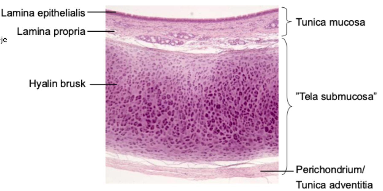
What type of cartilage is found in the trachea?
Hyaline cartilage

What is the main collagen type in hyaline cartilage?
Type II collagen.

Why does hyaline cartilage look glassy in histological sections?
Type II collagen fibrils are very fine and embedded in a hydrated extracellular matrix.
What is the function of tracheal cartilage?
To keep the airway open while remaining somewhat flexible.

What shape do tracheal cartilage rings have?
They are C-shaped: open posteriorly.

Why are tracheal cartilage rings open posteriorly?
The oesophagus lies behind the trachea and needs room to expand during swallowing

Which muscle bridges the posterior gap in the tracheal cartilage?
The trachealis muscle, a band of smooth muscle.
What is the function of the trachealis muscle?
It can adjust tracheal diameter and contribute to forceful expiration or coughing

How does cartilage change from trachea to bronchi?
Tracheal C-rings become irregular cartilage plates in bronchi.

Is cartilage present in bronchioles?
No. Bronchioles lack cartilage

Why are bronchioles more prone to collapse or constriction?
They lack cartilage and have a relatively prominent smooth-muscle component.

What are the major layers of the tracheal wall, from lumen outward?
Mucosa → submucosa → cartilage/perichondrium with posterior trachealis muscle → adventitia.

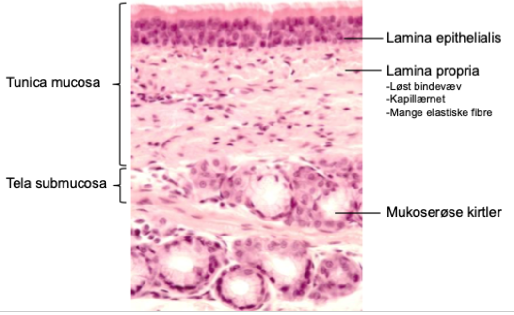
What is included in the tracheal mucosa?
Respiratory epithelium, basal lamina, and lamina propria
What is the lamina propria?
A vascular, elastic, loose connective-tissue layer beneath the epithelium.

Why is the lamina propria rich in elastic fibres?
Elastic fibres permit repeated stretching and recoil during breathing
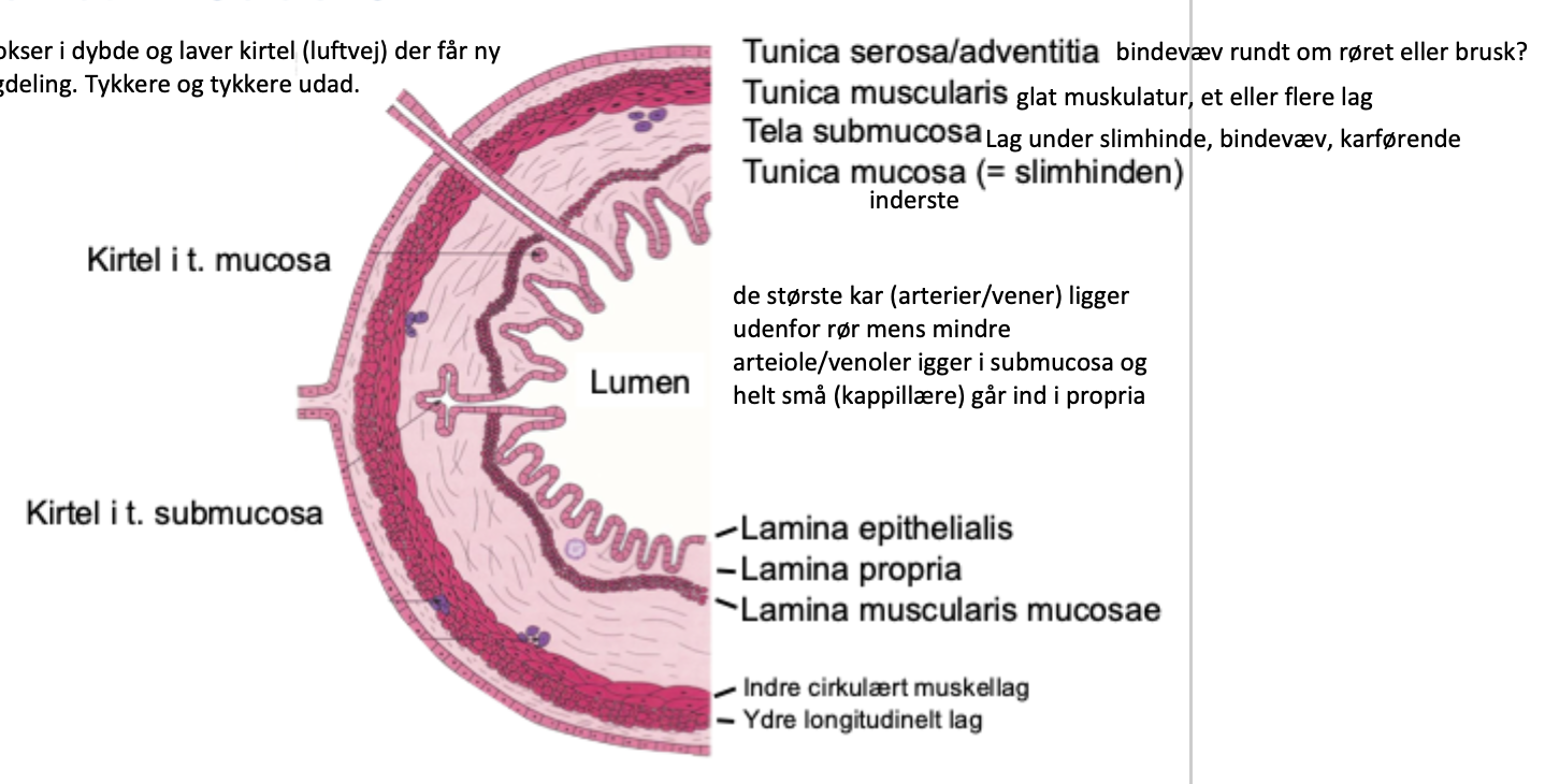
What is the submucosa of the trachea?
Connective tissue containing vessels, nerves, and mainly seromucous glands.

What are seromucous glands?
Glands that produce both watery serous secretion and viscous mucous secretion.
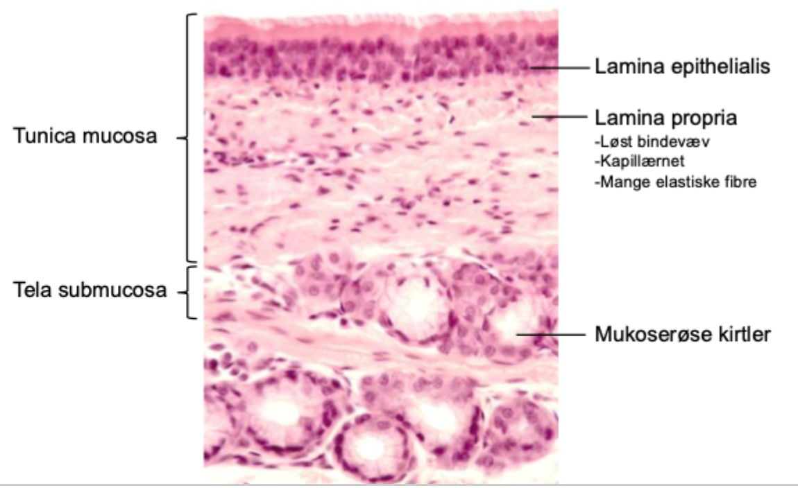
Why are submucosal glands important?
They add fluid and mucus to the airway surface, helping humidification and mucociliary clearance.

What is the perichondrium?
Dense connective tissue surrounding cartilage; it supports nutrition, growth, and repair of cartilage.

What is the adventitia?
The outer connective-tissue layer that anchors the trachea to surrounding structures

(Does the trachea have a typical gastrointestinal-style muscularis externa?)
No. Its key smooth muscle is the posterior trachealis muscle, not a continuous two-layer muscularis externa.
(Does the trachea have a prominent muscularis mucosae like the GI tract?)
No. This is not a prominent, standard tracheal feature
What is the basal lamina?
A specialised extracellular-matrix layer directly beneath epithelial cells.
What are important components of the basal lamina?
Laminin, type IV collagen, nidogen, and heparan sulfate proteoglycans.
What is the basement membrane?
In light microscopy, this term often includes the basal lamina plus the supporting reticular lamina beneath it.
What is the reticular lamina mainly associated with?
Connective tissue and mainly type III collagen fibres.
Why is the basement membrane important?
It anchors epithelium, influences cell polarity and differentiation, and acts as a selective barrier.

Why is airway basement membrane clinically relevant in asthma?
Thickening of the reticular basement membrane is a feature of airway remodelling in asthma.
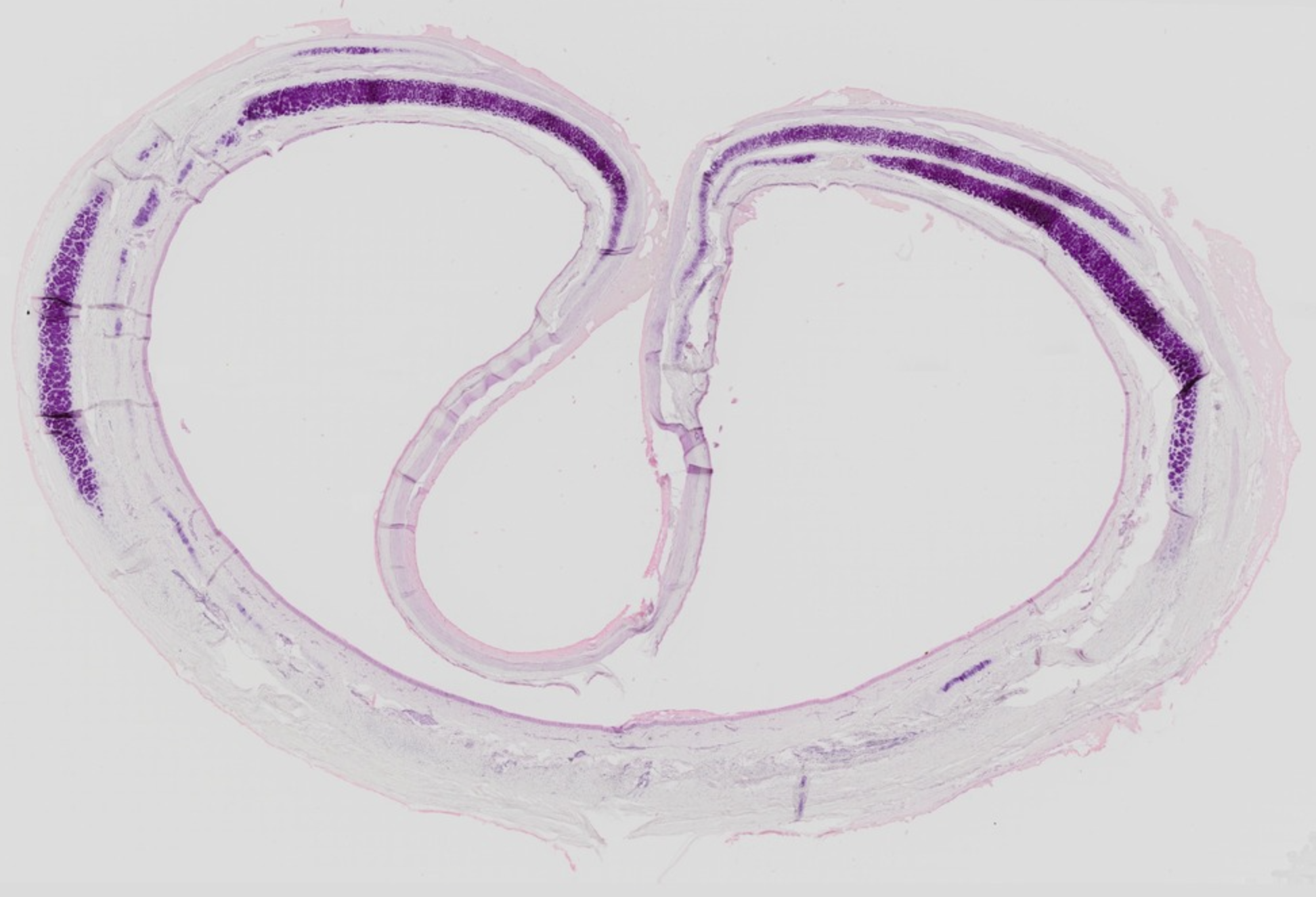
How do you identify trachea in a histological section?
Look for pseudostratified ciliated columnar epithelium, goblet cells, submucosal seromucous glands, hyaline cartilage, and posterior trachealis muscle.

How can you identify goblet cells in H&E?
They are pale, mucus-filled cells with nuclei displaced toward the basal part of the cell.

How can you identify ciliated cells in H&E?
Look for tall columnar cells with a fuzzy apical ciliary border.


How can you identify hyaline cartilage in H&E?
It has a smooth, glassy extracellular matrix with chondrocytes in lacunae.

How can you distinguish the lumen from the outer wall in trachea?
The lumen faces respiratory epithelium; cartilage and adventitia are further outward.
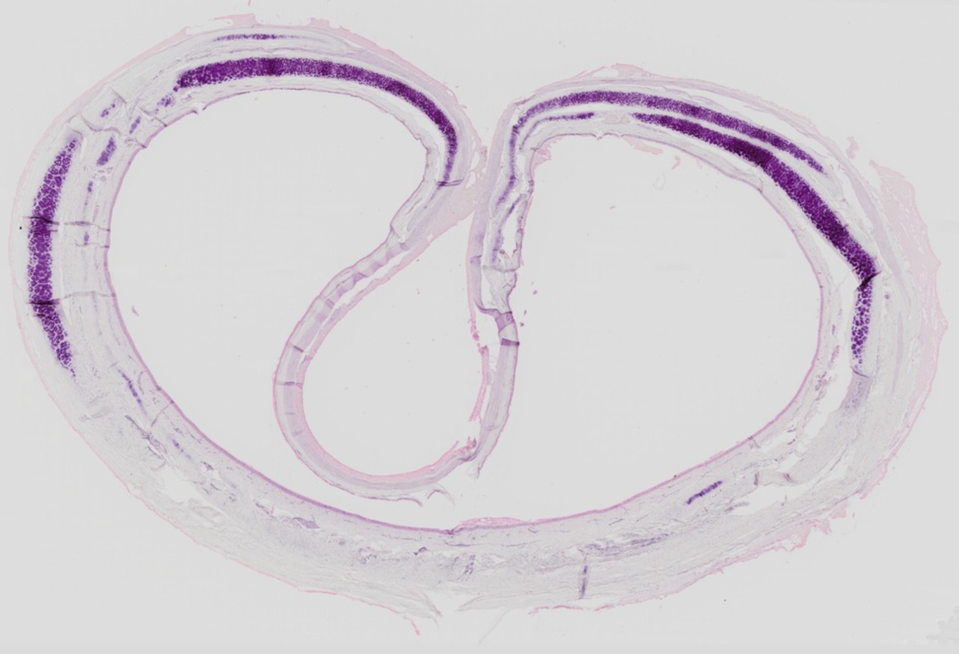
Where are tracheal glands usually found?
Mainly in the submucosa.

Which structure should make you think “bronchiole” rather than “bronchus”?
Absence of cartilage and submucosal glands.
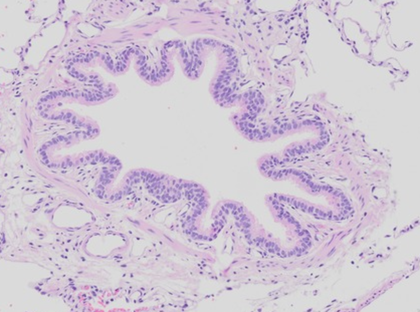
Which structure should make you think “respiratory bronchiole”?
Bronchiolar wall interrupted by alveoli.

Which structure should make you think “alveolus”?
Very thin walls lined mainly by simple squamous type I pneumocytes.