NROSCI 0080: Neurotransmitters
 Call Kai
 Learn
 Practice Test
 Spaced Repetition
 Match
 Flashcards
 Knowt Play
1/23

There's no tags or description
Looks like no tags are added yet.
24 Terms
Classes of Neurotransmitters
Small-molecule transmitters: synthesized from dietary nutrients and packaged ready for use in axon terminals
Amino Acids: Glutamate (Glu), Gamma-aminobutyric acid (GABA), Glycine (Gly), Acetylcholine (ACh)
Amines: Dopamine (DA), Norepinephrine (NE), Serotonin (5-HT)
Peptide transmitters (= neuropeptides)
Small proteins
Synthesized in the soma based on the genetic information from the nucleus
E.g. Endogenous opioids
Lipid transmitters
E.g. Endocannabinoids
Nucleotide-related transmitters
E.g. Adenosine
Glutamate
Main excitatory neurotransmitter in the brain and spinal cord (CNS). Amino Acid. Is a component of many proteins that we consumed as food.
Receptors: Ionotropic, metabotropic.
Ionotropic glutamate receptors depolarize the postsynaptic cell membrane, which leads to excitatory response
3 types of ionotropic glutamate receptors
AMPA, NMDA, Kainate

How Does Glutamatergic Synapse Work?
1. Action potential depolarizes presynaptic
membrane
2. Depolarization opens voltage-gated Ca2+
channels and Ca2+ ions go inside the
presynaptic terminal along their
electrochemical gradient.
3. Ca2+ ions stimulate glutamate
(transmitter) release from synaptic vesicles
into the synaptic cleft.
4. Glutamate binds to the glutamate
receptors and opens transmitter-gated ion
channels on postsynaptic membrane.
5. Na+ and Ca2+ ions go inside the dendritic
spine (postsynaptic cell) along their
electrochemical gradient and generate
depolarizing response on the postsynaptic
membrane, EPSP, that propagates along
dendritic membrane towards axon hillock.
6. Excessive glutamate is re-uptaken onto
presynaptic terminal and transported into
astrocytes

Glutamate: Excitotoxicity
High levels of glutamate can be toxic to nerve cells.
Excitotoxicity hypothesis: prolonged depolarization of receptive neurons leads to their eventual damage or death
may occur with brain ischemia (interruption of blood flow) like from stroke
massive release of glutamate occurs in the affected area
GABA (gamma-aminobutiric acid)
Main inhibitory neurotransmitter in the brain.
If either GABA or glycine is blocked, convulsions and death can result.
Amino acid.
GABA is made from glutamate.
GABA binds to GABA receptors.
Has two receptors: ionotropic, metabotropic
GABA receptor channels allow Cl- to move from outside to inside the cell.
Causes hyperpolarization and inhibition of the postsynaptic cell
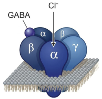
How does GABAergic Synapse Work?
1. Action potential depolarizes presynaptic
membrane
2. Depolarization opens voltage-gated Ca2+
channels and Ca2+ ions go inside the
presynaptic terminal along their
electrochemical gradient.
3. Ca2+ ions stimulate GABA (transmitter)
release from synaptic vesicles into the
synaptic cleft.
4. GABA binds to the GABA receptors and
opens transmitter-gated Cl- ion channels
on postsynaptic membrane.
5. Cl- ions go inside the postsynaptic cell
along their concentration gradient and
generate hyperpolarizing response on the
postsynaptic membrane, IPSP, that
propagates along dendritic membrane
towards axon hillock.
6. Excessive GAB is taken up into the presynaptic terminal and transported to the
astrocytes → this is the deactivation process for GABA

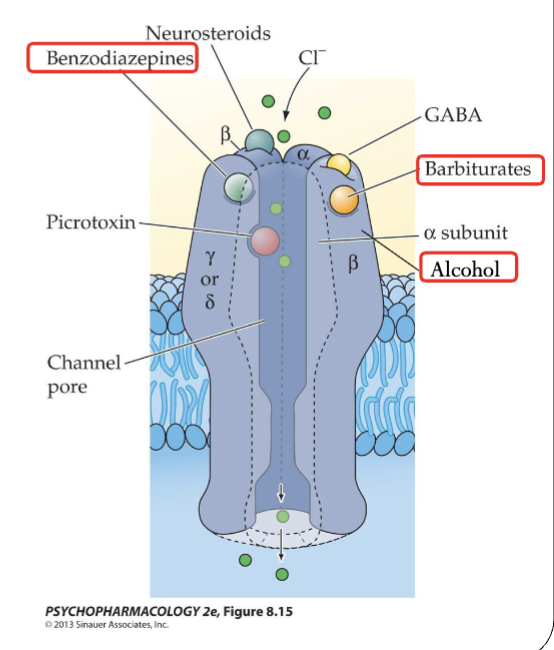
GABA: Benzodiazepines, Barbiturates, Alcohol
GABAA receptors are sensitive to some CNS-depressent frugs with antianxiety, sedative, and anticonvulsant profiles.
Benzodiazepines and barbiturates activate GABAA receptor.
Alcohol has similar properties (is a GABA agonist)
GABA and Epilepsy
Since GABA is involved in neuronal inhibition, it might play a role in epilepsy.
Some forms of epilepsy are linked to mutations in GABA receptor subunits.
Many epileptic drugs enhance GABA response.
Glycine
Acts as inhibitory neurotransmitter in the spinal cord, brain stem.
Amino acid synthesized from serine.
Bonds to Ionotropic receptors. These receptors produce IPSPs by opening Cl- ion channels and allowing Cl- into the cell. Can be blocked by strychnine.
Deactvation: Can be re-uptaken onto presynaptic terminal and transported into astrocytes.

Mechanisms of action of glycine at the synapse
Bonds to Ionotropic receptors. These receptors produce IPSPs by opening Cl- ion channels and allowing Cl- into the cell. Can be blocked by strychnine.
Mechanisms of action of acetylcholine at the synapse
ACh is formed from choline and acetyl CoA, and there are 2 types of receptors:
Nicotinic ACh receptors are ionotropic and respond to nicotine (agonist), which is an alkaloid found in the tobacco plant → these mediate fats excitatory responses in the NMJ, brain, and autonomic nervous system
When ACh binds, the channel opens and Na+ and Ca2+ enter, depolarizing the membrane
Muscarinic ACh receptors are metabotropic and respond to muscarine (agonist), which is an alkaloid first found in fly agaric mushrooms
What drugs potentiate (make something more powerful) GABA receptors?
GABA-A receptors are sensitive to some CNS depressant drugs used for antianxiety, sedatives, and anticonvulsants
things like benzodiazepines and barbiturates. These are GABA agonists (a substance or muscle that triggers or enhances a specific action or biological response), and alcohol has similar properties
What is strychnine?
A glycine receptors blocker. It causes muscular convulsions and eventually death through asphyxia or sheer
exhaustion. Was used as a pesticide.
What are the mechanisms of deactivation of glutamate, GABA?
GABA: Excessive GAB is taken up into the presynaptic terminal and transported to the astrocytes
Glutamate: Excessive glutamate is re-uptaken onto presynaptic terminal and transported into astrocytes
How is acetylcholine deactivated? What is acetylcholinesterase?
ACh is broken down by acetylcholinesterase (AChE, an enzyme) into acetic acid and choline in the synaptic cleft
Mechanisms of botulinum toxin
Potentially deadly illness caused by a toxin produced by the Clostridium botulinum bacteria, and this toxin blocks the release of ACh leading to skeletal muscles not being able to activate
This toxin is used (called botox) to treat facial wrinkles and chronic migraines → injection of the toxin causes localized paralysis, reducing wrinkles for several months
Mechanisms of curare
Has been used historically as a paralyzing poison by South American indigenous people.
blocks acetylcholine receptors at the neuromuscular junction
Mechanisms of sarin
Irreversible inhibition of AChE, leading to ACh accumulation and overstimulation of cholinergic synapses in the CNS and PNS. Very toxic varieties are “nerve gas”. ACh accumulation and overstimulation of cholinergic synapses throughout both the CNS and PNS lead to muscle paralysis and death by asphyxiation.
Dopamine
Produced from amino acids in foods like phenylalanine and tyrosine. It is a precursor to both norepinephrine
and epinephrine (adrenaline, mostly acts as a hormone).
There are 3 Dopamine (DA) pathways:
Nigrostriatal: axons from neurons in the substantia nigra project to the basal ganglia (SN→ striatum) for motor control
Mesolimbic: axons from neurons in the VTA projects to the nucleus accumbens (VTA→ NAc) mainly for reward processing and action
Mesocortical: axons from the VTA project to the prefrontal cortex (VTA→PFC) for selective attention, working memory, memory consolidation
There are 5 metabotropic DA receptor subtypes (D1 to D5)
that all interact with G proteins and function via second
messengers
Drugs that increase DA response stimulate behavior: cocaine, amphetamine
Drugs that decrease DA response (DA receptor antagonists) like antipsychotics such as haloperidol and clozapine suppress exploratory and locomotor behavior
Norepinephrine
Receptors are metabotropic, and the noradrenergic system is involved in many behavioral functions like arousal, cognition, and consolidation of memories and emotional experiences
NE neurons in the brain are in the pons and medulla in the brainstem → they project to most areas of the forebrain, the cerebellum, and the spinal cord
NE is used in the periphery in the sympathetic nervous system, where their projections target organs throughout the body causing increased heart rate, blood pressure, and breathing (stimulatory)
Serotonin
Synthesized from the amino acid tryptophan and is associated with eating behavior, anxiety, pain sensitivity, and learning and memory. It activates behaviors
Almost all serotonergic neurons in the CNS are found along the midline of the brainstem in the raphe nuclei
The 5-HT receptors are the receptors for serotonin, and they are mostly metabotropic (5-HT3 forms a transmitter gated channel→ionotropic)
Dopamine, Norepinephrine, Seratonin
Dopamine + Norepinephrine: DA and NE move
from the synaptic cleft into
the nerve terminal via specific
membrane transporter
proteins. #6
The molecules are then
repackaged into vesicles or
broken down.
2. Breakdown of catecholamines
by catechol-O-
methyltransferase
(COMT) and monoamine
oxidase (MAO). ##7,8,9
Serotonin is deactivated through reuptake and degradation by MAO
What are COMT and MAO?
MAO (monoamine oxidase) and COMT (catechol-O-methyltransferase) are two major enzymes in the human body that break down and clear out key neurotransmitters—such as dopamine, norepinephrine, and serotonin—to keep our nervous system balanced
What chemicals are used as neurotransmitters in the autonomic nervous system?
The main neurotransmitters of the autonomic nervous system (ANS) are acetylcholine (ACh) and norepinephrine (NE), which regulate involuntary body functions through the sympathetic and parasympathetic divisions.