NESC 4265: Exam 1
 Call Kai
 Learn
 Practice Test
 Spaced Repetition
 Match
 Flashcards
 Knowt Play
1/111

There's no tags or description
Looks like no tags are added yet.
Name | Mastery | Learn | Test | Matching | Spaced | Call with Kai | Chat |
|---|
No analytics yet
Send a link to your students to track their progress
112 Terms
What is a neural circuit?
population of neurons interconnected by synapses to carry out a specific function

Structure of a neuron

Neurological cells
1) Astrocytes
2) Oligodendrocytes
3) Microglial cell

Evolution of types of retinal ganglion cells
-1892 (100 years ago): 3
-2002 (20 years ago): 14 types
-2015 (10 years ago) : 35-40 types
Single-cell sequencing of RTC's
can label individual cells and sort them based on their features (established 46 types of ganglion cells)
Neural circuit - Stretch reflex (Knee-jerk response)
1) sensory neuron (afferent) excites the motor neuron (efferent) in the spinal chord which projects to flexor and extensor muscles
2) the synaptic connections between the sensory afferents and extensor efferents are excitatory causing the extensor muscles to contract
3) the interneurons activated by the sensory afferents are inhibitory and cause flexor muscles to become less active or extend

Electrophysiological recording of stretch reflex
1) firing of sensory neuron increases first upon hammer tap
2) this triggers higher frequency of action potentials in extensor motor neuron and interneuron
3) the inhibitory synapses made by the interneurons onto the flexor motor neurons cause the frequency of action potentials in these neurons to decline which extends the leg

Intracellularly recorded responses of stretch reflex
Intracellular recordings can detect the smaller graded potential changes that serve to trigger action potentials. These graded triggering potentials can arise at either sensory receptors or synapses and are called receptor potentials or synaptic potentials

Nervous system organization
1.Neurons never function in isolation; they are organized into ensembles or circuits that process specific kinds of information;
2.The direction of information flow in any particular circuit is essential to understanding its function;
3.Neurons show enormous variety;
4.Information is carried by electrical signals;
5.Neuronal connections are either excitatory or inhibitory;
6.Neuronal connections are selective and precise.
Connections in developing neural circuits can be shaped in an ______ -dependent manner
experience dependent (babies mature with more experience and learning)
Synaptic pruning
The formation of new synapses are are rapid and relatively nonselective connections at early development; then gradually the brain will remove the ones that aren't used often enough. Life experience is like the pruning process.

Critical period
a time period where a given behavior requires specific environmental influences to develop properly
example:
language acquisition: language acquisition must occur before the onset of puberty in order for language to develop fully
(ex: genie/feral child ), a girl who was deprived of language and social interaction until her discovery at the age of thirteen

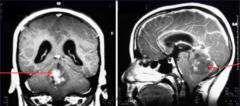
Medulloblastoma
The Purkinje cells produce the mitogen, Shh, while the granule cell progenitors express the Shh receptors, patched.
Mutations in patched results in uncontrolled cell production of granule cells in cerebellum.
TB Authors
Dan H. Sanes, Thomas A. Reh, William A. Harris
Animal models used to study the brain
1) C Elegans: have 302 neurons, 56 glia help to study communication between neurons
2) Chicks: embryo used in the Hamburger Hamilton stages of development
3) Monkey: higher cognitive function, drug discovery
4) Birds: development of language
Golgi silver staining vs Nissl staining
golgi: stains a small number of neurons randomly shows dendrites and axons
nissl: stains nucleic acids blue or purple

Immunohistochemistry
localizing antigens or proteins in tissues using labeled (colored or fluorescent) antibodies
1) primary: customized, specific to recognize the antigen
2) secondary: flexibility, species-specific, attached to gfp-monoclonal: binds to one part of antigen,
polyclonal: different antibodies binds to one antigen

GFP (green fluorescent protein)
a gene cloned from jellyfish and used in live imaging by "tagging" a protein with a fluorescent molecule

Conventional Vs Confocal Imaging
1) Conventional: light is spread out
2) Cofocal: has a little narrow hole that allows signals to pass through creating a more accurate signal

Two-photon Imaging
Make use of relatively low-energy red light to excite fluorochromes; can trace cellular activity in real time (ex: used to detect amyloid aggregates)
1. Cells are stained with fluorochrome dyes
2. Two photons of long-wavelength (red) light are used to excite the dyes
3. Can study living cells up to 1 mm deep

How are new born neurons labeled?
1) 3H-Thymidine: Compound used in autoradiography to label DNAwhen progenitor cells are actively dividing, they synthesize DNA and incorporate isotopically labeled thymidine-cells born within a day after injection will have heavily labeled nuclei
2) 5-bromo-2-deoxyuridine (BrdU): a thymidine analog that is incorporated in newly synthesized DNA

How to label human newborn neurons?
Retrospective birth dating:
-testing of nuclear weapons resulted in increase of radioactive C14 in the atmosphere and this can be related to level of 14C is genomic DNA allowing for tracing of new cells in the cortex of humans
Neuron labeling vs connection labeling

Anterograde and Retrograde Tracers
Anterograde: Cholera Toxin B dye injected into cell bodies and carried to axons
Retrograde: Fluorogold dye taken up by axon terminal and carried back to cell bodies (going against the flow)
both used to analyze transport pathways in neurons

Viral Labeling (AAV and Gene Therapy)
1) AAV vectors are made by splicing a single gene and putting a new gene inside
2) virus is converted into a recombinant vector
3) the packaging and vector constructs are introduced into the packaging cell by transfection
5) both packaging constructs and vector constructs express their proteins and the proteins are encapsulated into virus particles to generate the recombinant viral vector

Gene Therapy
-replacing a mutated gene that cause disease with a healthy copy
-inactivating a mutated gene that functions improperly
-introducing a new gene to fight a disease
-commonly used in retroviral vectors
Action Potential
1. Stimulus starts the rapid change in voltage or action potential.
2. Depolarization: a rapid rise in membrane potential opening of sodium channels in the cellular membrane, resulting in a large influx of sodium ions.
3. Repolarization: a rapid sodium channel inactivation as well as a large efflux of potassium ions from activated potassium channels.
4. Hyperpolarization: a lowered membrane potential caused by the efflux of potassium ions and closing of the potassium channels.
5. Resting state is when membrane potential returns to the resting voltage that occurred before the stimulus occurred.

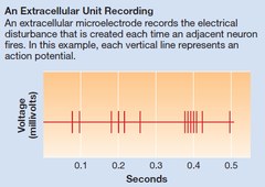
Single-unit recording
recording of the electrical activity of a single neuron

Multiple-unit recording
use macroelectrodes to record the electrical activity of many neurons
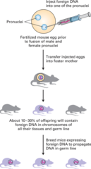
Transgenic organism
organism that is genetically engineered by inserting a gene from another organism and bred until DNA is stabilized to get transgenic line
Conditional knockout animals
spatial and temporal control using the Cre-Lox system to delete of gene of interest

Cre-lox system
uses the enzyme Cre-recombinase to delete a sequence flanked by lox sites (specific target sequence for the enzyme)

CRISPR system and Cas9
1) CRISPR allows to edit parts of genome by removing or editing sections of the DNA sequence
2) recognizes foreign DNA sequences that have previously entered the cell and directs the Cas proteins to destroy them.
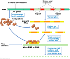
Sufficient vs Necessary
Sufficient: overexpression of protein A = expansion of structure B
Necessary: knockout protein A = structure B disappeared

Optogenetics
Transgenic technique that combines genetics and light to control targeted cells in living tissue and can control specific behaviors (like movement)

Development of vertebrate central nervous system
1. Cell division: series of cleavage where cells divide and move around randomly
-forms an animal pole (becomes the embryo) and a vegetal pole (provides nutrition)
2. Blastula forms: a collection of cleaved cells
3. Blastopore forms after involuting marginal zone (IMZ) cells grow into the interior of the embryo
4. Gastrulation: the involuting cells form mesodermal tissues and induce cells of the overlaying ectoderm to develop into neural tissue/neurogenic region (layer formation)
5. After neural induction, the neurogenic region becomes the neural plate and only gives rise to neural tissue and the neural plate generates the neurons and glia in the adult brain and spinal chord

First steps in neuronal differentiation - Primary Induction
Primary Induction: the interaction between mesodermal cells and overlaying ectodermal cells
-the neural plate on the ectoderm folds at its midline to form the neural groove, separated from the ectoderm and lies above the notochord (spine) and flanked by somites (skeletal muscles)
-the involuting cells condense to form a rod-shaped notochord
-neural plate rolls up and fuses to dorsal margins to form neural tube (becomes CNS) and at the same time the neural crest (a group of cells that migrate to become PNS)

Neural lineage experiment
Pregastrula: if animal cap is isolated from rest of embryo before gastrulation the cells develop as the epidermis
Gastrula: if animal cap is isolated during gastrulation, it will develop into neural tissue
This experiment led to idea that neural lineage arises during gastrulation
Spemann organizer
1) Spemann and Mangold transplanted the dorsal lip of blastopore from a pigmented embryo to nonpigmented host embryo
2)second axis was induced by transplanted tissue, included some pigmented and some nonpigmented tissue
3) dorsal lip (organizer) could induce cells to form a new body axis
They concluded that the dorsal lip cells could "organize" the host cells to form a new body axis, and they named this special region of the embryo the organizer.

Neural Induction requires the inhibition of ____
BMP signal (Bone-morphogenic proteins): neural fate is suppressed by cellular associations in the ectoderm
-if intact animal caps are put into culture = they develop as epidermis
-intact + BMP inhibitors = develop into neuron
-if animal cap is dissociated = develop into neurons
-if animal cap is dissociated + BMP added: develops into epidermal cells

What are the three BMP signal inhibitors?
chordin, noggin, and follistatin; released by involuting mesodermal cells and interfere with activation of BMP receptor to induce development of neural tissue (brain, spinal chord, and peripheral nervous system)

BMP signal transduction pathway
1) BMP Activation: BMPs are created in a form that needs to be processed and activated. Once active, they form pairs called dimers.
2) Receptor Binding: The active BMP dimers bind to a specific set of receptors on the cell surface. These receptors form a group of six parts, with two types of receptors (type I and type II).
3) Receptor Activation: The type II receptors activate the type I receptors by adding a phosphate group to them (phosphorylation). The type I receptors then do the same thing to a set of proteins called R-Smads.
5) Smads Move to the Nucleus: Once the R-Smads are activated, they join with another protein called Smad-4. Together, they move into the nucleus (the control center of the cell) to activate the genes they are targeting.
6) Regulation by BMP Antagonists: Sometimes, BMP antagonists (proteins that block BMPs) can prevent the signal from working by forming a complex with the BMPs before they bind to the receptors, stopping the process before it starts.

Loss of noggin and chordin in mice
1) loss of noggin = defective ear
2) loss of noggin and chordin = extreme deletion of forebrain, eye, nose and face feature
*only mild effect with loss of noggin, but extreme defects in head/no head when both noggin and chordin are knocked out

Brachydactyly (short fingers and toes) is caused by
mutations in Noggin

BMP vs Wnt signaling pathways
BMP: neural induction process
Wnt: important in regional specification of the nervous system

Current model of neural induction
Epidermal: BMP activates receptor, receptor activates intracellular Smad, Smad inhibits Zic 1, inhibiting activation of neural genes (this is the default condition)
Neural:
1) n/c/f inhibits BMP, inactivating Smad and no longer inhibiting Zic 1
2) FGF binds ERK, which activates Zic 3, which leads to neural gene expression
*both Zic 1 and Zic 3 activate downstream neural progenitor genes

Notch/Delta pathway
Delta binds Notch = cleavage of Notch with gamma-secretase = releases Notch ICD to intracellular domain = turn on expression of downstream target genes (SuH) = transcription of E(spl) genes (these are repressors of asc gene transcription and block further neural differentiation and reduce level of delta expression)
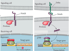
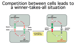
Lateral inhibition of Notch and Delta
1) Initial State: In the proneural cluster, all the cells can potentially become nerve cells. Each of these cells produces (Asc) and components of the Notch signaling pathway.
2) Activation of Neuroblast Genes: The Asc proteins trigger the production of neuroblast genes (which are important for a cell to become a nerve cell) and a signaling molecule, Delta. This sets up a positive feedback loop: if one cell starts producing more Asc, it also starts producing more Delta.
3) Notch and Delta Interaction: Delta from the first cell activates the Notch pathway in its neighboring cells. When the Notch pathway is more active in these neighboring cells, it causes them to produce more E(spl).
4) Inhibition of Neighbors: E(spl) inhibits the production of Asc in the neighboring cells, so these cells stop producing Delta and can't become nerve cells. This ensures that only the first cell (with higher Asc and Delta levels) will turn into a neuroblast, while the surrounding cells will not.
In amphibian embryos, Chordin, Noggin, and Follistatin are secreted by ____ to interfere with the activation of BMP-BMP receptor which results in neural induction.
Involuting mesodermal cells
Brain sections
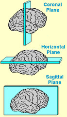
Brain axes
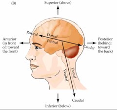
Forebrain, Midbrain, Hindbrain
forebrain: prosencephalon, telencephalon and diencephalon (forms cortex)
midbrain: mesencephalon (forms colliculus)
hindbrain (rhombencephalon): metencephalon and myelencephalon, divided into rhombomeres (forms cerebellum)

Development of the nervous system depends on the expression of particular genes at a particular ____ and ____
place and time (spatio-temporal factors = certain molecules may be turned off on on at a different time or place)
Graded concentrations
graded concentrations of antagonizing diffusible molecules are involved in setting up patterns and gives extra power for generation by creating an antagonizing gradient
*signaling molecules act to restrict expression of certain transcription factors which regulate the expression of downstream target genes for specific regional identity
Anterior-Posterior Patterning
Division between rostral brain and hindbrain
Otx2 only expressed in anterior neural plate (head) and remains expressed in most of brain through development
(Fibroblast growth factor FGF low)
Gbx2 only expressed in posterior neural plate (hindbrain and spinal chord) (FGF high)

Hox gene in drosophila and mouse
Difference: Hox gene in drosophilla are only in one chromosome and on four different chromosomes in mouse
Similarity: overall order of hox genes is the same its 3' to 5' anterior to posterior (spatial organization and A-P axis specification)
Orthologs: retain same function during evolution
Paralogs: different genome in 1 species (a3, b3, d3)

Hindbrain Rhombomere
hindbrain rhombomere identity determined by Hox code (3' to 5')
trigeminal nerve = jaw
abducens nerve = eye
glossopharyngeal nerve = swallowing
vagus nerve = digestive tract

Hox gene knockout affect on rhombomere
removal of Hoxa1 results in loss of rhombomere 5 (no abducens motoneurons) and reduction of rhombomere 4 (reduced facial motorneurons)

How is Hox regulated?
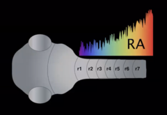
Retinoic acid (RA): important for A-P pattern pf Hox gene
1) RA (a metabolite of vitamin A) binds to RAR (RA receptor)
2) RARE binds to promoter region to activate Hox region

Fetal retinoid syndrome
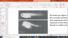
physical and mental birth defects resulting from retinoid usage showing that increase in retinoic acid during early development creates teratogens and disrupts head development
Prosomeric model of forebrain development
there are longitudinal and transverse (time and place)patterns gene expression that subdivide the neural tube into a grid of different regional identities
-Emx 1 and 2: anterior and posterior cerebral hemisphere

RA gradients
less RA in anterior (head) region, more in hindbrain and spinal chord region
Dorsal-Ventral Patterning
Shh: expressed in notochord and later floorplate and induces ventral differentiation in neural tube (going up)
BMP, Wnt: expressed in ectoderm over neural tube and later in dorsal neural tube forming roofplate (going down)
*These two signals antagonize one another, and through this mutual antagonism they set up opposing gradients that control both the polarity of spinal cord differentiation and the amount of spinal cord tissue that differentiates into dorsal, ventral, and intermediate cell fates.

Differentiation of neural tube
-depends on factors from adjacent, non neural tissues
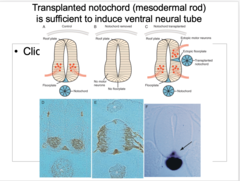
1) Control: notochord expressing Shh so motor neurons stick to ventral side
2) Notochord removed: No floorplate = no motor neurons (NECESSARY)
3) Notochord transplanted (2 of them): a second notochord induces a second floorplate region = more motor neurons (SUFFICIENT)
Shh in intermediate explant for ventral region
adding Shh to explants of the neural tube is enough to induce them to differentiate as ventral neural tissues including floorplate and motor neurons (ventral genes Olig2 and Nkx2.2 increase with increasing Shh)
Pax7 expressed in intermediate and dorsal regions

BMP in intermediate explant for dorsal region
Antagonism by dorsal BMP and ventral Shh pattern the expression of dorsal-ventral genes in a dose dependent manner
BMP also activates HNK1 and slug
Regional identity of brain

What two transcription factors are critical for specification of regional identities in the cortex?
1) pax6 (anterior; motor cortex) and emx2 (posterior; visual)
2) loss of pax 6 = visual cortex expanded and motor cortex reduced
3) loss of emx2 = motor cortex expanded and visual cortex reduced

pax6 role in eye development
1) overexpression of pax6 = more eye structures
2) pax6 mutation (lose one pax6) = aniridia (iris-less)
3) lose both pax6 = lethal

Fgf8 patterns in cerebral cortex
normally FGf8 is expressed anteriorly and BMP posteriorly (creating an antagonizing gradient)
1) increasing FGF8 causes a caudal shift (somatosensory moves down)
Placing another FGF8 in caudal cortex (posteriorly) causes formation of duplicated somatosensory map

interkinetic nuclear migration
process by which the nucleus of a radial glial cell migrates away and back towards the ventricular zone; necessary for DNA replication and ultimate cell division
- the neural tube contains the progenitors of all the neurons and glia in the mature brain and spinal chord
- when cells are ready to become neurons, they will move away from ventricular zone (G2 and M-phase) and migrate up (S-phase)

Multipotent progenitor cells
Clonal analysis in chick neural tube: one progenitor cell labeled with a virus
1) the single infected progenitor cell goes though single-cell division to produce two daughter cells after 1 day
2) if embryo survives neurons and glial cells can be identified in the mature spinal chord
3) multiple different cell types arise like motorneurons, astrocytes and oligodendrocytes
This shows that viral genes are inherited in all the daughter cells of the infected progenitor cells

Cerebral cortex histogenesis
1) apical progenitor cells in ventricular zone generate postmitotic cells (note: stem cells migrate upward from VZ to SVZ and divide symmetrically to generate more progenitors)
2) newly generated neurons migrate along the radial glial cells that generated them to form cortical plate

Inside-out pattern of cerebral cortical histogenesis through birthdating
1) pregnant rat injected with H-thymidine at later intervals stages of gestation and pups brains analyzed for labeled neurons
2) E11: postmitotic neurons, closter to subplate/VZ
3) E13: neurons in deep cortical layers (moving upwords)
4) E15: neurons in superficial cortical layer (surface)

Generation of neurons and glia
dissociated progenitor cells from developing CNS studied in cell culture
-unipotent: 1 type of cell (oligodendrocyte)
-tri-potent
-bi-potent

Cell Cycle
During M phase
-Karyokinesis: nuclear division
-Cytokinesis: cellular divison (2 daughter cells)

The overall length of progenitor cells cycle _____ during embryogenesis
increases; this increase in cell-cyle period is mainly due to increase in G1 phase (where DNA synthesis prep/growth happens so it requires more time)

Cerebral cortex growth

P/Q ratio
ratio of progenitors to quit (post-mitotic) cells (total = P +Q)
Starts out having higher P, then goes to having higher Q
p27 and Rb cause cells to go into Q (post-mitotic), quit cell cycle
cyc D and Cdk cause cell to continue in cell cycle
increased Q = thinner ctx (more cells leaving)
increased P = thicker ctx (this is done by removing inhibition of Rb and p27)

What genes and pathway regulate neurogenesis?
-the proneural genes and notch pathway regulate neurogenesis
-proneuroal genes segregtes neural tissue from epidermis by inducing Notch pathway which activates Hes1 expression that lateraly inhibits adjacent proneural genes making them epidermal tissue instead (cells expressing higher level of proneural genes differentiate as neurons)
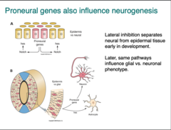
Regulating neurogenesis vs gliogenesis
FGF2 and Neurotrophin3 help cells become neurons.
EGF and CNTF push cells to become astrocytes.
BMPs work with CNTF to encourage glial development.
Notch pushes cells to become astrocytes while blocking them from becoming neurons.
Hes gene maintenance of radial glial cells
Hes = maintenance of neural stem cells and promotion of astrocyte formation by Hes genes (maintains projenitor cells)
increase Hes = more cells stay in ventricular zone and produce more cells that become astrocytes/radial glia instead of neurons
Neurogenesis in adults (C14 Birth dating)
-most cell growth occurs early on in life
-cerebellar cells born about 2 years
-cortex cells grown about 7 years
-hippocampus keeps growing! 700 new neurons per day, average date of birth = 30 years
-low C14 levels at birth vs high C14 levels after show that some cells are born after birth (neurogenesis throughout life)
4 steps of cerebral cortex histogenesis
First stage, progenitor "stem" cells, which occupy the ventricular zone (VZ).
Second stage:
Postmitotic cells -> preplate: superficial Cajal-Retzius cells and the subplate cells.
Third stage: newly generated neurons (red) migrate along radial glial fibers to form a layer between the Cajal-Retzius cells and the subplate. This layer is called the cortical plate, and the majority of the neurons in the cerebral cortex accumulate in this layer.
Forth Stage: layers appear in cortical plate; inside-out pattern. (neurons migrate from inside or neural tube to outside with earlier born neurons staying in deep layers and later born neurons migrating to superficial outer layers)
ex: in neurogenesis: pyramidal neurons are born first and migrate to subcortical regions
local interneurons born after are in middle and pyramindal cells born last are in cortical areas (superficial)
cell determination vs differentiation
cell determination: stem cells becomes committed to develop into one type of cell
cell differentiation: the process by which cells become different types of cells after determination (muscle cell, nerve cell, skin cell)

Neurons of cerebral cortex derive from _____
intrinsic and extrinsic sources
-ventral-derived interneurons (MGE)
-cortical-derived interneurons
cells can grow vertically and laterally
What is reelin and what does it do?
an extracellular matrix glycoprotein that controls cerebellum growth
Mutation in this gene results in lissencephaly cerebellar hypoplasia (LCH) = smaller cerebllum + problems with walking/balance
Function of reelin in cerebral cortex
A. in normal cortical development, the newer generated neurons bypass the older ones to produce inside-out developmental ordering (newer cells on top and older on bottom)
B. in reeler mice, this inside-out order is disrupted and the newly generated neurons cannot pass the older ones so layers are reversed (newer cells on bottom and older on top)

Reelin Pathway
1) Reelin is expressed in Cajal-Rezius cells in superficial layer of the cortex
2) Reelin binds to ApoER2 and VLDLR resulting in phosphorylation and ubiquitinylation of Dab1 leading to degredation and inhibition of migration/cells stop moving
(activation of Dab1 promotes migration)
3) Reelin can trigger differnet types of signal molecules downstream leading to other affects than migration inhibition like dendrite and spine development and synaptic function
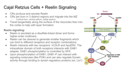
Are all neurons generated before adulthood?
No, in adult mice newly generated hippocampal neurons are functionally integrated with preexisting neurons
EphrinB receptor
inhibitory signaling cascade
Brainbow technique
used the Cre-lox system to insert short DNA sequences into sequence that express a different color (need 2 pairs of lox so cre recognized where to cut)
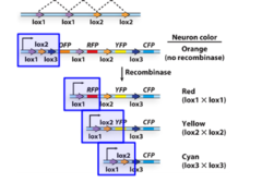
Ti1 pioneers experiment
-growth of Ti1 pioneers is aided by guidepost or stepping-stone cells
-ablation of guidepost cells leads pioneers to become lost
-shows that environmental factors are important

Salamander Mauthner cells
-rotate part of the forebrain and replace it, then neurons originally grow in the opposite direction (anteriorly), but when the axon reaches the boundary of the hindbrain, adjusts trajectory --mauthner cells grow posteriorly due to local cues

Cutting retina experiment
-when optic nerve is cut, axon of retinal ganglion cell still can navigate to the tectum using their growth cones to guide them -shows the importance of intrinsic cues
Growth cones are made up of ___
actin filaments and microtubules
How do growth cones grow?
1) microtubules can reversibly assemble and disassemble (microtubules added to growing end) and there is also interstitial growth
2) this requires alot of energy so when filamentous actin is assembled in the filopodia of the growth cone, some of this is left behind in the submembranous cortex of the axon as part of the axon's cytoskeleton.
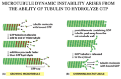
structure of growth cone
-microtubule = backbone of axon
-filopodia: bounded by membranes with cell adhesion and recpetors and poke out at terminal end and retract at bottom end
-lamellipodia: sheets that extend forward between the filopedia

a growth cone is both a ____ and ____ structure
sensory and motor
Follower axon and leader axon
leader axons growth cone does most of work to sense positive and negative midline cues and interpret them correctly so its progress is slow and spends more time at midline wheras follower axons that grow along the leader are less exposed to midline and have more simple growth cones that grow faster