protozoa: class lobosea (the amebae)
 Call Kai
 Learn
 Practice Test
 Spaced Repetition
 Match
 Flashcards
 Knowt Play
1/38

There's no tags or description
Looks like no tags are added yet.
39 Terms
ameba
single-celled parasite that moves by means of cytoplasm extensions called pseudopods (false feet). two forms: trophozoites, cysts.
trophozoite
motile feeding stage of amebas that multiply by binary fission. actively multiply by asexual binary fission if in intestinal tract. disintegrate in outside environment. 1 nucleus
cyst
non-motile, non-feeding stage of amebas that are capable of survival outside their hosts and of initiating human infection. initiation of human intestinal infection if ingestion. in environment, continue to develop for subsequent entry into unsuspecting human & new cycle begins. can have more than 1 nucleus
karyozome
central mass of chromatin inside select amebic nuclei
peripheral chromatin
ring of chromatin that surrounds the nuclei of select amebas
chromatid bar
oblong structures containing condensed RNA material in the cytoplasm of select amebic cysts
glycogen mass
cytoplasmic area without defined borders that is believed to represent stored food, usually visible in young cysts
excystation
transformation of amebic cysts into amebic trophozoites. ileocecal area of intestine.
encystation
transformation of amebic trophozoites into amebic cysts. changes in intestinal enviornment
intestinal species
entamoeba: histolytica/dispar, hartmanni, coli.
iodamoeba butschlii, endolimax nana
extraintestinal species
naegleria fowleri, acanthamoeba spp.
what happens when cysts &/or trophozoites do not/cannot make conversion in time?
they are found in the stool
specimen of choice for amebas
intestinal: stool.
extraintestinal: varies by species but include mouth scrapings, CSF/brain tissue, corneal scrapings.
ameba pathogenesis & clinical symptoms
most intestinal infections are asymptomatic. diagnosis of nonpathogenic amebas important finding suggests ingestion of contaminated food/drink may have occurred. E. histolytica only intestinal ameba considered pathogenic with characteristic symptoms, can also expand beyond intestinal tract & cause extraintestinal symptoms

entamoeba histolytica/dispar trophozoite
motility: progressive, finger-like pseudopodia.
karyozome: small & central.
peripheral chromatin: fine & evenly distributed.
cytoplasm: finely granular.
cytoplasmic inclusions: ingested RBCs (diagnostic)


entamoeba histolytica/dispar cyst
shape: spherical to round.
nuclei: immature have 1-3, mature has 4.
karyosome: small & central.
peripheral chromatin: fine & evenly distributed.
cytoplasm: finely granular.
cytoplasmic inclusions: chromatid bars, rounded ends in young. diffuse glycogen mass in young.

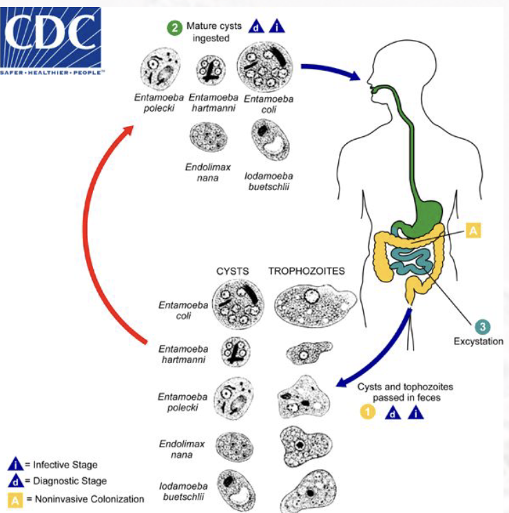
entamoeba histolytica/dispar life cycle
transmitted via contaminated food/water, hand-to-mouth, unprotected sex. transmitted by circulating flies & cockroaches who carry the parasite on their legs. dispar is asymptomatic, carrier. histolytica trophozoites are symptomatic.
cysts found in formed stool, trophozoites found in diarrheal stool. excystation occurs in small intestine & trophozoites are released and migrate to large intestine. they can migrate out of the intestinal tract into liver.

entamoeba histolytica/dispar
found in tropic, subtropic, & cold climates.
intestinal amebiasis: diarrhea, abdominal pain & cramping, weight loss & anorexia, chronic fatigue, flatulence, toxic megacolon.
extraintestinal amebiasis: varies w/ location of parasite; liver/lung, pericardium, venereal, spleen, brain.
people at risk: individuals who live in areas of poor sanitation, mentally ill hospitalized patients, prisoners, MSM. prevented by proper water treatment & food handling, protected sex, protection of food from insects, avoidance of using human stool as fertilizer.
nonpathogenic intestinal amebae
E. hartmanni, E. coli, E. nana, I. buetschlii

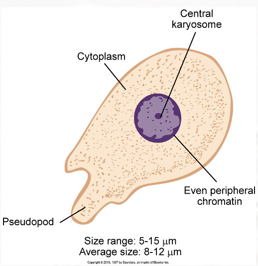
entamoeba hartmanni trophozoite
motility: nonprogressive, finger-like psuedopods
karyosome: small & central.
peripheral chromatin: fine & evenly distributed.
cytoplasm: finely granular.
cytoplasmic inclusions: ingested bacteria may be present.

entamoeba hartmanni cyst
shape: spherical.
nuclei: immature have 1-3, mature have 4.
karyosome: small & central.
peripheral chromatin: fine & evenly distributed.
cytoplasm: finely granular.
cytoplasmic inclusions: chromatoid bars, rounded ends in young. diffuse glycogen mass in young.

entamoeba hartmanni
found in metropolitan areas. transmitted by consuming contaminated food/water or via insects like E. histolytica. usually asymptomatic. prevented by good sanitation & personal hygiene practices, protection of food from insects.

entamoeba coli trophozoite
motility: nonprogressive, blunt psuedopods.
karyosome: large, irregular shape, eccentric.
peripheral chromatin: unevenly distributed.
cytoplasm: coarse & granulated.
cytoplasmic inclusions: vacuoles containing bacteria often visable.


entamoeba coli cyst
shape: round to spherical.
nuclei: 1-8.
karyosome: large, irregular shape, eccentric.
peripheral chromatin: unevenly distributed.
cytoplasm: coarse & granulated.
cytoplasmic inclusions: diffuse glycogen mass present in young, may displace nuclei (often seen w/ 2 nuclei) to opposite ends of cyst. thin chromatoid bars w/ pointed to splintered ends in young
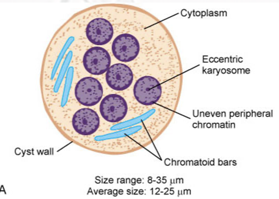
entamoeba coli
found worldwide. transmitted by consuming contaminated food & water or via insects like E. histolytica. usually asymptomatic. prevented by good sanitation & personal hygiene practices, protection of food from insects

endolimax nana trophozoite
motility: sluggish, nonprogressive, blunt psuedopods.
karyosome: large, irregular, blotlike.
peripheral chromatin: absent.
cytoplasm: granular & vacuolated.
cytoplasmic inclusion: bacteria.


endolimax nana cyst
shape: spherical, ovoid, ellipsoid.
nuclei: 1-4 (4 most common).
karyosome: large, blotlike, usually central.
peripheral chromatin: absent.
cytoplasm: granular & vacuolated.
chromatin inclusions: chromatin granules. nondescript small masses. diffuse glycogen mass in young

endolimax nana
found in warm, moist regions. transmitted by consuming contaminated food & water or via insects like E. histolytica. usually asymptomatic. prevented by good sanitation & personal hygiene, protection of food from insects.

iodamoeba butschlii trophozoite
motility: sluggish, usually progressive.
karyosome: large, usually central; refractive achromatic granules may be present.
peripheral chromatin: absent.
cytoplasm: coarsely granular & vacuolated.
cytoplasmic inclusions: bacteria, yeast cells, other debris.


iodamoeba butschlii cyst
shape: ovoid, ellipsoid, triangular, other.
nuclei: 1.
karyosome: large, eccentric; achromatic granules on one side may be present.
peripheral chromatin: absent.
cytoplasm: coarsely granular & vacuolated.
cytoplasmic inclusions: well-defined glycogen mass (diagnostic), granules may be present.

iodamoeba butschlii
use iodine prep (iodine lover) for lab diagnosis. found in tropic regions. transmitted by consuming contaminated food/water, hand-to-mouth. usually asymptomatic, diarrhea if any. prevented by practicing good personal hygiene & sanitation practices.

naegleria fowleri trophozoite
motility: sluglike, blunt pseudopods.
karysome: large & usually centrally located.
peripheral chromatin: absent.
cytoplasm: granular, usually vacuolated.


naegleria fowleri cyst
do not form in human tissue. spherical shape. smooth, single-layered wall. single nucleus.

naegleria fowleri
found in warm bodies of water (lakes, streams, ponds, swimming pools). grows best at higher temps. saline/iodine wet preps of CSF, giemsa stain of CSF. trailing affect on culture plate. laboratory-confirmed (nucleic acid, organisms, antigen) = diagnostic.
may be asymptomatic. primary amebic meningoencephalitis (PAM. initial signs & symptoms of PAM begin 1-14 days after infection. death within 3-7 days. typical treatments infective but investigational anti-leishmania drug miltefosine.
naegleria fowleri life cycle
3 morphological forms: ameboid trophozoite (only form known in humans), flagellate form, cyst. entire life cycle appears to occur in external environment. ameboid trophs enter nasal mucosa and then may migrate to brain via olfactory nerves.


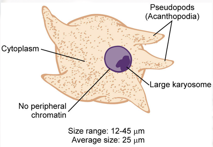
acanthamoeba spp trophozoite
motility: sluggish, spinelike pseudopods (diagnostic).
karyosome: large.
peripheral chromatin: absent.
cytoplasm: granular & vacuolated

acanthamoeba spp cyst
shape: roundish w/ ragged edges.
nuclei: 1.
karyosome: large & central.
peripheral chromatin: absent.
cytoplasm: disorganized, granular, sometimes vacuolated.
other features: double cell wall, smooth inner cell wall & outer jagged cell wall

acanthamoeba spp
CSF, brain tissue, corneal scraping specimens. tracks on culture plate using confocal microscopy, calcofluor white staining, RTPCR
immunocompromised/debilitated individuals at greatest risk for CNS infections, soft contact lens wears also 85% of cases. causes granulomatous amebic encephalitis (GAE): headaches, seizures, stiff neck, N/D, granulomatous lesions of the brain (characteristic), parasites may invade other body areas. causes keratitis: ocular pain/vision problems, loss of vision.
acanthamoeba spp life cycle
humans contact parasite in 2 different ways. 1. aspiration or nasal inhalation of the organisms through nasal mucosa &/or skin — serious CNS infection. 2. direct invasion into the eye — serious eye infection possible. 10 spp that are known to infect humans. life cycle poorly understood.
