Complex Needs Exam 3
 Call Kai
 Learn
Learn Practice Test
 Spaced Repetition
 Match
Match Flashcards
 Knowt Play
1/129

There's no tags or description
Looks like no tags are added yet.
Name | Mastery | Learn | Test | Matching | Spaced | Call with Kai | Chat |
|---|
No analytics yet
Send a link to your students to track their progress
130 Terms
Depolarization
electrical charge changes → cell does to WORK
Repolarization
electrical charge return to “normal” cell is resting.

How may seconds are these big boxes
.20 seconds
P wave
Atrial depolarization → if this is upright → the beat originated from the SA node → first name of rhythm will be sinus.
QRS
ventricular depolarization means the ventricles are ready to work (beat/pump)
T wave
ventricular repolarization - the ventricles is resting

Measure of the PR interval this is time for atria to
Contract (pump the last bit of blood into the ventricles)
Allows the electrical impulse (wave) to be sent on down to the ventricles
Last 0.12 -0.20 sec

How long should the QRS complex be
< .12 sec
Is the QRS is narrow <.12 then the impulse originated from the
Atrium
YES there is a P wave but the PR wave is prolonged and fixed measuring same for every beat then this is a.
First Degree AV block
In this cardiac rhythm there is not always a p wave before every QRS. The P-R interval is longer and longer and longer—then there is a drop
Second Degree Av block (Mobitz type 1) Wenkeback
In this AV block the P wave is upright; there is a not a p wave before every QRS, the PR interval is the same(fixed) this rhythm is a
Second degree AV block Mobitz type 2
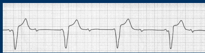
In this rhythm the P wave looks weird, there is not always a P wave before QRS. The QRS is not measurable and is (WIDE) bc impulse came from ventricles.
Third Degree AV block
If wave is identified as Normal sinus rhythm but the patient has no pulse, meaning there is no mechanical activity the heart is not pumping blood, the patient is dead. then this a
PEA
Regardless of the monitor you must treat every
Symptomatic pt
Examples of symptomatic pt complaints to TREAT
chest pain, dyspnea, dizzy, diaphoretic, cool/clammy, palpitations
Signs of hemodynamically unstable pt to TREAT
Low B/P, low LOC, Low Oxygenation, pulse/pulseless
Pt is hemodynamically stable BUT had symptoms how do you treat
Pt may need IVP meds
Pt is hemodynamically unstable AND has symptoms how do you treat
May be need IVP meds OR cardioversion/defib or BLS
All dysrhythmias have potential to decrease
Cardiac output
If ventricles generate major portion of the cardiac output which kind of dysrhythmias are mor concerning/higher priority
Ventricular dysrhythmias
After electricity is delivered from Cardioversion or defibrillation what normally appears after
Flat line →normal rhythm
This is used when pt has a pulse; lower amounts of energy.
MUST hit synchronize button, electricity must be delivered on R wave.
Cardioversion
What is cardioversion is delivered on T wave
sends pt into V fib
Used when pt does not have pulse, higher energy used in comparison, pt does not have pulse to deliver shock on.
Defribrillation

If the symptomatic pt with this rhythm is dizzy, feels like he's going to pass out, B/P is 80/40, O2 sat 98%. how would u treat this AV block/sinus bradycardia
Adequate airway and give 100% oxygen
Atropine 1mg I push
Epinephrine IV infusion or dopamine IV infusion (drip)
Pacemaker (transcutaneous, transvenous, epicardial)
How many times can I repeat atropine in Heart blocks/Sinus bradycardia
3 times.

If man presents with B/P is 80/40, O2 sat 98%. and several doses of atropine has been administered and there is no change in rhythm or condition; what is the treatment ?
Pace the patient

Nursing student up studying all night for her critical care exam, and drinking red bull, feels like heart is going to jump out of chest, pt has palpitations: B/P 130/70, O2 sat 100%; what is her rythym
Supra-ventricular tachycardia SVT
Non medication interventions for the treatments for SVT (atrial tachycardia)
Ensure patent airway give 100% oxygen
Valsalva or vagal maneuvers
Medication interventions for the treatments for SVT (atrial tachycardia)
Adenosine 6 mg rapid IV push
Second dose 12 mg rapid IV push
What kind of shock can be given to a pt who has symptomatic SVT and is hemodynamically unstable
Synchronized cardioversion

70 y.o man presents with palpitations. The feeling began suddenly while on the golf course. denies "pain" just feels like "my heart is about to jump out of my chest". A, A O X3. B/P 130/70, O2 sat 100%: this rhythm is classified as
Atrial fibrillation
What meds are giving during symptomatic Atrial fibrillation (ABCD
Amiodarone, beta blockers, cardioversion (synchronized) diltiazem (cardiazem), digoxin
Some medications convert the rhythm to normal sinus rhythm these are
Amiodarone(2fnct) and adenosine.
Some drugs simply slow the rate of the dysrhythmias these are:
Diltiazem, beta blockers , digoxin, amiodarone (2fnct)
Which meds increase the rate of the heart
Atropine and epinephrine

What would be the treatment of a pt with symptomatic Atrial fibrillation AND also hemodynamically unstable, after all medications have been given:
Synchronized cardioversion
A fib with RVR (rapid ventricular rate) the pt is still hemodynamically stable, but this is recurring for the pt, what would the cardiologist consider
ablation
What kinda cells cause A fib
Rogue atrial cells that initiate an electrical signal and the ventricles respond by depolarizing and beating.
An ablation may be performed to eliminate the arrhythmia, what occurs during ablation.
A radio frequency(burning) or cryo energy(freezing) is used to destroy the rogue cells stopping the abnormal rhythm while leaning the normal/sinus pathway intact.
Atrial dysrhythmias should always include a_____ and never be treated with ______
ALWAYS PULSE !!
NEVER CPR !!!
How would u treat a pt who is in PEA
CPR
Secure airway; give 100% oxygen
epinephrine
Treat underlying causes
Review H and Ts
NO SHOCK
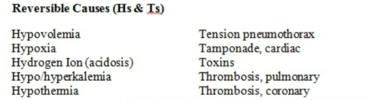
How would you ID this ECG and how would U treat it

Asytole:
CPR
Secure airway: give 100% oxygen
epinephrine
Treat underlying cause:
H and Ts
NO SHOCK

Identify this rhythm how do u treat it. Pt denies pain, but feels like his heart is about to jump out of his chest.
V tach with pulse.
Amiodarone 150 mg IVP over 10 min (1st choice, repeat as needed if VT recurs)
(procainamide 20-50mg/min, sotalol 100mg over 5 min, or adenosine 6/12mg)
What kind of shock would be delivered to a pt in VT with a pulse
who is unstable; after all medication interventions have been given.
Synchronized cardioversion
Pt having V Tach, meds have been given, cardioversion provided when pt was unstable, pt now does NOT HAVE A PULSE, what is the next intervention
CPR/ Defibrillation as soon as available
Medications for Ventricular Fibrillation or Pulseless Ventricular Tachycardia
Epinephrine 1mg IV/IO every 3-5 min
Amiodarone 300 mg IV/IO may be additional 150 mg once
Lidocaine 1-1.5 mg/kg IV/IO
Magnesium IV if torsades de pointes
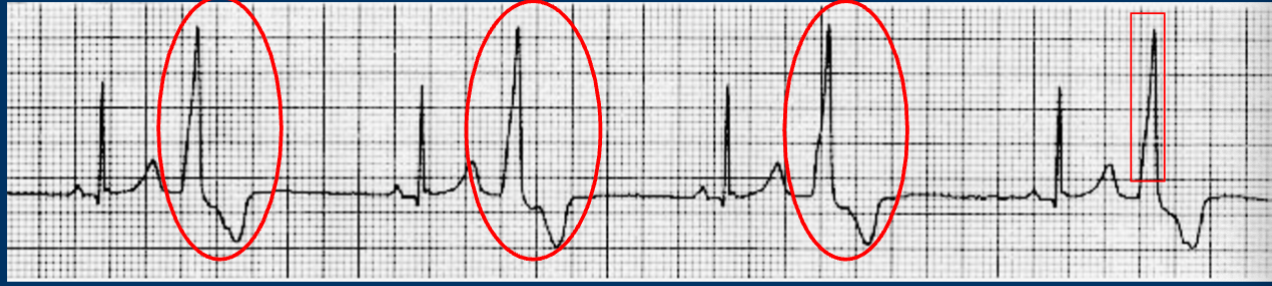
Premature ventricular contractions if the pt is symptomatic what medication and nursing responsibilities must take place:
If pt is symptomatic
Ensure patent airway and adequate oxygenation
Amiodarone or Procainamide IV
look for causes H and Ts

What causes (TdP) and what specific med is used in torsades de pointes.
A prolonged QT interval: •
• Antiarrhythmics (e.g., quinidine, sotalol, dofetilide, procainamide)
• Antibiotics (e.g., macrolides like erythromycin, fluoroquinolones)
• Antipsychotics and neuroleptics (e.g., haloperidol, thioridazine)
• Antidepressants (tricyclic antidepressants)
• Electrolyte Imbalances: Low levels of key minerals destabilize cardiac repolarization:
• Hypokalemia (low potassium)
• Hypomagnesemia (low magnesium)
• Hypocalcemia
Fix with Magnesium IV
This pacing system is non invasive, applied externally through the chest,
flow from defib machine to the pads though the skin to the heart “listen and respond”.
A short-term emergent type of pacing. Pacing the ventricles only
preferred if the patient is overly anticoagulated (ex. PT / INR way out of normal range).
Transcutaneous Pacing
An invasive method of pacing in which a special pacing catheter is inserted via the
right or left femoral vein, subclavian vein, or internal jugular vein and advanced into
the right side of the heart.
• Pacemaker tip rests in the RV apex and provides single chamber pacing only, paces the ventricle only.
• Connected to a pacing generator (the battery and brains) outside of the patient
that is set and monitored by the nurse.
Transvenous pacing
What are temporary pacemaker electrodes (wires) and where are they placed?
They are temporary pacing wires sutured or hooked to the outside of the heart (epicardium) and pulled out through the chest incision.
How do temporary wires actually pace the heart?
The wires are connected to an external pacemaker box (generator) to send electrical signals if needed.
Not readily available for an acute/emergent situation, must be implanted under strict sterile technique.
The generator/battery lasts 10 years. Can be externally programmed allowing for non-invasive adjustments of settings. NOTE: a magnet can disrupt permanent pacemaker functioning
Permanent pacing
Synchronous pacing
Consider the pacemaker as “on demand” pacemaker fires only when needed and in “harmony” with the patient’s own electrical rhythm, allows spontaneous beats (the person’s own electrical impulse to stimulate depolarization) without competition from the pacemaker.
What does it mean when doctor ordered pacing rate at 60 :
setting the pacemaker rate at 60, ensures that if the patient’s own heart rate falls below 60 beats per minute, the pacemaker will then deliver an electrical current to stimulate the cardiac cell to depolarize.
Asynchronous pacing:
fixed-rate pacing. delivers an electrical stimulus regardless of the patient’s spontaneous cardiac activity. Used only with no spontaneous beats – asystole

When would a pacemaker spike appear:
When the pacemaker fires an electrical impulse.
What happens if the pacing device doesn’t sense or “see” any electrical activity within a certain time period by the patient’s own electrical wiring also known as (pacing)
The pacing generator/pacemaker is activated, the device will “send out” an electrical impulse a set amount of energy, measured in milliamps, will be delivered from the pacemaker through the pacing pads/wires to the myocardium stimulating depolarization of the cardiac cells.
Capture
Refers to the successful stimulation of the myocardium by the pacemaker impulse resulting in
depolarization. It is seen on the ECG by a pacemaker spike closely followed by a ventricular complex (QRS).
The ability of the myocardium to “hear and respond “ is controlled (in part) by the amount of :
Current – measured in miliamps (mAs) –used to deliver the electrical impulse.
Sensitivity (pacemaker sensing)
How sensitive is the pacemaker to sense and "read" the patient's own native (intrinsic) heart
rate. If the pacemaker “sees or senses” the heart’s own electrical activity the pacemaker will
“stand down” and not fire an electrical impulse. The pacemaker is on demand and in tune or in sync with the heart’s own electrical activity.
Over sensing (the fence is toooo low)
Seeing ALL the electrical activity might cause the pacemaker to "misinterpret" the P waves and T waves as being QRSs too the pacemaker would then not pace thinking the P, QRS and T are 3 beats instead of 1 beat.
How to fix Oversensitive pacemaker:
The fence/sensitivity must be “raised” /increased to “hide” more of the electrical complex
Pacemaker sensitivity is a setting on the pacemaker “box” where the lower the number :
The more sensitive the pacemaker
Under sensing (the fence is tooooooo high – all the way up)
Pace maker ignores the patient's own native heart beat (cannot sense or see any electrical activity of the patient), and paces the patient when the pacer should not, fire spikes will occur when they should not = spikes all over the place
How to fix under sensing
The fence/sensitivity must be “lowered”/decreased to “show” more of the electrical complex
Pacemakers pace the heart while ICDs offer
Pacing and internal cardioversion or "shock therapy" when they sense Ventricular Tachycardia or Ventricular Fibrillation.
A nurse is reviewing the medical records of several clients to determine who meets the criteria for secondary prevention with an implantable cardioverter-defibrillator (ICD). Which client finding meets these criteria
• A history of survival from ventricular tachycardia (VT)/V FIb cardiac arrest with no identifiable reversible causes.
• Why it's right: Secondary prevention means protecting someone who already experienced the danger. Because they survived VT/VF and it can't be fixed by simple means (like fixing an electrolyte imbalance), they need an ICD to protect them from the next one.
Incorrect options to watch out for on a test:
• Incorrect: A patient who had a heart attack 10 days ago (Must be at least 40 days).
• Incorrect: A patient with an ejection fraction of 45% (Must be less than 35%).
• Incorrect: A patient whose cardiac arrest was caused by a temporary drug overdose (This is a reversible cause, so they don't get an ICD).
Patient Teaching Points - if the ICD delivers a shock
The patient should sit or lie down immediately.
Contact their healthcare provider ASAP and seek emergency medical care as directed.

Failure to Pace (no spikes, bradycardia or a rate that is less than the pacemaker rate setting).
Nursing interventions for failure to pace
Make sure dials are set correctly and the generator is on
Check heart rate, check sensitivity
Make sure connections are secure.
May need to shave the patient if the pacing pads are not adhering to the skin (transcutaneous pacing =TCP).
Limit the tension on the wire (physician will have to change the wire if damaged).
Change battery or generator (perm. Pacing)
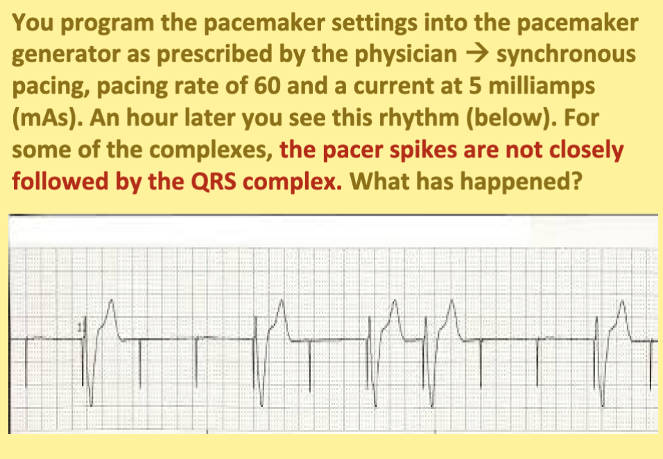

Nursing interventions for Failure to Capture
Increase the mAs (miliamps).
Check all the connections, change battery/ generator.
Move patient’s arm inward (adduct) or turn on side in case the transvenous pacing wire has been displaced – is not touching the muscle of the right ventricle
A nurse is caring for a patient who has a temporary transvenous pacemaker inserted via the right subclavian vein. The patient reaches across their bedside table with their right arm to grab their cell phone. Suddenly, the telemetry monitor shows pacemaker spikes, but they are not followed by a QRS complex (loss of capture). After instructing the patient to return their arm to a neutral position, what is the nurse's immediate priority action?
Correct Answer: Check the patient's pulse, blood pressure, and level of consciousness to assess hemodynamic stability.
Rational/Why: When a temporary wire pulls away from the myocardium, the electrical impulse cannot "swim" through the blood to stimulate a heartbeat, causing loss of capture. Before troubleshooting the machine or calling the physician, the nurse must immediately assess if the patient is stable or symptomatic from the sudden lack of pacing.
When a new permanent pacemaker/ICD is implanted, the patient must be instructed NOT to move their left arm outward (abduct = arm moved away from their body) for the first 24-48 hours WHY:
Allows time for the wire to “set” into place in the right ventricle. The wire must be touching the myocardium (inner layer of muscle) of the right ventricle for proper functioning/pacing to occur
You program the pacemaker settings into the pacemaker generator as prescribed by the physician synchronous pacing, pacing rate of 60 and a current at 5 milliamps (mAs). An hour later you see this rhythm (below). The pacer spikes occur where they should not OR the pacer spikes do not occur in synchrony with the patient’s own heart beats/rhythm. What has happened?
Failure to Sense spikes occur when they should not

Notice when the pacemaker fired (green circle/circle #2), why is this dangerous
Pacer spike fires randomly and lands directly on the T-wave of a patient’s natural heartbeat (known as the R-on-T phenomenon), it can throw the patient into a lethal, chaotic heart rhythm like Ventricular Fibrillation (V-Fib).
Nursing interventions for Undersensing
• Decrease the sensitivity dial setting (lower the mV number so the machine can see the heart beats).
• Reposition the patient: Turn them on their side or move their arm in case the wire is floating.
• Replace the generator if the machine is faulty.
• Monitor closely for dangerous ventricular arrhythmias (V-fib/V-tach).
Probe w/ultrasound placed on chest (non-invasive) and sound waves used to assesses the movement of the heart structures and can calculate the ejection fraction.
Echo
Probe w/ultrasound transducer is swallowed; may involve contrast medium. Pt must be NPO at least 6 hours prior, remove dentures. IV sedation and local anesthetic, maybe bite block. Will need to be driven home
Transesophageal Echocardiogram (TEE)
This test evaluate effect of exercise tolerance on cardio function. 3-min stages at sets speeds and elevation on treadmill or exercise bike. Terminated at peak exercise tolerance or if there is chest discomfort, significant change from baseline VS, or if ECG indicates ischemia. Monitoring continues after exercise to see if disturbances occur or if return to baseline.
Exercise Stress Test and Nuclear Testing
Coronary angiogram (cardiac catherization)
Evaluates the patency of coronary arteries, gives info about oxygen levels and pressure
readings within the heart chambers
Pt Teaching and Nursing care for coronary angiogram (cardiac catherization)
Uses contrast medium (dye) check for iodine or shellfish allergy AND renal function
as the dye/contrast is cleared by the kidney
NPO 6-12 hrs before procedure
Pre-op check assess circulation of the peripheral pulses, color, sensation of
extremities
Use of conscious sedation versed and fentanyl during the procedure
Assess puncture site for hematoma and bleeding
Assess breathing (conscious sedation)
Chronic stable Angina
Intermittent chest pain that occurs over a long period with the same pattern of onset, duration, and intensity of symptoms
Pain usually lasts 3 to 5 minutes
– Subsides when the precipitating factor is relieved.
– Pain at rest is unusual.
– Transient myocardial ischemia occurs during event but there is no permanent myocardial damage
Precipitating Factors CAD, Chronic Stable Angina
Include physical exertion, temperature extremes, emotional upset, heavy meals
and smoking.
Critical teaching points for pt with Chronic stable angina diagnosis.
Seek immediate medical attention if:
Onset of pain changes.
Has a worsening pattern
Occurs at rest or during sleep
Life-style changes (diet, weight management, ETOH use, tobacco, salt restriction, exercise, sleep, etc
Follow medical/medication regimen.
Must understand that if she does not do the above she is at risk for the development of unstable angina or an acute myocardial infarction.
A patient with a history of chronic stable angina is walking in the hallway and begins to experience chest tightness and shortness of breath. Which action should the nurse instruct the patient to take first?
Correct Answer: Stop walking immediately and sit down to rest.
Rationale: In chronic stable angina, physical activity increases the myocardium's demand for oxygen beyond what the narrowed coronary arteries can supply. The immediate priority at the onset of symptoms is to stop the activity to lower the heart's oxygen workload.
A nurse is providing discharge teaching to a patient newly prescribed sublingual nitroglycerin for acute angina attacks. Which statement by the patient indicates a correct understanding of how this medication works?
Correct Answer: "This medicine dilates my heart arteries so that more oxygen can get to the heart muscle."
Rationale: Nitroglycerin works by dilating the coronary arteries. This directly increases blood flow and delivers the extra oxygen needed by the myocardium to relieve ischemia and chest pain.
A patient with chronic stable angina reports that they are still experiencing regular chest pain during minimal daily activities despite using their prescribed sublingual nitroglycerin and a transdermal nitro patch as directed. What should the nurse anticipate as the next step in the medical management of this patient?
Correct Answer: The physician will likely prescribe additional maintenance medications (such as beta-blockers or calcium channel blockers) to control the symptoms.
Rationale: If a patient's angina symptoms are not adequately controlled with short-acting and long-acting nitrates alone, other classes of cardiac medications will most likely be prescribed to manage the myocardial oxygen demand.
A nurse is reviewing a patient's medication administration record (MAR) and notes prescriptions for accupril and enalapril. The nurse recognizes these medications belong to which drug class, and what is their primary hemodynamic effect?
ACE Inhibitors (end in "-pril"); they inhibit the formation of Angiotensin II, leading to vasodilation and a reduction in afterload, which decreases myocardial oxygen demand.
A nurse is preparing to administer an ACE inhibitor to a patient. Which two assessment findings would require the nurse to hold the medication and notify the healthcare provider?
Correct Answer: A low blood pressure or an elevated potassium level increase potassium
Key Fact: ACE inhibitors block aldosterone, causing the body to excrete sodium and water but retain potassium.
A patient taking an ACE inhibitor develops a dry, persistent cough and swelling of the face and lips (angioedema). What is the nurse's understanding of these symptoms, and what medication change is anticipated?
These are signs of a drug allergy/intolerance; the patient will likely be switched to an Angiotensin Receptor Blocker (ARB) instead.
Safety Note: Use extreme caution or hold the drug if the patient is suffering from hypovolemia or dehydration.
A nurse administers metoprolol to a patient with cardiac disease. How does this drug class (ending in "-olol") lower blood pressure and protect the heart muscle?
They decrease the heart rate and the force of contraction. This lowers overall cardiac output, which reduces blood pressure and decreases the myocardium's oxygen demand.
A patient taking carvedilol complains of feeling unusually tired when walking up a flight of stairs. Why does this occur, and what are the implications if this patient is scheduled for an exercise stress test?
Beta-blockers prevent the heart rate from rising normally during exertion, causing early fatigue. This makes an exercise stress test invalid because the heart rate cannot achieve the required target zone. The patient is typically evaluated regarding the timing of administration before a stress test.
A patient with severe coronary artery disease continues to experience chest pain despite taking regular nitroglycerin. The physician adds amlodipine or nifedipine to the regimen. What is the clinical reasoning for adding these drugs?
Correct Answer: These CCBs (ending in "-pine") cause arterial vasodilation, which prevents arterial spasms and reduces afterload. They are utilized when additional management is needed for chest pain.
Assessment Parameter: Clinical evaluation includes monitoring blood pressure prior to administration.
A patient taking atorvastatin (Lipitor) calls the clinic complaining of severe, unexplained muscle pain. What life-threatening complication must the nurse suspect, and what diagnostic/laboratory follow-ups are required?
Correct Answer: Rhabdomyolysis (severe breakdown of muscle tissue).
Key Assessment Red Flags: Watch for dark/amber-colored urine (myoglobinuria), which can lead to kidney failure. The nurse must also monitor the patient's liver enzymes.
Scenario: A patient is prescribed Niacin to lower their triglycerides and LDL levels. To minimize the common and uncomfortable side effect of flushing, what clinical strategy is utilized?
Pretreatment with agents like aspirin (ASA) or an NSAID is commonly evaluated to help manage the flushing sensation associated with Niacin therapy.
The nurse identifies gemfibrozil on a patient's home medication list. The nurse knows this medication is prescribed to treat what condition?
High cholesterol and elevated lipid levels.