ANAT215 Knowledge Block 1
 Call Kai
 Learn
Learn Practice Test
 Spaced Repetition
 Match
Match Flashcards
 Knowt Play
1/25

Earn XP
Description and Tags
L2-8, from scratch
Name | Mastery | Learn | Test | Matching | Spaced | Call with Kai | Chat |
|---|
No analytics yet
Send a link to your students to track their progress
26 Terms
Connective Tissue (CT) functions (5 main but go in depth)
structural framework for body
medium for transport/exchange of nutrients/waste
Defence - inflammation, antibodies, phages, immune response, phagocytes
storage - fat cells, stores electrolytes + water
repair - scars
Ground Substance of CT: what is it, what are its characteristics, and what is it made of
holds things tgthr: CT with cells
high water content, transparent, colourless, viscous
made of glycosaminoglycans and structural glycoproteins
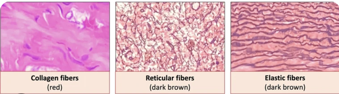
compare and contrast the fibres of CT
Collagen: type 1, 3 subunits, very strong, most abundant, long unbranched, tightly packed, flexible
Reticular: type 3 (reticulin), also strong but less than ^, branched network → makes it resistant to stress from multiple directions, smaller diameter
Elastic: elastin, can stretch up to 150% of resting state, complex system to form it, rubber-like
What are the fixed cells of CT and brief function description
fibroblasts - most common in CT, synth and maintenance of fibres in ground substance, active cell, and when its job is completed it matures in to a fibrocyte
fibrocytes - monitors the environment and if sm happens it can differentiate back to a fibroblast
macrophages - phagocytic, scattered throughout
mesenchymal cells - stem cells in extracellular matrix (ECM) of CT, respond to infection by dividing and specializing (mostly into fibroblasts)
adipocytes - stores fat
What are the wandering cells of CT and brief function description
free macrophages - phagocytic, derived from monocyte that has left tissue and differentiated, gets signalled when there’s an injury
mast cells - regulate inflammatory response (histidine)
plasma cells - produce antibodies
lymphocytes -
microphages - neutrophils, eosinophils, blood cells involved in immune response
What are the 3 main types of CT? (and their subcategories)
CT proper - loose, dense regular/irregular
Supportive CT - cartilage/bone
Special CT - fluid, adipose, elastic, reticular
What are the characteristics of loose and dense CT
Loose
most abundant, packing material (fills space), supports epithelium, blood vessels, nerves
ground substance is most of its volume
looks like lots of cells, no bundles
Dense
collagen fibre, can see bundles
regular: fibres very tightly packed, nuclei are thin lines
irregular: inconsistent bundles, can resist forces from all sides

What kind of CT (loose/dense/regular/irregular) are each of the special CTs, and any additional characteristics?
fluid - blood
adipose - loose CT
elastic - dense regular, parallel bundles of elastic fibres, fibroblasts and fibrocytes that can be stretched, walls of large blood vessels, some ligaments, under epithelium
reticular - loose, macrophages and fibroblasts, structure, stained with silver
What are the 3 classifications of neurons? (+ characteristics)
Multipolar - 3+ cell processes, most common, esp in CNS
Bipolar - 2 cell processes (1 axon, 1 dendrite), not common, for specialized sensory organs
Unipolar - 1 cell process, in PNS ganglia, pseudounipolar: short and divides to form axon
What kind of signal does a neuron send?
Electrical
Contrast the terminology used for cell body clusters and cell processes in the CNS vs PNS.
CNS: cell body clusters: nuclei; cell processes: the track
PNS: cell body clusters: ganglia (s:ganglion); cell processes: the nerve
Map out the general organization of the nervous system.

Describe the functionality of the neuron classifications.
Sensory/afferent neurons: unipolar with cell bodies in ganglia of PNS (think, it’s going → CNS from PNS, so cell body starts there)
Motor/efferent neurons: multipolar with cell bodies in CNS (going out from CNS, so starts there)
Interneurons: multipolar, in between afferent and efferent, in the CNS
What are the supporting cells of the CNS and PNS and their main function/characteristics?
CNS (C.AMEO):
Astrocytes - large, most abundant, structure of neural tissue
Microglia - small, not abundant, monitor health, can destroy antigens
Ependymal cells - cuboidal/columnar with cilia, lines cavities, CSF production/movement
Oligodendrocytes - myelinate (up to 60) axons by wrapping around
PNS
Schwann cells - myelination in PNS, only 1 portion of 1 axon per cell
Satellite cells - surround soma, chemical environment control
What are the hypo/epi/thalamus role and location?
Thalamus: It is small and egg-shaped in the centre of brain, close to brain stem, its function is to recieve sensory info (except smell) and redirect that info
Hypothalamus: Hypo=under, under and to the front of thalamus, has pituitary gland connected, endocrine system
Epithalamus: on other side of thalamus (to back of brain), has pineal gland → melatonin, role in sleep-wake cycle
What are peduncles? And specifically what do the middle cerebellar peduncles do?
Peduncles are thick nerve fibers (myelinated axons) that structurally + functionally connect brain parts
The middle cerebellar peduncles connect the pons to the cerebellum
What are the components of the brain stem (in order from top to bottom) and their main role(s)?
Midbrain - has 4 colliculi: reflex + sensory (auditory) processing
Pons - 4th ventricle is behind it, also has middle cerebellar peduncles that attach to cerebellum
Medulla oblongata - has pyramids, olives, cardiovascular control (heart rate, blood pressure, breathing depth, coughing, sneezing, vomiting, hiccuping, etc.), foramen magnum separates medulla oblongata and spinal cord.
What are the meninges (in order from out to in), and main structure/function/characteristics?
Dura mater - hard, has outer periostial layer and inner meningeal layer that are almost always in contact, except for separations = dural venous sinuses, dense irregular CT
Arachnoid membrane - adhered to dura mater, loosely covering brain, loose collagen and elastic fibre, has subarachnoid space with CSF
Pia mater - tightly clings to brain/spinal cord, thin membrane, thin elastic fibres from squamous/cuboidal cells, arteries and veins to supply nutrients + O2 and take away CO2, denticulate ligaments anchor it to arachnoid membrane and stabilize horizontally
List all the parts of the ventricles (and picture where they are)
lateral ventricles: anterior horn, body, posterior horn, inferior horn
3rd ventricle
interventricular foramen
cerebral aquaduct
4th ventricle
central canal
choroid plexus
What is the choroid plexus and what does it do?
network of blood vessels and ependymal cells
produces CSF
List (and visualize location) the arteries of the Circle of Willis/brain blood supply in order-ish of the direction of blood flow, including any relevant info on how they enter the skull.
vertebral artery (enters skull from foramen magnum)
basilar artery
posterior cerebral artery
posterior communicating artery
anterior cerebral artery
anterior communicating artery
internal carotid artery (enters skull from carotid canal)
Falx**
What are the spinal cord englargements?
cervical enlargement
lumbar enlargement (from T9-T12, not actually in lumbar region of vertebrae cuz they’re not perfectly lined up)
What are the endings of the spinal cord called
conus medullaris
cauda equina
filum terminale
List (and visualize) the parts of the internal topography of the spinal cord.
dorsal root
ventral root
dorsal root ganglion
central canal - CSF through it
dorsal (posterior) horn (sensory/afferent)
ventral (anterior) horn (motor/efferent)
lateral horn (only in thoracic and lumbar spinal cord region L1-L2/L3, autonomic functions)
gray commissure
anterior commissure
anterior median fissure
posterior median sulcus
Grey/White matter?
Grey matter: unmyelinated axons, soma, and dentrites
White matter: myelinated nerve fibres (axons?)