Cases 2012

0.0(0)
Studied by 0 people
call kaiCall Kai
learnLearn
examPractice Test
spaced repetitionSpaced Repetition
heart puzzleMatch
flashcardsFlashcards
GameKnowt Play
Card Sorting

1/14

flashcard set

Earn XP

Description and Tags

ahh

15 Terms

1
New cards
<p>Cases #337</p><p><span>A five-year-old boy developed loose, watery stools approximately one week after attending a birthday party for a friend. The parents took the child to their health care provider for medical attention. The parents told the physician that at the party, the child played in splash pads. A stool specimen was collected in 10% formalin and submitted for laboratory testing. A formalin-ethyl acetate concentration was performed and a wet mount of the sediment was examined for ova and parasites. Objects shown in </span><strong>Figures A</strong><span> and </span><strong>B</strong><span>, measuring 5-6 micrometers on average, were observed in moderate numbers at 1000x magnification. The laboratory recommended to the attending physician that a modified acid-fast stain be done. It was ordered and </span><strong>Figure C</strong><span> shows what was observed.</span></p>

Cases #337

A five-year-old boy developed loose, watery stools approximately one week after attending a birthday party for a friend. The parents took the child to their health care provider for medical attention. The parents told the physician that at the party, the child played in splash pads. A stool specimen was collected in 10% formalin and submitted for laboratory testing. A formalin-ethyl acetate concentration was performed and a wet mount of the sediment was examined for ova and parasites. Objects shown in Figures A and B, measuring 5-6 micrometers on average, were observed in moderate numbers at 1000x magnification. The laboratory recommended to the attending physician that a modified acid-fast stain be done. It was ordered and Figure C shows what was observed.

Cryptosporidium spp.

2
New cards
<p>Case #338</p><p><span>A 42-year-old pig farmer presented to his local medical facility with intermittent diarrhea and abdominal cramping. A stool specimen was collected in 10% formalin for laboratory testing. </span><strong>Figures A</strong><span> and </span><strong>C</strong><span> were captured at 200x magnification and show what was found in high numbers. </span><strong>Figures B</strong><span>, </span><strong>D</strong><span>, and </span><strong>E</strong><span> show a higher magnification (400x) of the same objects.</span></p>

Case #338

A 42-year-old pig farmer presented to his local medical facility with intermittent diarrhea and abdominal cramping. A stool specimen was collected in 10% formalin for laboratory testing. Figures A and C were captured at 200x magnification and show what was found in high numbers. Figures B, D, and E show a higher magnification (400x) of the same objects.

A,B and C: Ascaris lumbricoides AND Balantidium coli

D: Ascaris lumbricoides

E: Balanridium coli

3
New cards
<p>Case #335</p><p><span>A state health laboratory received Wright-stained blood films for malaria confirmation and identification. The patient had most-recently traveled to Pakistan (but had been in the U.S. for the past nine months without additional international travel). The following day, the state health lab received additional blood films on the same patient, but from a different hospital. In this second set of specimens, the slides were stained with Giemsa. Images from both sets were captured and sent to the CDC-DPDx for diagnostic assistance. </span><strong>Figures A</strong><span>–</span><strong>E</strong><span> were from the earlier specimen stained with Wright stain; </span><strong>Figures F</strong><span>–</span><strong>I</strong><span> were from the later specimen stained with Giemsa.</span></p>

Case #335

A state health laboratory received Wright-stained blood films for malaria confirmation and identification. The patient had most-recently traveled to Pakistan (but had been in the U.S. for the past nine months without additional international travel). The following day, the state health lab received additional blood films on the same patient, but from a different hospital. In this second set of specimens, the slides were stained with Giemsa. Images from both sets were captured and sent to the CDC-DPDx for diagnostic assistance. Figures A–E were from the earlier specimen stained with Wright stain; Figures F–I were from the later specimen stained with Giemsa.

Plasmodium vivax

4
New cards
<p>Case #334</p><p><span>During the laboratory component of the DPDx Diagnostic Parasitology Workshop on Intestinal Parasites, a participant made an observation on a trichrome-stained fecal smear and wanted to share with the class. The images in </span><strong>Figures A</strong><span> and </span><strong>B</strong><span> were captured at 1000x oil magnification. The two images show the same object in different focal planes.</span></p>

Case #334

During the laboratory component of the DPDx Diagnostic Parasitology Workshop on Intestinal Parasites, a participant made an observation on a trichrome-stained fecal smear and wanted to share with the class. The images in Figures A and B were captured at 1000x oil magnification. The two images show the same object in different focal planes.

Entamoeba coli with ingested cyst of Giardia duodenalis

5
New cards
<p>Case #331</p><p><span>A 35-year-old woman, who had emigrated from Azerbaijan five years previous, presented to her health care provider with abdominal pain. A CT scan revealed multiple pancreatic cysts. The patient noted that she was diagnosed with pancreatic cysts while still living in Azerbaijan. The cysts were aspirated and the fluid was sent to Pathology for histological work-up. </span><strong>Figures A</strong><span>–</span><strong>D</strong><span> show what was observed on a slide made from the aspirate, stained with trichrome. The objects of interest measured 12-15 micrometers on average.</span></p>

Case #331

A 35-year-old woman, who had emigrated from Azerbaijan five years previous, presented to her health care provider with abdominal pain. A CT scan revealed multiple pancreatic cysts. The patient noted that she was diagnosed with pancreatic cysts while still living in Azerbaijan. The cysts were aspirated and the fluid was sent to Pathology for histological work-up. Figures A–D show what was observed on a slide made from the aspirate, stained with trichrome. The objects of interest measured 12-15 micrometers on average.

Entamoeba histolytica

6
New cards
<p>Case #332</p><p><span>A 55-year-old man presented with complaints of abdominal pain, constipation, intermittent diarrhea with blood and mucus, and blurry vision accompanied by headaches. The patient had been taking prednisone and amoxicillin for other medical issues. A stool examination was performed for parasites and objects resembling tapeworm proglottids were observed and sent to the state health laboratory for diagnostic assistance. The specimens were in turn forwarded to the CDC-DPDx for additional assistance. Two objects were received at the CDC, measuring 30 and 15 millimeters in length. The object in </span><strong>Figure A</strong><span> shows part of the larger object. </span><strong>Figure B</strong><span> shows a close-up of one of the ends of the same object. </span><strong>Figure C</strong><span> shows the same object under higher magnification after clearing in lactophenol for two hours. </span><strong>Figure D</strong><span> shows the smaller of the two objects.</span></p>

Case #332

A 55-year-old man presented with complaints of abdominal pain, constipation, intermittent diarrhea with blood and mucus, and blurry vision accompanied by headaches. The patient had been taking prednisone and amoxicillin for other medical issues. A stool examination was performed for parasites and objects resembling tapeworm proglottids were observed and sent to the state health laboratory for diagnostic assistance. The specimens were in turn forwarded to the CDC-DPDx for additional assistance. Two objects were received at the CDC, measuring 30 and 15 millimeters in length. The object in Figure A shows part of the larger object. Figure B shows a close-up of one of the ends of the same object. Figure C shows the same object under higher magnification after clearing in lactophenol for two hours. Figure D shows the smaller of the two objects.

Artifact

7
New cards
<p>Case #330</p><p><span>A 36-year-old man sought medical attention for sharp recurring episodes of fevers, night sweats, loss of appetite and weight-loss. He was HIV-positive but was currently on medication. He had been working in Liberia for some time and reported that he had taken malaria prophylaxis. A bone marrow aspirate was obtained due to his HIV status to evaluate its productive capacity. Smears were prepared from the aspirate, stained with Wright-Giemsa and examined microscopically. It was noted by the attending pathologist that the aspirate was diluted with peripheral blood and appeared to be positive for malarial parasites. The aspirate smears were forwarded to the DPDx Team for diagnostic assistance. The objects in </span><strong>Figures A</strong><span>–</span><strong>D</strong><span> were observed by CDC microbiologists.</span></p>

Case #330

A 36-year-old man sought medical attention for sharp recurring episodes of fevers, night sweats, loss of appetite and weight-loss. He was HIV-positive but was currently on medication. He had been working in Liberia for some time and reported that he had taken malaria prophylaxis. A bone marrow aspirate was obtained due to his HIV status to evaluate its productive capacity. Smears were prepared from the aspirate, stained with Wright-Giemsa and examined microscopically. It was noted by the attending pathologist that the aspirate was diluted with peripheral blood and appeared to be positive for malarial parasites. The aspirate smears were forwarded to the DPDx Team for diagnostic assistance. The objects in Figures A–D were observed by CDC microbiologists.

Plasmodium malarie

8
New cards
<p>Case #337</p><p><span>A three-year-old boy with no known international travel history was taken to his pediatrician with diarrhea and abdominal cramping. The patient was one of several children in his day care presenting with diarrhea. Stool specimens were collected in polyvinyl alcohol (PVA) and 10% formalin and sent to the State Health Lab for ova-and-parasite (O&amp;P) examination. </span><strong>Figures A</strong><span>–</span><strong>D</strong><span> show what was observed in high numbers on a slide stained with trichrome made from the PVA-preserved stool. The objects of interest measured on average 10-13 micrometers long at their greatest length.</span></p>

Case #337

A three-year-old boy with no known international travel history was taken to his pediatrician with diarrhea and abdominal cramping. The patient was one of several children in his day care presenting with diarrhea. Stool specimens were collected in polyvinyl alcohol (PVA) and 10% formalin and sent to the State Health Lab for ova-and-parasite (O&P) examination. Figures A–D show what was observed in high numbers on a slide stained with trichrome made from the PVA-preserved stool. The objects of interest measured on average 10-13 micrometers long at their greatest length.

Dientamoeba fragilis
9
New cards
<p>Case #326</p><p><span>A 65-year-old female, who lives in northern California, had a colonoscopy performed as part of a routine screening. A foreign body was observed and subsequently removed. The specimen was sectioned, stained with hematoxylin and eosin (H&amp;E), and examined by the attending pathologist. Digital images were captured and sent to the DPDx Team for diagnostic assistance. </span><strong>Figure A</strong><span> was captured at 2x; </span><strong>Figure B</strong><span> at 20x; and </span><strong>Figures C</strong><span> and </span><strong>D</strong><span> at 40x magnification.</span></p>

Case #326

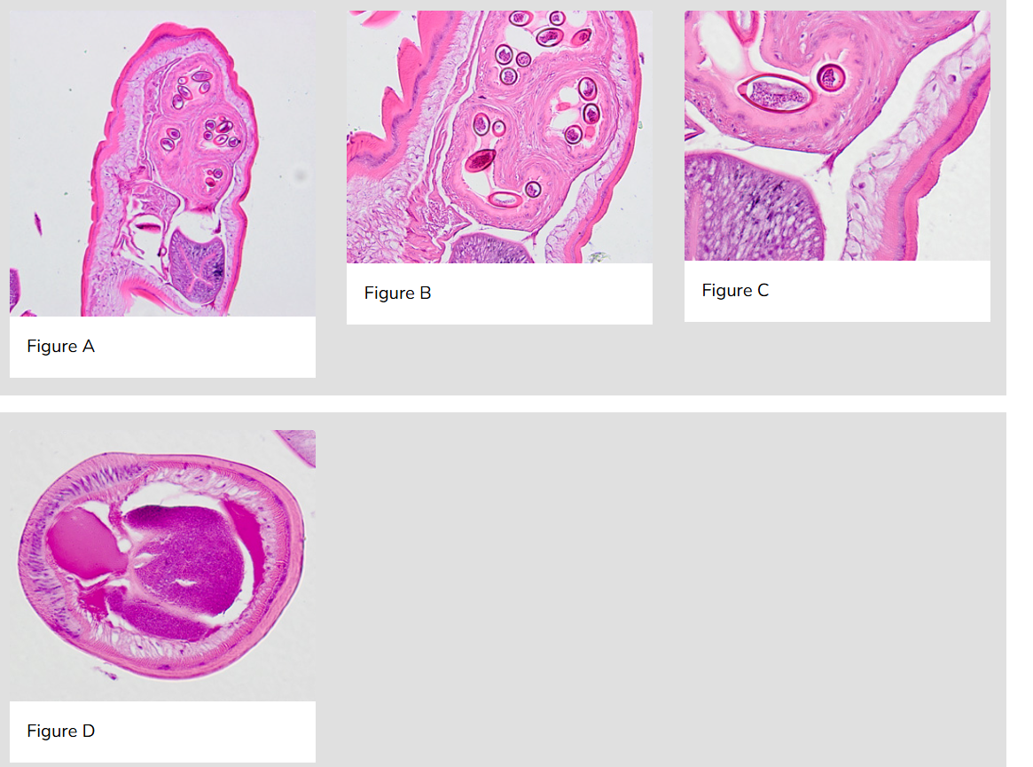
A 65-year-old female, who lives in northern California, had a colonoscopy performed as part of a routine screening. A foreign body was observed and subsequently removed. The specimen was sectioned, stained with hematoxylin and eosin (H&E), and examined by the attending pathologist. Digital images were captured and sent to the DPDx Team for diagnostic assistance. Figure A was captured at 2x; Figure B at 20x; and Figures C and D at 40x magnification.

Trichuris trichura

10
New cards
<p>Case #323</p><p><span>A 50-year-old man from Cambodia was admitted to the hospital for recurrent fevers. Blood specimens were collected in EDTA and sent to Hematology for routine work-up. </span><strong>Figures A</strong><span>–</span><strong>E</strong><span> show what was observed on a thin smear stained with Wright-Giemsa. The case was originally reported as </span><em>Plasmodium falciparum</em><span> and the patient was treated accordingly. One month later, the patient returned to the hospital, again with recurrent fevers. There was no documented international travel between hospital visits. A second blood specimen was collected in EDTA and sent to Hematology for work-up. Images </span><strong>F</strong><span>–</span><strong>I</strong><span> show what was observed on a thin smear stained with Wright-Giemsa. After the second examination, digital images from both collection dates were sent to DPDx for diagnostic assistance.</span></p>

Case #323

A 50-year-old man from Cambodia was admitted to the hospital for recurrent fevers. Blood specimens were collected in EDTA and sent to Hematology for routine work-up. Figures A–E show what was observed on a thin smear stained with Wright-Giemsa. The case was originally reported as Plasmodium falciparum and the patient was treated accordingly. One month later, the patient returned to the hospital, again with recurrent fevers. There was no documented international travel between hospital visits. A second blood specimen was collected in EDTA and sent to Hematology for work-up. Images F–I show what was observed on a thin smear stained with Wright-Giemsa. After the second examination, digital images from both collection dates were sent to DPDx for diagnostic assistance.

A-E: Plasmodium falciparum

F-I: Plasmodium vivax

11
New cards
<p>Case #324</p><p><span>A 58-year-old female who had emigrated from Africa was seen by her health care provider for complications with squamous cell carcinoma of the cervix. CT scans of the pelvis revealed a small mass in the bladder. Biopsy specimens of the bladder and subsequent bladder washings were sent to Pathology for histological work-up. Microscopic examination of the specimens by the attending pathologist revealed structures suggestive of a parasite. Digital images were sent to the DPDx Team for diagnostic assistance. </span><strong>Figures A</strong><span>–</span><strong>D</strong><span> show what was observed in the biopsy specimens, stained with hematoxylin and eosin (H&amp;E). </span><strong>Figures E</strong><span> and </span><strong>F</strong><span> show what was observed in the bladder washings, stained with Papanicolaou stain. The object in Figure E measured 150 micrometers in length.</span></p>

Case #324

A 58-year-old female who had emigrated from Africa was seen by her health care provider for complications with squamous cell carcinoma of the cervix. CT scans of the pelvis revealed a small mass in the bladder. Biopsy specimens of the bladder and subsequent bladder washings were sent to Pathology for histological work-up. Microscopic examination of the specimens by the attending pathologist revealed structures suggestive of a parasite. Digital images were sent to the DPDx Team for diagnostic assistance. Figures A–D show what was observed in the biopsy specimens, stained with hematoxylin and eosin (H&E). Figures E and F show what was observed in the bladder washings, stained with Papanicolaou stain. The object in Figure E measured 150 micrometers in length.

Schistosoma haematobium

12
New cards
<p>Case #322</p><p><span>A 28-year-old man presented to his health care provider with intermittent diarrhea, mild abdominal cramping, and low-grade fever. Travel history included camping in the northwestern United States. Stool specimens were collected in formalin and polyvinyl alcohol (PVA) and sent to a commercial lab for ova-and-parasite (O&amp;P) examination. </span><strong>Figures A</strong><span> and </span><strong>B</strong><span> show what was observed in moderate frequency on a trichrome-stained smear made from the PVA-preserved stool. The objects of interest measured 12-15 micrometers on average in length. The images were captured at 1000x magnification.</span></p>

Case #322

A 28-year-old man presented to his health care provider with intermittent diarrhea, mild abdominal cramping, and low-grade fever. Travel history included camping in the northwestern United States. Stool specimens were collected in formalin and polyvinyl alcohol (PVA) and sent to a commercial lab for ova-and-parasite (O&P) examination. Figures A and B show what was observed in moderate frequency on a trichrome-stained smear made from the PVA-preserved stool. The objects of interest measured 12-15 micrometers on average in length. The images were captured at 1000x magnification.

Pentatrichomonas hominis

13
New cards
<p>Case #320</p><p><span>Stool specimens were collected from Haitian refugees as part of a screening program for intestinal parasites. Stool was collected in polyvinyl alcohol (PVA) and 10% formalin and sent to the county public health lab for routine ova-and-parasite (O&amp;P) examination. </span><strong>Figures A</strong><span>–</span><strong>D</strong><span> show what was observed in moderate numbers from one of the patients on a trichrome-stained slide from the PVA-preserved stool. All images were captured at 1000x magnification.</span></p>

Case #320

Stool specimens were collected from Haitian refugees as part of a screening program for intestinal parasites. Stool was collected in polyvinyl alcohol (PVA) and 10% formalin and sent to the county public health lab for routine ova-and-parasite (O&P) examination. Figures A–D show what was observed in moderate numbers from one of the patients on a trichrome-stained slide from the PVA-preserved stool. All images were captured at 1000x magnification.

Endolimax nana

Blastocystis hominis

14
New cards
<p>#318</p><p><span>A patient with travel to Mexico presented to his local public health facility with general abdominal discomfort. A stool specimen was collected and processed using the formalin-ethyl acetate (FEA) procedure. A wet mount was prepared and examined. Digital images were captured at 1000x oil magnification and sent to DPDx for consultation. </span><strong>Figures A</strong><span>–</span><strong>D</strong><span> show the objects of interest, which measured 12-15 micrometers in length.</span></p>

#318

A patient with travel to Mexico presented to his local public health facility with general abdominal discomfort. A stool specimen was collected and processed using the formalin-ethyl acetate (FEA) procedure. A wet mount was prepared and examined. Digital images were captured at 1000x oil magnification and sent to DPDx for consultation. Figures A–D show the objects of interest, which measured 12-15 micrometers in length.

Artifact

15
New cards
<p>Case #315</p><p><span>A 31-year-old man, originally from Ecuador, vomited what appeared to be a long worm-like object. The object (</span><strong>Figure A</strong><span>) measured 27 centimeters in length. The object was sent to a pathology laboratory for sectioning and staining with hematoxylin and eosin (H&amp;E). </span><strong>Figures B</strong><span>–</span><strong>D</strong><span> show what was observed microscopically by the attending pathologist.</span></p>

Case #315

A 31-year-old man, originally from Ecuador, vomited what appeared to be a long worm-like object. The object (Figure A) measured 27 centimeters in length. The object was sent to a pathology laboratory for sectioning and staining with hematoxylin and eosin (H&E). Figures B–D show what was observed microscopically by the attending pathologist.

Ascaris lumbricoides



