PLTW: HBS - 1.1 - 2.1.2 up to midterm
 Call Kai
 Learn
 Practice Test
 Spaced Repetition
 Match
 Flashcards
 Knowt Play
1/83

There's no tags or description
Looks like no tags are added yet.
84 Terms
Adipose Tissue
Connective tissue in which fat is stored and which has the cells distended by droplets of fat.

Appendicular Skeleton
Bones of the limbs and limb girdles that are attached to the axial skeleton.

Axial Skeleton
The skeleton of the trunk and head.

Connective Tissue
Animal tissue that functions mainly to bind and support other tissues, having a sparse population of cells scattered through an extracellular matrix.

Epithelial Tissue
Sheets of tightly packed cells that line organs and body cavities.

Femur
The proximal bone of the hind or lower limb that is the longest and largest bone in the human body, extends from the hip to the knee.

Forensic Anthropology
The branch of physical anthropology in which anthropological data, criteria, and techniques are used to determine the sex, age, genetic population, or parentage of skeletal or biological materials in questions of civil or criminal law.

Humerus
The longest bone of the upper arm or forelimb extending from the shoulder to the elbow.

Pelvis
A basin-shaped structure in the skeleton of many vertebrates that is formed by the pelvic girdle together with the sacrum and other various coccygeal and caudal vertebrae and that in humans is composed of the two hip bones bounding it on each side and in front while the sacrum and coccyx complete if behind.

Skull
The skeleton of the head forming a bony case that encloses and protects the brain and chief sense organs and supports the jaws.
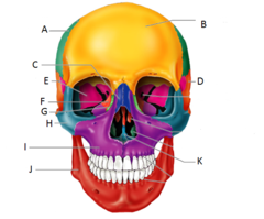
Tibia
The inner and usually larger of the two bones of the leg between the knee and ankle that articulates above with the femur and below with the talus - called also shinbone.

Tissue
An integrated group of cells with a common structure and function

Mandible
The lower jaw, which causes the compressions during breastfeeding helping to milk the breast

Maxilla
Forms most of the upper jaw, and part of the hard palate, contains upper molars, premolars, and canines

Zygomatic Process
A bridgelike projection joining the zygomatic bones (checkbone) anteriorly. Together these two form the Zygomatic arch

Frontal Bone
the large cranial bone forming the front part of the cranium: the forehead and the upper part of the orbits

Temporal Bone
bone that forms parts of the side of the skull and floor of the cranial activity. There is a right and left temporal bone.

Occipital Bone
forms the posterior floor and walls of the cranium

Parietal Bone
either of two skull bones between the frontal and occipital bones and forming the top and sides of the cranium

Orbicularis Oculi
Muscle that closes the eye

Orbicularis Oris
lip muscle to close lips/kiss

Temporalis
Elevates and retracts mandible

Coccyx
forms the end of the spine and is actually made up of four small vertebrae that are fused together (tailbone)

Sacrum
One of three bones (sacrum and two pelvic bones) that make up the pelvic ring; consists of five fused sacral vertebrae.

Eye Orbit
Eye socket

Zygomatic Bone
cheekbone

Cervical Vertebrae (C1-C7)
The first seven bones of the vertebral column (forming the neck) are the

Thoracic Vertebrae (T1-T12)
second set of 12 vertebrae; they articulate with the 12 pairs of ribs to form the outward curve of the spine

Lumbar Vertebrae (L1-L5)
third set of five larger vertebrae, which forms the inward curve of the spine

Fibula
The lateral and smaller bone of the lower leg
Radius
lateral bone of the forearm
Ulna
Inner and larger bone of the forearm, attached to the wrist and located on the side of the little finger.
Patella
knee bone
Clavicle
collar bone
Metacarpals
bones of the hand
Metatarsals
foot bones
transverse fracture
bone break with fracture line straight across shaft of bone
sprial fracture
Fracture which twists around the bone, may occur from a twisting force
comminuted fracture
bone breaks into many fragments
MRI
a technique that uses magnetic fields and radio waves to produce computer-generated images that distinguish among different types of soft tissue; allows us to see structures within the brain
ultrasound imaging
technique for using high-pitched sound waves to form an image
Intramedullary nailing
how are femoral shaft fractures in an adult repaired, with metal put into the medullary cavity of the bone to reinforce the bone from within. Never has to be removed but takes the place of bone marrow.
Fracture plating
An invasive procedure used to treat most bone fractures involving a permanent plate being placed parallel to the fracture on the outside of the bone, and screws driven through the plate and into the bone to keep the fracture stable and allow healing. Commonly used on younger patients so as to not affect their red marrow.
osteoblasts
Bone building cells with many projections (arms) that are found in the periosteum
osteoclasts
large cells that resorb or break down bone matrix, come from the bone marrow
bony callus
step 3 of 4 of fracture healing: when the fibrocartilage callus is converted to spongy bone
Angiogenesis
formation of new blood vessels
hematoma
a solid swelling of clotted blood within the tissues
growth plates (epiphyseal plates)
Areas of cartilage at the end of long bones; where ossification takes place until the bone is full grown
Frontal Lobe
Responsible for problem-solving, reasoning, personality, and motor skills (movement).
Parietal Lobe
Contains the area where bodily sensations occur, such as touch and pain.
Temporal Lobe
Contains areas important to hearing and understanding speech/language.
Occipital Lobe
Contains areas important to vision.
Medulla Oblongata
Part of brain stem that controls vital autonomic processes like regulation of breathing, heart rate and blood pressure.
Cerebellum
Control of finely coordinated movements. Coordination center, voluntary movement and balance.
Cerebellum image

Cerebrum image

Frontal Lobe image

Medulla Oblongata image

Occipital Lobe image

Parietal Lobe image

Brain Stem image

temporal Lobe image

Spinal Cord
Sends signals between brain and rest of body

Brain Stem
Oldest part of the brain, responsible for survival functions (e.g. breathing, heart beat, etc.); includes medulla and pons; aka "the lizard brain"

Pons
Responsible for coordination and movement (relays messages between cerebrum and cerebellum); involved in sleep, dreams, and attention.

Thalamus
Relays information from the senses and transmits the information to the cerebral cortex.

Limbic System
Hippocampus, Amygdala, Pituitary Gland, Hypothalamus; involved in emotion

Hippocampus
Helps to form memories ("hippo never forgets")

Amygdala
Fight or flight, emotional center, anger or fear, happy or sad

Hypothalamus
Controls body temperature (the body's thermostat), circadian rhythms, and thirst and hunger, and smell.

Pituitary gland
The "master" endocrine gland, responsible for growth.

Corpus Callosum
Thick band that connects left and right hemispheres of the brain.

Cerebrum
Information processing, outer layer of brain.
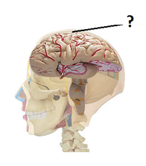
Lobes of the cerebrum
Occipital, Temporal, Parietal, Frontal

motor cortex
area at the rear of the frontal lobes that controls voluntary movements
sensory cortex
area at the front of the parietal lobes that registers and processes senses, body touch, and movement
Broca's area
controls language expression - an area in the frontal lobe, that directs the muscle movements involved in speech.
Role of the nervous system
-coordinates all body systems
-detects and responds to stimuli
-brain and spinal cord act as switching centers
-nerves carry messages to and from centers
CNS
central nervous system
PNS
peripheral nervous system
how do central and peripheral nervous system work together?
The nerves of the PNS extend from the central nervous system to the outermost areas of the body. The peripheral system allows the brain and spinal cord to receive and send information to other areas of the body, which allows us to react to stimuli in our environment.
How did Henry Molasion's surgery pave the way for further exploration?
While the surgery was successful, it led to H.M. suffering from anterograde amnesia, in which he could no longer form or keep new memories. His case was important as scientists figured out the function of the hippocampus and studied memory and cognitive abilities of the portions of the brain responsible for memory.
Why is the story of Phineas Gage considered so extraordinary? What does his story teach us about the brain?
He survived a pole through his head and his changed personality was linked with his head injury