Psychology Chapter 2 - Nervous System Functioning
 Call Kai
 Learn
Learn Practice Test
 Spaced Repetition
 Match
Match Flashcards
 Knowt Play
1/104

There's no tags or description
Looks like no tags are added yet.
105 Terms
What is the nervous system and its main roles?
The body's information processing system that allows us to receive, interpret and respond to stimuli.
What are the two main divisions of the nervous system
Central NS - brain and spinal cord
Peripheral NS - nerves (outside CNS) and sensory receptors in the body
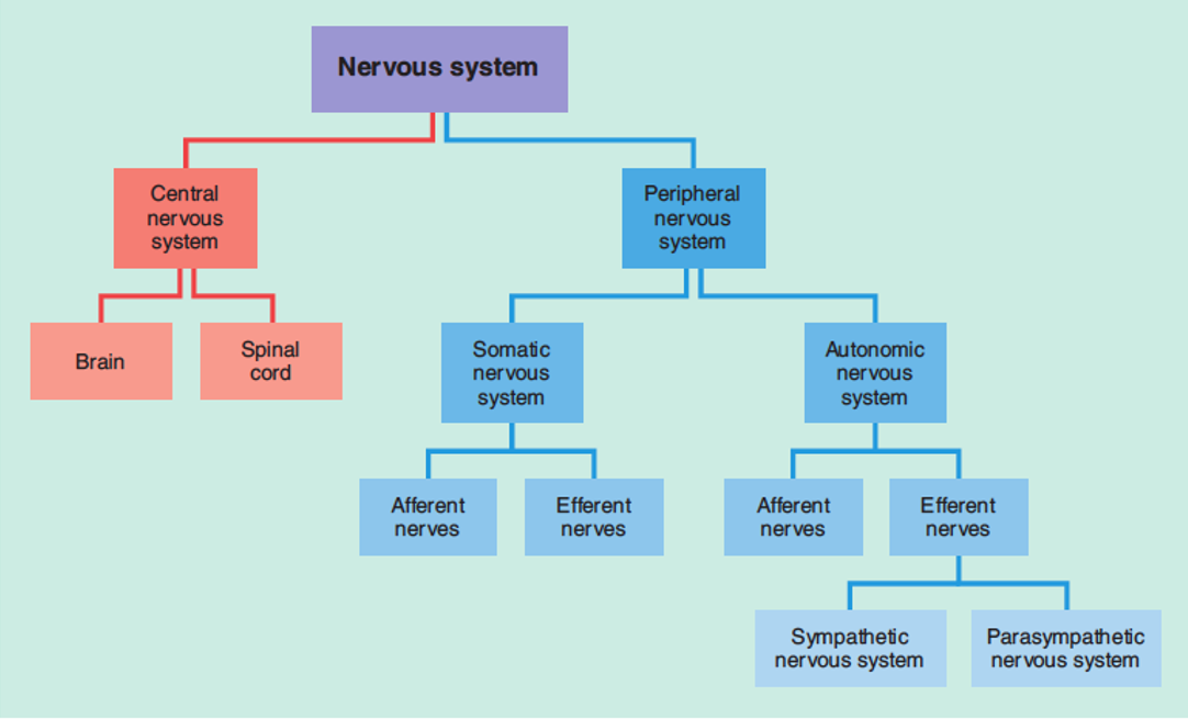
Full hierarchical diagram of the nervous system
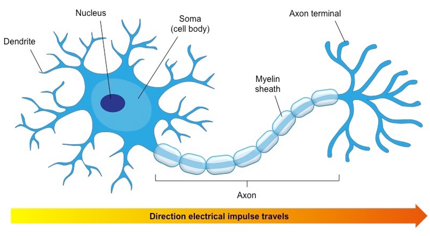
Neuron diagram
3 types of neurons
Afferent (sensory) - carry signals towards the CNS
Efferent (motor) - carry signals away from the CNS
Interneurons (relay/association) - connect afferent and efferent pathways (found only in CNS)
Neural pathway
A neural route for connection within brain and between the brain and other areas of the NS
3 main roles of the Central NS (and what they mean we can do)
- Receive bodily messages
- Process information
- Send messages to the body
Means that we can perceive and respond to the world, experience consciousness and have subjective mental experiences
Brain
Command centre for almost all NS responses. The cerebral cortex is the only part in the NS that has consciousness.
3 main roles of the spinal cord
- Convey bodily sensory information (from PNS) to the brain
- Convey motor information to the body's muscles, glands and organs (via PNS)
- Spinal reflex
How does paralysis occur?
When the spinal cord is damaged, sometimes nerves below damaged area can be disconnected from the CNS, leading to paraplegics and quadriplegics.
2 main functions of the Peripheral NS
To carry sensory/afferent information from the body's organs, muscles glands and receptor sites to the brain, and to carry motor/efferent information back.
Somatic NS
Peripheral NS subdivision that connects skeletal muscles and sensory organs with the brain
2 main functions of the Somatic NS
- Detect external stimuli and transmits it to the brain
- Allows voluntary movement of skeletal muscles
Autonomic NS
Peripheral NS subdivision that connects the CNS to internal organs, cardiac muscles, smooth muscles and glands
The 4 different types of muscles
Skeletal - Voluntary movement
Cardiac - Heart functions
Smooth - Autonomic functions in hollow organs like intestines, blood vessels and bladder
2 main functions of the Autonomic NS
- Detect internal stimuli, mostly in the cardiac and digestive system
- Facilitates and REGULATES (not initiates) involuntary movement of cardiac and smooth muscles, as well as the release of hormones from glands
Where is information processed from the Somatic NS and Autonomic NS
Somatic information is mainly processed in the in the cerebral cortex (sensory and motor cortices) where conscious awareness is experienced.
Autonomic information is mainly processed in the hypothalamus (homeostasis) and brainstem.
How autonomous is the autonomic nervous system?
While many of its organs, glands and processes are self-regulating and aren't under voluntary control, it is linked to the cerebral cortex, meaning we can choose to control some of its functions with conscious effort.
Three Autonomic NS subdivisions
- Sympathetic
- Parasympathetic
- Enteric
How do the sympathetic and parasympathetic NS function in relation to each other?
They counterbalance each other's activities without conscious effort, with one system usually dominant at any given time.
Somatic NS vs Autonomic NS
Somatic:
Afferent = five senses
Efferent = skeletal muscles
= conscious perception of external environment and voluntary movement
Autonomic:
Afferent = Internal organs
Efferent = cardiac and smooth muscles, glands
= self-regulating internal systems.
A conscious response involves...
1. awareness of environmental stimuli received through sensory systems
2. formation of an intention to respond
3. a response involving deliberate physical movement
Sensory receptors
Where external stimuli is detected so an afferent signal can be generated
Effector sites
Where an efferent signal attaches to a muscle or gland so a function can be executed
5 steps of a conscious response
1. Sensation - Stimulation from environment is detected in sensory receptors in somatic NS
2. Afferent signal - Sensory signal generated and sent via afferent pathways in somatic NS to CNS
3. Processing - Brain receives signal and consciously perceives it, making a decision and initiating a motor signal
4. Efferent signal - Motor signal travels via efferent pathways in the SNS to motor organs
5. Response - Motor signal received at effector site causing motion
Unconscious responses and 2 locations they occur
External or internal actions carried out in response to sensory stimuli that involve little/no awareness. Two places they occur are in autonomic NS and in somatic NS with spinal reflex.
An unconscious response usually involves
1. Internal stimuli without our awareness or a threatening external one
2. Feedback in lower parts of brain to regulate bodily functions
3. Involuntary cardiac or smooth muscles movement or hormone secretion from a gland in order to stimulate or supress bodily functions.
Sympathetic Nervous System
Autonomic NS subdivision that increases physiological arousal, putting the body in a state of biological and behavioural preparedness.
Roles of sympathetic NS
- increase heart rate, dilate pupils, increase oxygen intake and glucose release
- Suppress parasympathetic NS functions (i.e. functions not related to immediate survival)
- Stimulates adrenaline release from adrenal medulla above kidney
- Stimulates noradrenaline release in brainstem to increase threat perception
- Triggers fight-or-flight response
Fight-flight-freeze response
Automatic physiological reaction to a perceived threat. Flight and fight responses are caused by sympathetic NS dominance and noradrenaline release, and are adaptive responses, but freeze response occurs due to parasympathetic NS dominance that 'locks' with sympathetic NS functions, leading to state of immobility, high arousal, alertness and tension
Adrenaline
Released from adrenal medulla above kidney in response to perceived threat (sympathetic NS). Increases physiological arousal.
2 main functions of adrenaline
- Stimulates functions that increase our ability to physically exert ourselves, mainly aids sympathetic NS with its functions.
- Suppresses functions unrelated to physical exertion, by relaxing bladder, decreasing salivation and slowing digestion.
Parasympathetic Nervous System
Autonomic NS subdivision that decreases physiological arousal and stimulates life-sustaining functions.
Roles of Parasympathetic NS
Returns body to homeostasis
Decreases physiological arousal
Allows life-sustaining functions to occur: e.g. digestion, salivation, reproduction, tears and pupil constriction in light environments
Contributes to 'freeze' response
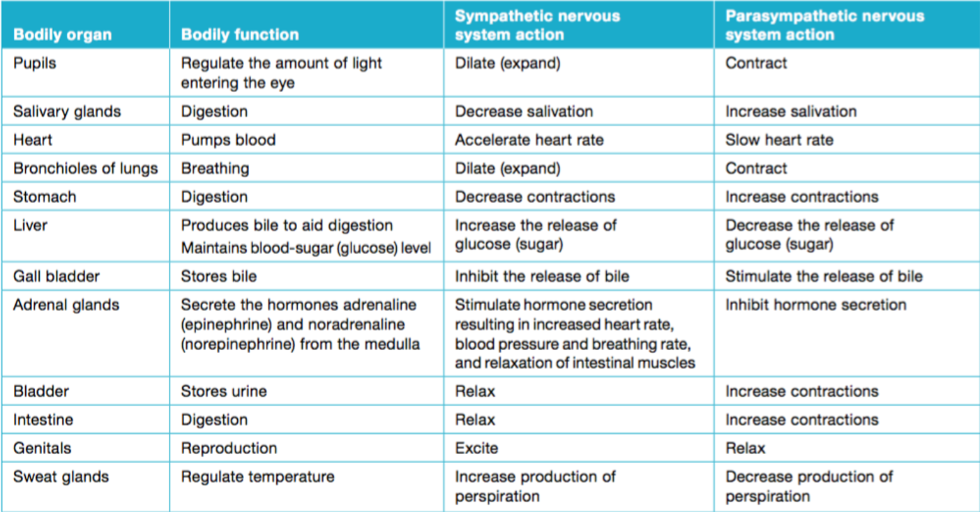
Table comparing Sympathetic vs Parasympathetic functions
Where is the Enteric Nervous System found
Embedded in walls of gastrointestinal tract (hollow digestion organs like mouth, oesophagus, stomach, intestines and rectum) is a mesh-like system of neuron clusters (ganglia). They also connect with non-hollow digestion organs like liver, pancreas and gallbladder.
Enteric NS roles
- Detect physiological state of gastrointestinal tract and controls its movement and functions.
- Works with CNS to control digestive system
Has some functions independent of other NS divisions, even some memory of past gut movements
Spinal reflex
Involuntary, nearly instantaneous motor response initiated by spinal interneurons in response to sudden pain or intense heat. The shorter pathway due to the spinal reflex arc increases efficiency and leads to pain perception after a response has already occurred. Brain still will make conscious decision about further movements afterwards.
Spinal reflex examples
- Patellar reflex (can be used to test NS functioning)
- Withdraw reflex
- Reflexes involving muscle length and tension during movement
Do all reflexes happen through the spinal cord?
No, some occur in the base of the brain such as the startle reflex
Which division of the peripheral nervous system is involved in the spinal reflex - SNS or ANS?
Both, but majorly the Somatic NS.
Steps in the spinal reflex
1. Sensation - Sensory receptors in SNS detect intense heat, pain, etc.
2. Afferent signal - Afferent neurons carry signal towards CNS
3. Processing - SPINAL INTERNEURONS in the CNS relay the message to a motor neuron to initiate reflexive movement
4. Efferent signal - Efferent neuron in SNS carries message to effector site
5. Response - Withdrawal reflex occurs involuntarily
6. Extra step - Message simultaneously conveyed to brain; conscious awareness of stimuli follows response
Dendrites
Branch-like structures reaching out from neuron. Contains neurotransmitter receptor sites, and stimulation received determines if neuron will 'fire'
Axon
Portion of neuron that conveys signal towards the next cell, passing electrical potential along the axon from the dendrites and soma to the synapse.
Myelin/myelin sheath
Fatty axon coating made of other cells that insulates the signal to speed up transmission. Prevents 'crossed wires' and means that the electrochemical signal can 'jump' more efficiently between nodes (gaps in myelin).
Axon terminals
At the ends of the axon, with small swellings called terminal buttons. Release chemicals (neurotransmitters).
What is an Action Potential (AP)?
The technical term for when a neuron 'fires', i.e. an electrochemical signal is propagated along the axon
How is electrical (action) potential created?
Ion channels 'gates' between myelin allow charged particles in and out, creating an electrical potential (a difference between the charge inside and outside the neuron).
Neuron's voltage (resting is at around -70mV) is changed by other neuron's stimulation. If threshold if -55mV is reached, cell depolarises (has a sharp spike in positive charge) and an AP is 'fired'. The neuron then resets its voltage to rest.
Why is action potential an electrochemical signal?
Because it involves the transfer of electrical charge via the flow of charged ions.
The 'all-or-none' principal
Action potentials either happen or they don't, there's no such thing as a big or small AP. If threshold is reached, AP will fire every time, otherwise it won't fire at all.
Neurons are...
The basic building blocks of the nervous system, specialised to communicate information around the body
Synaptic gap/cleft
Physical gap at the synapse between the axon terminal of one neuron and the receptors of the next neuron
Pre-synaptic neuron
Neuron passing on the signal
Post-synaptic neuron
Neuron receiving the signal
Neural Synapse
Typical point of communication between neurons, includes pre-synaptic axon terminal (and its terminal buttons), synaptic cleft and post-synaptic dendrite with neurotransmitter receptor sites
Neurotransmission
Communication between neurons, more than the action potentials within the neuron but the events that occur at the synapse that change the outcome.
Neurotransmitter
Chemical substance produced by a neuron that carries a message to other neurons or cells in other tissue.
Lock-and-key process
Keys - Neurotransmitters with unique molecular structure (shape and size)
Locks - Post-synaptic receptor sites, which needs to have a complementary chemical structure (shape and size) to bind with a neurotransmitter.
This 'binding' process changes the post-synaptic neuron, making it more or less likely to fire AP.
Reuptake
Neurotransmitter that doesn't bind to receptors in the post-synaptic neuron are absorbed back into the presynaptic terminal buttons.
What are the two different effects a neurotransmitter can have?
Excitatory - stimulates or activates postsynaptic neuron to perform functions/ increases voltage closer to threshold and makes it more likely to fire
Inhibitory - blocks or prevents postsynaptic neuron from firing/ decreases voltage and makes it less likely to fire
What determines the effects of a particular neurotransmitter?
The location and properties of the receptor the neurotransmitter binds with, along with the actual chemical itself
Summative neurotransmitter effects
A single neuron can have both excitatory and inhibitory receptor sites, therefore the excitation and inhibition adds together, and the overall charge determines if the cell reaches the threshold. Excitatory and inhibitory effects only last for a short time.
Do neurons only communicate through neurotransmitters?
No, neurons can also communicate electrically by transmitting electrical signals directly between different parts of neurons.
What are the 3 types of neurochemicals?
Neurotransmitters, neuromodulators, neurohormones
Neurohormones
Some neurotransmitters can also occur as hormones, like such as noradrenaline which is secreted as a hormone by the adrenal glands and as a neurotransmitter from neurons
Glutamate
The main excitatory neurotransmitter in the CNS. (2nd most abundant NT in the brain after GABA). Serves to depolarise post-syn neuron. It is involved in most normal brain functions including learning, memory, perception, thinking and movement. Its excitatory effects promote the growth and strengthening of synaptic connections, representing memory (synaptic plasticity).
GABA
Gamma-amino butyric acid
Primary inhibitory neurotransmitter in the CNS. Serves to further polarise post-syn neuron. Helps to fine tune neurotransmission in the brain to ensure it is at an optimal level. Without it seizures would be overly prevalent, and low levels (GABA dysfunction) are related to phobia and anxiety.
Neuromodulator
A type of neurohormone that can influence the effects of other neurotransmitters by modifying and regulating signal transmission, increasing or decreasing the excitability/responsiveness of neurons to neurotransmitter signals across a widespread brain region or brain tissue.
Some ways neuromodulators can influence other neurotransmitters' effects include...
- Changing receptor reactivity to particular neurotransmitters to enhance their responses
- 'Teaming up' with another neurotransmitter in the synapse to make it more or less potent
- Controlling amount of neurotransmitter released
- Increasing likelihood of neurotransmitter being received
Where do neuromodulators release their chemical messengers?
Affects up to 100,000 neurons or synapses (unlike neurotransmitters with only 1). This may be a whole neural tissue, brain area, pathway or pathways. Effects also take longer to establish and are more long-lasting
Dopamine
Neuromodulator with important roles in voluntary movements, pleasure, motivation, appetite, reward-based learning and memory.
Dopamine effects as neurotransmitter vs neuromodulator
NT - Primarily excitatory
NM - Excitatory or inhibitory depending on location and receptors present
Mental conditions implicated by dopamine
Parkinson's, depression, addiction, schizophrenia
What are the brain's two main dopamine pathways?
- Nigrostriatal pathway
- Mesolimbic (and mesocortical which overlaps) pathways
Forms dopaminergic system
Nigrostriatal pathway
Dopaminergic pathway originating in midbrain structure called substantia nigra. Dopamine produced by neurons in this structure carry messages to allow smooth voluntary muscle movement coordination.
What happens if the substantia nigra is damaged?
Brain structures linked to movement receive less dopamine messages as a result of lower dopamine production in nigrostriatal pathway, leading to extreme muscle rigidity (like in Parkinson's disease)
Mesolimbic pathway
Dopaminergic pathway that, along with mesocortical pathway, forms dopamine reward system responsible for pleasure and reward-based learning (with help from limbic system). Originates in ventral tegmental area in midbrain.
Reward-based learning
Pleasurable consequences make us more likely to repeat behaviour. Even the anticipation or thought of receiving a rewarding stimulus can release dopamine.
What causes schizophrenia?
High levels of dopamine in the mesolimbic pathway are associated with hallucinations and delusions
Serotonin
Neuromodulator with important roles in mood stabilisation, wellbeing, emotional processing, sleep onset, reproductive drive, appetite and pain perception.
Serotonin effects as neurotransmitter vs neuromodulator
NT - Inhibitory and excitatory depending on location
NM - Only inhibitory
Mental conditions implicated by serotonin
Depression, anxiety disorders and sleep disorders
Where is serotonin produced in the body?
10% in brain, mostly in Raphe Nuclei comprising clusters of cell soma in the brain stem.
Over 80% is in the enteric NS within the gastrointestinal tract.
Makes up serotonergic system
What chemical process is linked to depression?
Low levels of dopamine are associated with depression (and seasonal affective disorder and OCD) leading to prolonged hopelessness and decreased reward from pleasurable experiences.
Antidepressants
SSRIs - Serotonin-Specific Reuptake Inhibitors block serotonin reuptake to increase its availability in the synapse.
Serotonin and the sleep-wake cycle
Helps regulate when, how and how much we sleep with the help of other neurotransmitters and the hormone melatonin. The brain uses serotonin to produce melatonin, explaining insomnia in those with depression.
Serotonin Syndrome
Excessive serotonin can cause fever, high heart rate, hallucinations, seizures and unconsciousness or death. Often caused by illegal drugs
Neuroplasticity
Ability of neurons in the brain to change
Synaptic Plasticity
Involves functional and structural changes at the synapse that alter communication between neurons, resulting in increased (LTP) or decreased (LTD) communication.
2 types of neuroplasticity (different to last year)
Developmental plasticity - Change due to physical maturation. Includes synaptogenesis, synaptic pruning and myelination
Adaptive plasticity - Changes due to environmental conditions. Includes learning (LTP and LTD) and recovery from brain trauma (rerouting and sprouting)
Learning
Relatively permanent change in behaviour that occurs as a result of experience
Memory
Processing, storage and retrieval of information acquired through learning. If we don't remember, we haven't learnt
How are memories stored in the brain?
A memory is a pathway in the brain, created through synaptic plasticity (learning)
Hebb's rule
Neurons that fire together, wire together.
Repeated transmission and joint activation between two neurons causes change in the synapse, increasing the chance of firing together again and leading to more forcibly and efficiently transmitted signals in the future
What are the fundamental mechanisms of learning and memory formation?
Long term potentiation, long term depression, sprouting, rerouting and pruning
LTP
Long-term potentiation. Experience- based, long-lasting strengthening of synaptic connections occurring through repeated co-activation of the relevant neurons. This increases the likelihood of future transmissions along this pathway, and increases their strength and efficiency. Involved in learning and memory. Includes sprouting and rerouting at a structural level.
LTD
Long-term depression. Experience-dependent, long-lasting weakening of little-used synaptic connections occurring through repeated sub-threshold activation. Includes rerouting and pruning
What changes at the synapse during LTD and LTP?
- Amount of neurotransmitter produced
- Number of post-synaptic receptors available to the neurotransmitter
- Amount of depolarisation that occurs in the post-synaptic neuron when stimulated
The role of glutamate in LTD and LTP
As the brain's main excitatory neurotransmitter, it's the main chemical involved in synaptic plasticity because it causes increased chances of threshold-level charges, causing neurons to ‘fire together’ and ‘wire together’. LTP and LTD take place in glutaminergic synapses in the hippocampus, cerebral cortex and other brain areas.
What is LTD useful for?
By weakening or eliminating underused synapses, only important connections strengthened through LTP remain. It also allows us to correct our thinking when problem solving, adjust movements in motor learning (e.g. learning to serve in tennis), and erase unwanted or inappropriate thoughts and behaviours.