Biology 1407 Lab Practical #1
 Call Kai
 Learn
Learn Practice Test
 Spaced Repetition
 Match
Match Flashcards
 Knowt Play
1/94

There's no tags or description
Looks like no tags are added yet.
Name | Mastery | Learn | Test | Matching | Spaced | Call with Kai | Chat |
|---|
No analytics yet
Send a link to your students to track their progress
95 Terms
Whole Mount
Representation of the whole organism

Cross Section
This represents a thin section of the organism or structure that is perpendicular to the long axis of the organism

Longitudinal Section
Represents a thin section of the length of the structure or organism

Identification
Refers to the taxonomic classification of the organism. Genus is required
Description
Denotes the organism's region, structure, generation, or slide type being observed. Put common name here
Labels
Straight line drawn from structure denoting what it is.
Annotations
Used to give information about the specimen that cannot be seen on the diagram (color, flagella that couldn't be seen, etc) Indicated alongside the diagram, not a part of the desrpition
Size
Total magnification or a metric scale bar.
Habit Sketch
Used to illustrate a portion of or an entire organism
Low Plan Drawings
Show the basic plan of regions or sections of specimens. (Cross-sections & Longitudinal-sections). Typically observed with the scanning objective
High Power Detailed Drawings
Used to show detailed cells within a section of a specimen. (cells within the vascular bundle of a stem section)
Ocular Lense
Located at the top of the microscope (10x)
Compound Light Microscope
Used for prepared slides or temporary wet mount slides (40X - 1000X)

Dissecting Microscope
Useful for viewing larger specimens. (7X - 45X)

Scanning Lens
4X
Low Power Lens
10X
High Power Lens
40X
Oil Immersion Lens
100X
Revolving Nose Piece
Function: Holds the three objective lense

Head
Houses the lenses in the upper part of the microscope
Arm
Supports the upper part of the microcope
Base
The bottom of the microscope
Stage
Flat platform below the objective lens

Stage Clips
Hold the slide in place

Coarse Adjustment Knob
Makes large adjustments to the stage in order to bring the slide into focus

Fine Adjustment Knob
Makes small adjustments to the stage in order to bring the slide into sharper focus

X-Y Stage Control Knobs
Moves the stage left and right

Illuminator
The light source of the microscope, located at the base.
Rheostat
Controls the amount of light released by the illuminator

Condenser
Focuses the light onto the slide

Iris Diaphragm
Adjusts the amount of light that hits the slide

Total Magnification
Ocular Magnification X Objective Magnification

Inversion
The image is flipped both horizontally and vertically
Parfocal
Image remains in focus when switching between objective lenses
Filed of View
Circular area that is visible when looking through a microscope
Depth of Field
The vertical distance that remains in focus on the slide (increases with magnification)
Gastrodermis
Inner layer of a cell

Epidermis
Outer layer of a cell

Transmitted Light Source
Used to shine light through a clear object
Reflective Light Source
Hits an opaque object and is reflected into the objective lens.
Homologous Structure
Structures in different species that are similar because of common ancestry.
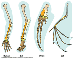
Divergence
The act of moving away in different direction from a common point
Analogous Structures
Body parts that share a common function, but not structure

Vestigial
Are little or no importance to organism, but remain from an ancestor.

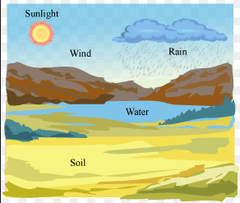
Biotic
Living

Abiotic
Non-living
Community
A group of populations in a particular area
Species Diversity
Number of different species that make up a particular area
Species Richness
Actual number of all species within the community
Relative Abundance
Proportion of a particular species in the community from the total number of individuals
Shannon Diversity Index
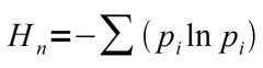
Relative Abundance (pᵢ)
Total # of Individuals of species A /
Total # of Individuals Sampled
Average Population Density (Per Quadrat)
Total # of Individuals of a species/
# of quadrats
Estimated Population Size
Average Population Density X (Total Area/Quadrat Size)
Haploid
Single set of chromosomes
Nucleoid
Tightly packed circular region containing a bacterium's chromosome

Ubiquitous
Present everywhere
Microbiota
Collection of all microbes living inside the gut
Decomposer
Organisms that break down dead organic matter
Domain Bacteria
prokaryotes, one of largest groups on earth, classified by shape & whether or not they use oxygen
Domain Archaea
Any of various single-celled prokaryotes genetically distinct from bacteria, often thriving in extreme environmental conditions
Phylum Cyanobacteria
Photosynthetic bacteria with gram-negative cell walls that consist of a thin layer of pepridoglycan
Phylum Proteobacteria
Largest and most diverse bacterial phylum. gram-negative.
Phylum Firmicutes
Gram-positive bacteria with cell walls that have a thick layer of peptidoglycan.
Culture
Bacteria that grow artificially on lab media
Broth Media
A liquid medium used for growing cells
Solid Media
Firm, does not move when tube moves. Contains a solidifying agent. May be liquefiable or nonliquefiable
Agar
a gel-like polysaccharide compound used for culturing microbes; extracted from certain red algae
Colony
Visible growth of bacteria on solid media.
Streak Plate Method
used to isolate pure cultures
Pure Culture
contains only one species or strain
Chlorophyll a
Only pigment that can participate directly in the light reactions.
Phycocyanin
Blue Pigment
Phycoerythrin
Rid pigment
Biofilm
A surface-coating colony of one or more species of prokaryotes that engage in metabolic cooperation.
Gas Vesicle
small, rigid structures that provide buoyancy to a cell
Unicellular
One cell
Glycocalyx
The external surface of a plasma membrane that is important for cell-to-cell communication
Filamentous
Having long threads or fibers;hyphae
Vegetative Cells
In filamentous colonies of cyanobacteria, the cells that perform photosynthesis
Heterocysts
a large transparent thick-walled cell that is found in the filaments of some cyanobacteria and is the site of nitrogen fixation
Nitrogen Fixation
The process of changing free nitrogen gas into a usable form
Akinete
Thick-walled dormant cells that are only produced from vegetative cells when environmental conditions are unfavorable
Coccus
A spherical bacterium.

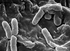
Bacillus
A rod shaped bacterium
Spiral Form
Spiral shaped bacteria
Comma-shaped, S-shaped, or spiral shaped

Vibrio
comma shaped

Spirochetes
Thin, flexible spiral shaped bacteria

Spirillum
Thick, rigid spiral shaped bacteria

Gram-Positive Bacteria
Bacteria that have a thick peptido glycan cell wall, and no outer membrane. They stain very darkly (purple) in Gram stain.

Gram-Negative Bacteria
Bacteria that have a thin peptidoglycan cell wall covered by an outer plasma membrane. They stain very lightly (pink) in Gram stain. Gram-negative bacteria are typically more resistant to antibiotics than Gram-positive bacteria.

Crystal Violet
Primary stain, stains Gram positive and negative cells both purple
Gram's Iodine
Mordant, that enters the cytoplasm of the cell and reacts with the crystal violet to form a larger complex that doesn't easily escape through the cell wall
Alcohol
Decolorizer, provides differentiation between Gram-positive and negative cells. Dehydrates the peptidoglycan of (+) cells causing it to shrink and trap the crystal violet-iodine complex inside the cell. Opposite effect happens in gram (-) cells
Safranin
Counterstain applied to stain the colorless gram (-) cells pink