PNS (Sensory)
 Call Kai
 Learn
 Practice Test
 Spaced Repetition
 Match
 Flashcards
 Knowt Play
1/63

There's no tags or description
Looks like no tags are added yet.
64 Terms
General (somatic) senses
Touch, temp, pressure, pain
Special senses
Sensory modalities including vision, hearing, taste, and smell, which involve specialized organs.
Subconsious processing
blood pressure, GI tract, blood glucose, internal body temperature, lung inflammation
Exteroreceptors
Sensory receptors located on the body's surface that detect external stimuli such as touch, temperature, and pain.
Interoceptors (visceroreceptors)
chemoreceptors, baroreceptors,and mechanoreceptors that monitor internal body conditions.
Interoceptors (proprioreceptors)
Sensory receptors that provide information about the body's position and movement by detecting changes in muscle length, tension, and joint angle.
Thermoreceptors
Sensory receptors that detect changes in temperature, allowing the body to sense heat and cold.
Mechanoreceptors
Sensory receptors that respond to mechanical forces such as pressure, vibration, and touch. They play a crucial role in sensing physical changes in the environment.
chemoreceptors
react to specific chemicals
Nociceptors
Sensory receptors that respond to potentially harmful stimuli by transmitting pain signals. They play a key role in protecting the body from injury.
Photoreceptors
Sensory receptors that respond to light, allowing us to perceive visual images. They are crucial for vision and are mainly found in the retina of the eye.
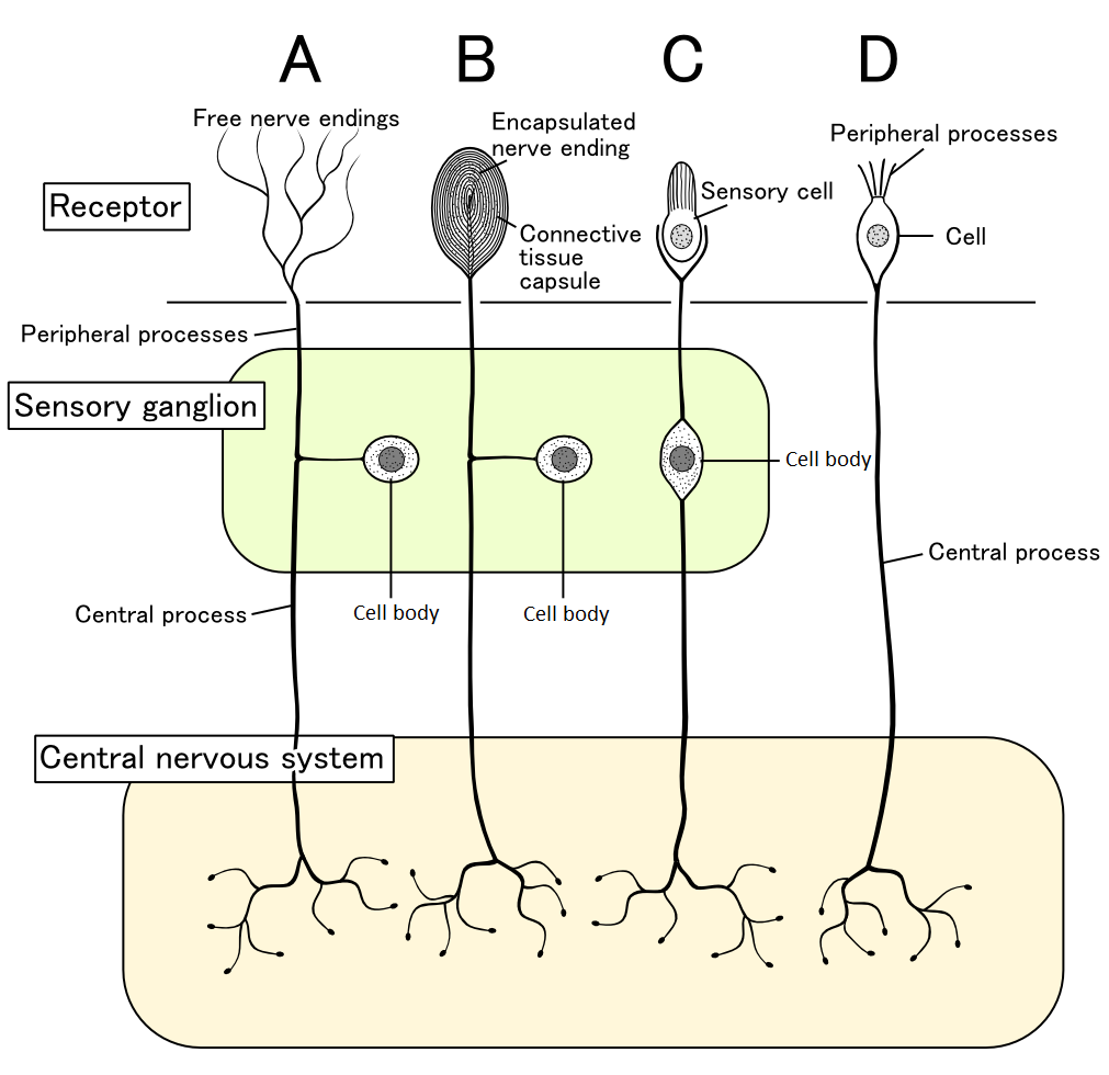
Free nerve endings
are sensory receptors that detect a variety of stimuli, including pain, temperature, and pressure. They are widely distributed throughout the body and play a vital role in the sensation of touch.

Encapsulated nerve endings
nerve endings are sensory receptors that are surrounded by a protective capsule, allowing them to detect specific types of stimuli such as pressure, vibration, and proprioception. They are important for providing detailed sensory feedback.
Rods and cones
are the two types of photoreceptors found in the retina, responsible for vision in low light conditions (rods) and color vision (cones). They convert light into electrical signals for the brain.
Hair cells
are specialized sensory receptors located in the inner ear, responsible for converting sound vibrations and head movements into electrical signals that the brain interprets as sound and balance. They play a crucial role in hearing and the vestibular system.
What kind of potential is receptor potential?
graded/synaptic.
Stronger stimulus will result in…
Higher frequency of action potentials from individual sensory receptors
Will activate receptors with lower and higher threshold recruitment
Phasic receptors
are sensory receptors that adapt quickly to a constant stimulus, allowing for the detection of changes in stimuli rather than the intensity of the stimulus itself.
Meissners
corpuscles are specialized mechanoreceptors located in the skin that respond to light touch.
phasic receptor
Pacinian
corpuscles are deep pressure and vibration mechanoreceptors found in the skin, joint capsules, and other tissues, known for their rapid adaptation to stimuli.
phasic receptor
Tonic receptors
are sensory receptors that adapt slowly to a constant stimulus, allowing for the continuous detection of the intensity of the stimulus over time.
Merkel Disks
are specialized mechanoreceptors in the skin that respond to light touch and texture, functioning as tonic receptors.
Ruffini
endings are slow-adapting mechanoreceptors found in the skin and joints, responding to skin stretch and the angle of joint position.
tonic receptors
Proprioceptors
are specialized sensory receptors located in muscles, tendons, and joints that provide information about body position, movement, and balance, allowing for proprioception.
tonic receptors
Receptive field
is the specific area of sensory space that a sensory receptor responds to, influencing the sensitivity and discrimination of sensory stimuli.
Sensory unit
An axon of the neuron and receptors on its dendrites that together detect sensory stimuli, functioning as a fundamental component of sensory perception.
Sensory discrimination
receptor fields are unevenly distributed throughout the body
Lateral inhibition
is a neural mechanism that enhances contrast in sensory perception by inhibiting neighboring neurons, allowing for more precise localization of stimuli.

Presynaptic inhibition
is a process that decreases neurotransmitter release from a presynaptic neuron, thereby reducing the activity of the postsynaptic neuron and modulating sensory information transmission.
axo-axonic synapses

First order neuron
is the neuron that transmits sensory information from the periphery to the central nervous system, carrying signals from sensory receptors to the spinal cord or brainstem.
Second order neuron
is the neuron that transmits sensory information from the first order neuron to higher brain centers, typically located in the spinal cord or brainstem, and is crucial for processing sensory signals.
Crosses over (decussation)
Synapses into thalamus
Third order neuron
is the neuron that carries sensory information from the thalamus to the appropriate cortical areas in the brain for further processing and perception.
Medial lemniscus (dorsal column) system
is a sensory pathway that transmits fine touch, vibration, and proprioceptive information from the body to the brain, traveling through the dorsal columns and medial lemniscus before reaching the thalamus.
Anterolateral system
is a sensory pathway that transmits pain, temperature, and crude touch information from the body to the brain, crossing over to the opposite side before reaching the thalamus.
Decussation
is the crossing over of nerve fibers from one side of the central nervous system to the other, often occurring in the brainstem or spinal cord, and is crucial for the contralateral processing of sensory information.
Gate theory of pain
activation of mechanoreceptors can modulate the sensation of pain
Ipsilateral
same side
Contralateral
opposite side
3 layers of the eye
outer fibrous ( sclera, cornea)
Intermediate vascular (choroid, ciliary body, iris)
Deep nervous (Retina)
Path of light
cornea
aqueous humor
pupil
lens'
vitreous humor
fovea
Emmetropia
normal vision
Myopia
near-sighted
focal point anterior to fovea
Hyperopia
far-sightedness - focal point posterior to fovea.
Neurons in the retina
rods/cones
bipolar cells
horizontal/amacrine cells
ganglion cells
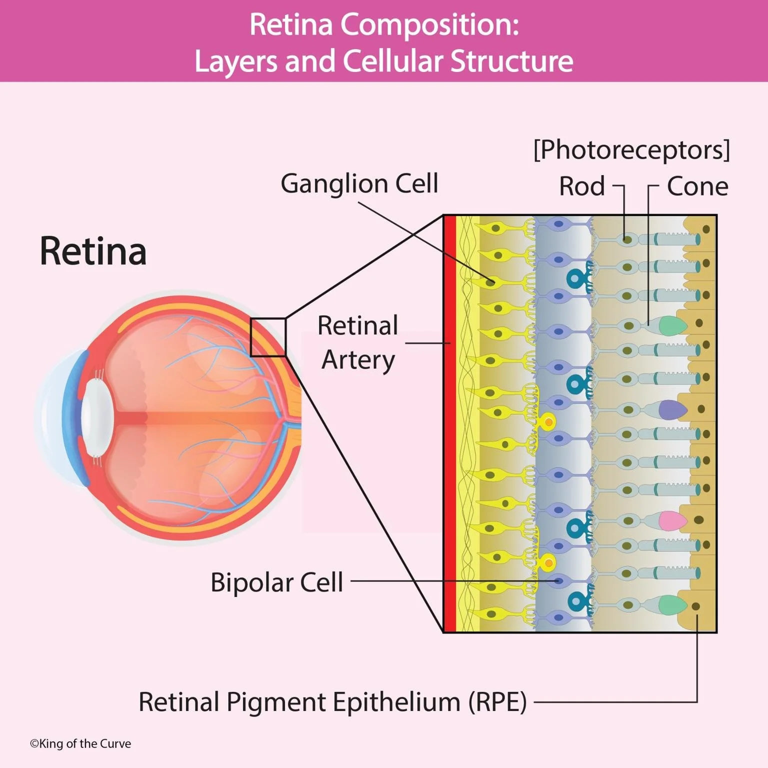
Lateral vs medial ganglion axons pathway to brain
lateral: stay on same side
medial: crosses over at optic chiasm
Optic chiasma
The X-shaped structure formed by the crossing of optic nerve fibers from the nasal half of each retina. This allows visual information from both eyes to be processed by both hemispheres of the brain.
External region of the ear
The external part includes the pinna (auricle) and the auditory canal, which collects sound waves and directs them toward the eardrum.
sound conduction
middle region of the ear
ossicles (malleus, incus, stapes) which amplify sound waves and transmit them to the inner ear.
sound waves converted into mechanical vibrations
Inner region of ear
contains the cochlea, convert sound vibrations into neural signals and maintain balance.
specialized receptors detect pitch and amplitude
Cochlear duct
a fluid-filled structure within the cochlea, critical for transforming sound vibrations into neural messages sent to the brain.
filled with endolymph
Scala vestibiuli and scala tympani
two fluid-filled chambers of the cochlea that play crucial roles in the transmission of sound vibrations; they surround the cochlear duct and contain perilymph.
Function of endolymph
High K+ concentration, important for generating electrical signals in hair cells of the cochlea.
Stapes
A small bone in the middle ear that transmits sound vibrations from the incus to the oval window of the cochlea, playing a crucial role in hearing.
Process of hearing
stapes vibrate perilymph and endolypmh in the cochlea, displaces basilar membrane, vibrating ciliated receptor cells (hair cells) which generate electrical impulses sent to the brain for sound perception.
How do cilia produce depolarization
Cilia on hair cells bend in response to sound vibrations, leading to the opening of ion channels, allowing K+ ions to enter the cell. This influx of positive ions causes depolarization, generating an electrical signal.
How does the ear detect changes in sound intensity
through chnages in the firing rate of afferent neurons connected to hair cells
How is pitch detected?
which region of the basilar membrane is displaced:
base= high frequency
apex: low frequency
afferent vs efferent
Afferent neurons carry sensory information to the central nervous system, while efferent neurons transmit signals away from the central nervous system to muscles or glands.
fasciculus gracilis
A bundle of nerve fibers in the spinal cord that carries sensory information from the lower body to the brain
MLS
fascisulus cuneatus
A bundle of nerve fibers in the spinal cord that carries sensory information from the upper body to the brain.
MLS
Spinotholmatic tracts
A set of nerve pathways in the spinal cord that transmit sensory information about pain and temperature to the brain.
part of Anterolateral system
Ascending vs Descedning pathways
Ascending pathways carry sensory information from the body to the brain, while descending pathways transmit motor commands from the brain to the body.
Ascending: MLS (Dorsal) and AS
Descending: Corticospinal and Corticobutal
What field of vision crosses over in optic chiasm?
The nasal retinas, which receive visual information from the outer portions of the visual field, cross over at the optic chiasm, allowing for the processing of visual information from both eyes in the opposite hemisphere of the brain.
MEDIAL GANGLION AXONS CROSS OVER