Topic 6: Cytoskeleton & Cell Movement
 Call Kai
 Learn
 Practice Test
 Spaced Repetition
 Match
 Flashcards
 Knowt Play
1/23

Earn XP
Description and Tags
Comprehensive flashcards covering Topic 6: Cytoskeleton & cell movement from BIOL 107, focusing on microtubules, microfilaments, intermediate filaments, motor proteins, cilia/flagella mechanics, and sarcomere contraction.
Name | Mastery | Learn | Test | Matching | Spaced | Call with Kai | Chat |
|---|
No analytics yet
Send a link to your students to track their progress
24 Terms
Which cytoskeletal functions are performed in eukaryotes but not in prokaryotes?
Moving things during regular physiological processes and propelling the cell or its surroundings.
What are the three broad functions of the eukaryotic cytoskeleton?
Maintain (and change) cell shape
Spatially organize the cell contents
Connect cells and environment
What are the three classes of cytoskeletal fibres in eukaryotes, along with their polarity and monomer types?
Microtubules (tubulin family): polar (directional), globular monomers
Microfilaments (actin family): polar, globular monomers
Intermediate Filaments: apolar, filamentous monomers
What are the structural characteristics, polarity, and mechanical strength properties of microtubules (MTs)?
They are stiff, hollow tubes made of tubulin heterodimers (α-tubulin and β-tubulin) with polarity (a plus side and a minus side). They are strong under compression and maintain cell shape under pressure.

How do the motor proteins kinesin and dynein differ in their direction of movement along microtubules? what do they have in common?
Kinesin moves toward the plus end (anterograde transport)
Dynein moves toward the minus end (retrograde transport).
Both are ATP-powered and walk in a hand-over-hand fashion.
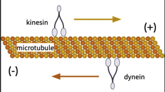

How do transport vesicles attach to microtubule motor proteins?
Kinesins and dyneins attach to transport vesicles via cargo adaptors, which are cargo-specific proteins on the vesicle membrane.

what are cilia made of? What are the specific microtubule doublet and triplet arrangements found in a cilium's basal body and motile portion? what do they have in common?
Made of microtubules
Basal body: 9 microtubule triplets with no central microtubule (9+0 configuration)
Motile portion: 9 microtubule doublets with a central microtubule pair (9+2 configuration)
ATP powered share the same configuration
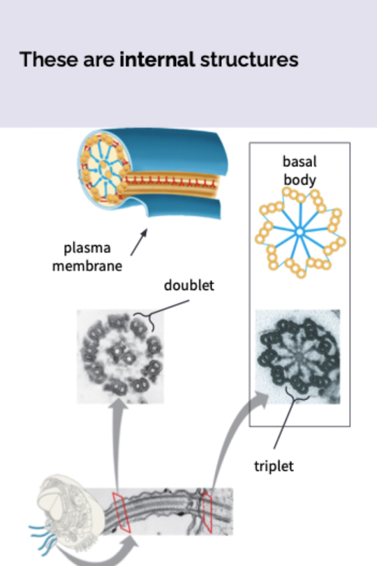

In the motile portion of a cilium, how do dynein motor proteins interact with adjacent microtubule doublets to produce movement?
Dynein arms extending from tubule A of one doublet attach to and walk along tubule B of the adjacent doublet, powered by ATP.

Why are cross-linking proteins and anchorage required for cilia to bend?
Without restriction, dynein movement causes microtubule doublets to slide unrestrained past each other
cross-linking proteins between outer doublets and anchorage in the cell restrict sliding to produce bending and wavelike motion.

How do cilia and eukaryotic flagella differ in their type of movement?
A flagellum undulates symmetrically side to side
a cilium beats asymmetrically with power stroke (fish the fluid) and recovery stroke (return to the starting point)

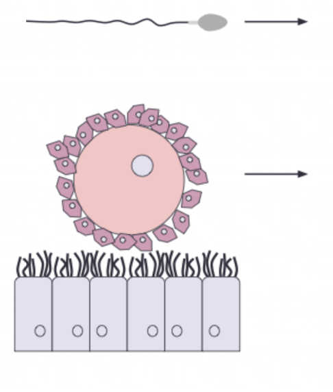
In reproductive physiology, how are the concepts of motility and mobility distinguished using sperm and ova?
Sperm are motile because they propel themselves using a flagellum.
Ova (eggs) are mobile because beating cilia on the epithelial cells of the oviducts ("Fallopian tubes") propel them and prevent backflow.

What paired cylindrical structures composed of microtubules are depicted in this image? What does this say about pants and fungi?
Centrioles ((centrosome)
Microtubule Organizing Center (MTOC)
How do plants and fungi organize their microtubules?
in the absence of centrioles other non-centriole structures serve the purpose of a Microtubule Organizing Center (MTOC).
During mitosis, what structure do microtubules and what do they do?
The spindle apparatus which is composed of microtubules
Form to guide chromosomes to opposite sides of the cell
What are the structural monomer, polymer form, diameter, and energy requirement of microfilaments?
The monomer is G-actin (globular actin)
Polymerizes into F-actin (filamentous actin)
Assembly into F-actin requires ATP.
What mechanical properties characterize microfilaments compared to microtubules? what is it’s main function?
Microfilaments provide tensile strength (how much pulling force a material can withstand before it breaks) to parts of the cell
Not strong under compression (unlike microtubules, which resist compression)
Help with cell shape, cell movement, and muscle contraction
What cellular specializations rely on microfilaments for internal structural support?
The cell cortex (sub-membrane shape support)
microvilli (outfolds on intestinal epithelial cells) to increase SA
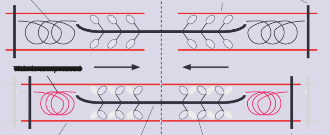
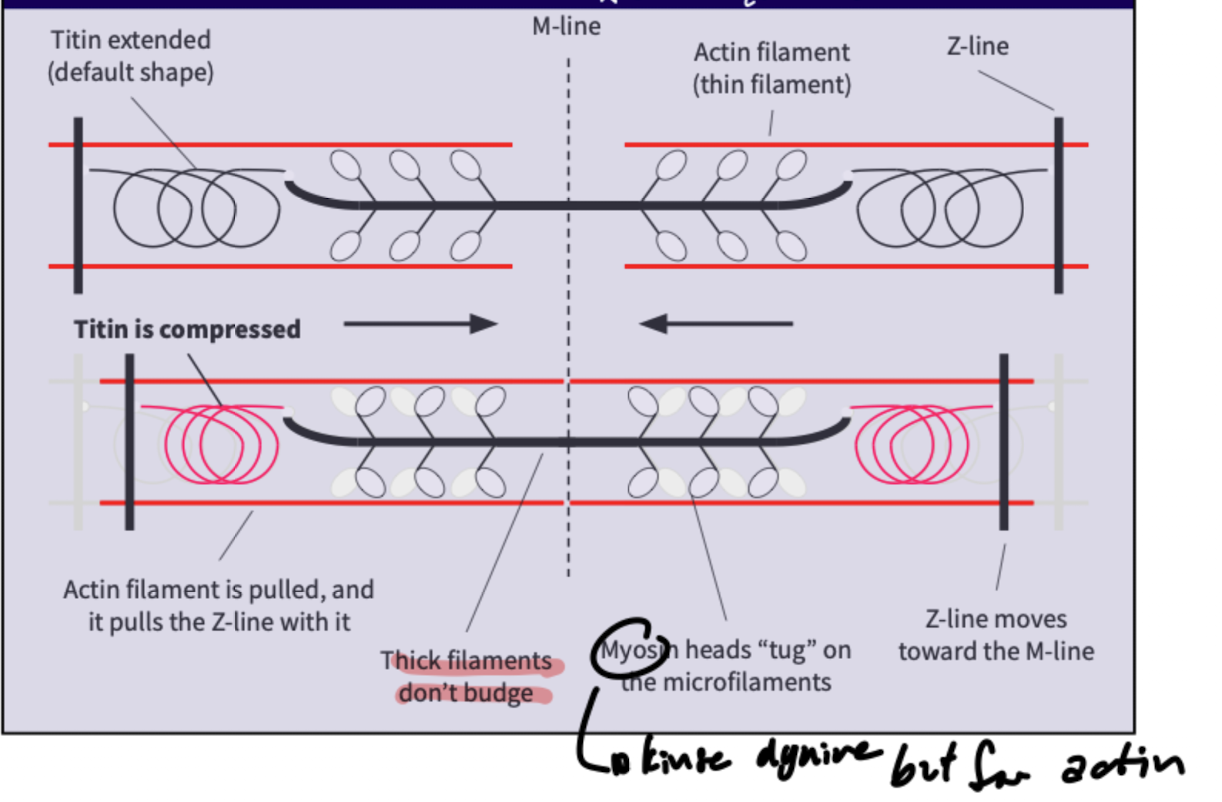
During sarcomere contraction, what happens to the thick filaments, actin, Z-line, and titin?
Thick filaments don't budge
Myosin heads tug on thin microfilaments; the actin filament is pulled which pulls the Z-line toward the M-line
Compresses titin from its default extended shape.
Which motor protein system drives amoeboid crawling locomotion and cytoplasmic streaming in plants?
The actin-myosin system (actin filaments interacting with myosin motor proteins).
actin filaments —> protein fibers help change shape
myosin—> use ATP to pull on actin filaments
amoeboid movement: cell extend Pseudopodia (temporary extension of cytoplasm) and moves its body forward like crawling
Which Gram-negative obligate intracellular parasite manipulates host actin-based cytoskeletons, what cells does it infect, and what disease does it cause?
Ehrlichia chaffeensis
Hijacks the cytoskeleton of monocytes and macrophages
is transmitted by the lone star tick
What are the key defining characteristics of intermediate filaments regarding what it’s made of, strength, and it does? what is it only in?
made of various proteins
high tensile strength
help toughen parts of cell to morph the cell into specific shape
animal structures only
What is the structural assembly hierarchy of intermediate filaments?
monomers intertwine via coiled-coil interactions and disulfide bridges
form dimers, dimers pair in a staggered fashion to form tetramers
tetramers assemble into protofilaments; protofilaments assemble into the final filament cable.
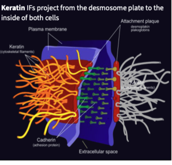
what does Keratin do? What’s an example? what do they also form?
Intermediate filament protein that provides high tensile strength to keep cells together
Velcro portion of desmosomes are made of Keratin
claws, horns, beaks, feather, hair and nails
Which specific intermediate filament protein forms a structural meshwork lining the inner nuclear membrane?
Lamin (which forms the nuclear lamina) —> in nucleus envelope