Chapter 12; Anatomy + Physiology
 Call Kai
 Learn
 Practice Test
 Spaced Repetition
 Match
 Flashcards
 Knowt Play
1/89

There's no tags or description
Looks like no tags are added yet.
90 Terms
cephalization
development of anterior (rostal) portion of brain
3 primary brain vesicles
prosencephalon; forebrain
mesencephalon; midbrain
rombencephalon; hindbrain
what primary brain vesicle gives rise to the telecephalon + diencephalon
prosencephalon
which secondary brain vesicle develops into the adult pons/cerebellum
metencephalon
which secondary brain structure develops into the epithalamus, thalamus, hypothalamus, and retina
diencephalon
what % of the diencephalon’s mass is formed by the thalamus
80%
what structures come from the mylencephalon
medulla oblongata
what major vital autonomic reflex centers are housed within the medulla oblongata
cardiovascular center
repiratory center
what non-cardiorespiratory autonomic reflexes are integrated within the medulla oblonata
vomiting hiccupping, swallowing, coughing, and sneezing
which adult neurla canal region corresponds to the mesencephalon
cerebral aquaduct

what physical constraint during fetal development forces the cerebral hempispheres to double back, envelope the diencephalon, and fold into gyri and sulci
the brain grows faster than the surrounding membranous skull
how does the amount of gray matter in the cerebrum and cerebellum differ from the spinal cord
in addition to the gray matter surrounded by white matter, they also possess:
an outer cortex of gray matter
inner islands of gray matter in the white matter

definition of ventricles in the CNS
fluid filled chambers that are continous to one another and to the central canal of the spinal cord
filled w/ cerebrospinal fluid + lined w/ ependymal cells
which anatomical structure separated the paired lateral ventricle of the cerebral hemispheres
septum pellucidum
what structure connects the third ventricle to the fourth ventricle
cerebral aquaduct
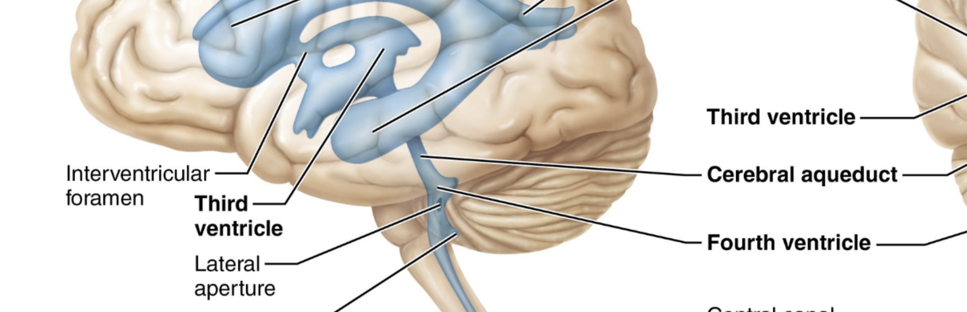
which passage does the lateral ventricle communicate w/ the third ventricle
interventricular foramen
what three openings allow cerebrospinal fluid to exit the fourth ventricle into the subarachnoid space
paired lateral apertures + a single median aperture

what surface landmark separates the left + right cerebral hemispheres
longtiudinal fissure
what fissure separates the cerebral hemispheres from the cerebellum
transverse cerebral fissue
3 main surface markings for the brain
gyri; ridges
sulci; shallow grooves
fissures; deep grooves

the sulci divde the hemispheres into 5 regions
frontal
temporal
parietal
occipital
insula
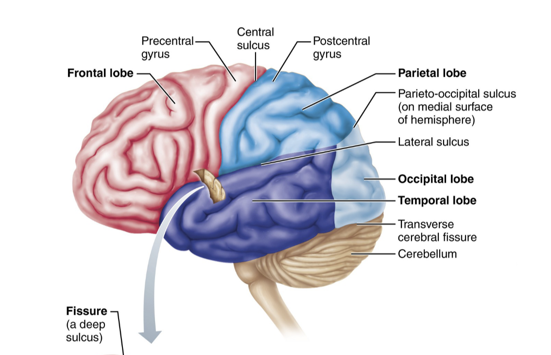
which central lobe is buried deep to the lateral sulcus and covered by parts of the temporal, parietal, and front lobes?
insula
central sulcus creates an anatomical boundary between which two gyri?
precentral gyrus + postcentral gyrus

what cellular components are present in the superficial gray matter layer of the cerebral cortex
4 main things
somas
dendrites
glial cells
blood vells
(no myelinated nerve axons
cerebral cortex function
executive site
part of brai nthat controls consciousness (awareness, sensory functions, motor initiation, etc)
3 main functional areas of the cerebral cortex
motor areas; voluntary movement
association areas; integrates diverse information
sensory areas; conscious awareness of information
where is the primary motor cortex located within the brain
precentral gyrus of the frontal lobe
premotor cortex is anterior to it
what is the function of the pyramidal tracts
they are formed by long axons of pyramidal cells in primary motor cortex + project down the spinal cord
allows conscious + precise control of skeletal muscles
in both motor homunculus + somatosensory homunculus, how are body regions organized across the cortex
organized as upsidedown representations (somatotopy)
the size of the cortical region corresponds to the motor control precision or sensory sensitivity rather than physical body size

primary role of the premotor cortex
acts as staging area for;
planning movements coordinating learned
repetitious/sequential motor skills based on sensory feedback
where broca’s area typically located + specific functional role
located anterior to the inferior premotor area (left hemisphere)
serves as a motor speech area that controls muscles involved in speech
what specific motor function is controlled by the frontal eye field
voluntary movement of the eyes
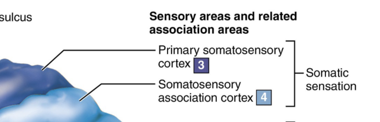
where is the primary somatosensory cortex located
what does it provide
located in the postcentral gyrus
reeceives somatic sensory input from skin; enables spatial discrimination

primary funcion of the somatosensory association cortex
integrates and analyzes sensory inputs from primary somatosensory cortex
determiens size, textture, and structural relationships of objects being felt
what clinical outcomes occurs from damage to the primary visual cortex vs. damages only to the visual association area
primary visual cortex damage; functional blindess
visual association; vision intact but cannot comprehend/recognize what is seen
where is primary auditory cortex located + what aspects of sound does it interpret
located on superior margin of temporal lobes
interprets pitch, loudness, sound location

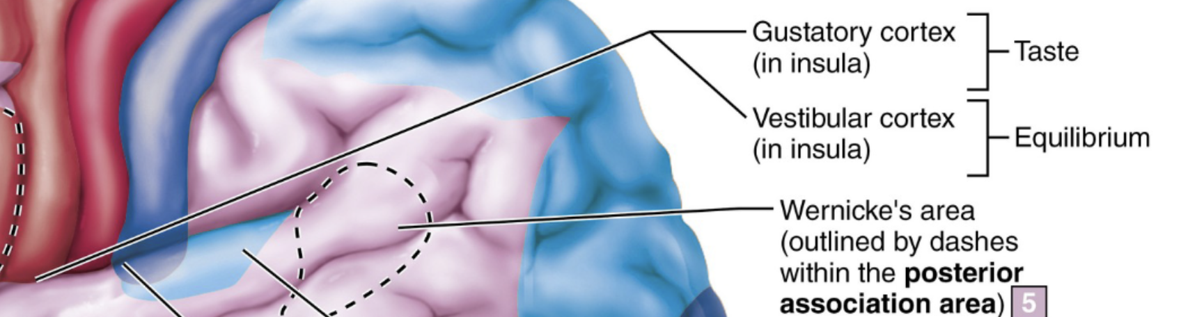
which cortical region is responsible for conscious awareness of balance and head position in space
vestibular cortex
where is the primary olfactory cortex located
medial aspect of temporal lobes

primary olfactory cortex location
on medial aspect of the temporal lobes
where are the gustatory cortex + visceral sensory area located relate to eachother
gustatory cortex; inside insule, deep to the temportal lobe
visceral sensory area; lies posterior to the gustatory cortex

what functional capability distinguishes multimodal association areas from primary/secondary sensory areas
multimodal associtation; recieve input from multiple sensory modalities + send output to multiple areas
integrating sensations, memories, and emotions to direct conscious action
directs conscious action
which multimodal associtation area is also called the prefrontal cortex
what cognitive function does it manage
anterior association area
manages; intellect, cognition, memory, etc
functional role of limbic association area
provides emotional context to experiences
which limbic system structure is critical for assessing danger recognizing fearful/angry facial expressions and eliciting fear
amygdaloid body (amygdala)
what fiber tract connects the various regions of the limbic system together
fornix
what role does the cingulate gyrus play within the limbic system
it plays a role in expressing emotions through physical gestures + resolving mental conflict
functional role of posterior associtation area
recognizing patterns/faces
when there is a turmor/lesion on the anterior associtation area, what clinical signs manifest
mental/personality disorders (loss of judgement/attentiveness)
when there is a tumor/lesion on the posterior association area, what are the symptoms
(spatial self-awareness center)
patient may deny ownership of the side of the body opposite of the lesion
cerebral dominance definition
(left/right minded people)
designates the hemisphere that is dominant for language
functions of the left cerebral hemisphere vs. the right hemisphere
left; language, math, logic
right; visual-spatial skills, intuition, emotion, artistic abilities
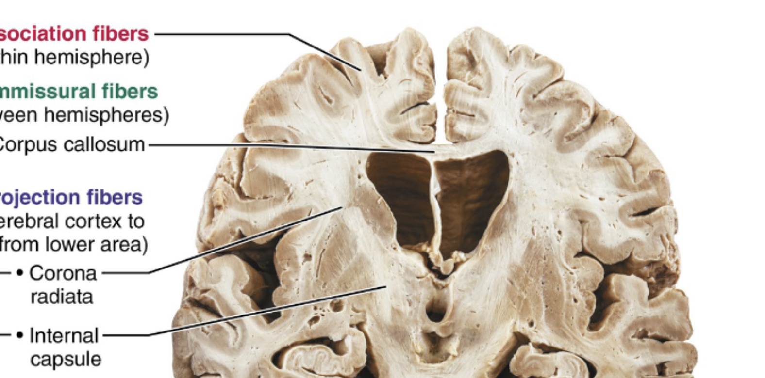
in cerebral white matter, what function division do associtation fibers travel
runs horizontally to connect parts of the same cerebral hemisphere
in cerebral white matter, what functional divison do comissionural fibers travel
runs horizontal to connect gray matter in two hemispheres
in cerebral white matter, what functional division do comissionural fibers travel
VERTICAL fibers that connect hemisphere with lower brain/spinal cord
what major tract forms the principal commissural fiber connection between the two cerebral hemispheres
corpus callosum
structural distinction between internal capsule and corona radiata
internal capsule; forms a compact band passing between the thalamus and basal nuclei
corona radiata; crownlike fan radiating through white matter to the cortex
which subcortical nuclei constitute the basal nuclei (third region fo the cerebellum)
candate nucleus, putamen, and globus pallidus
which two basal nuclei collective form the striatum
caudate nucleus and putamen
what are the key functional roles attributed to the basal nuclei
influence and regulate the intensity of slow/sterotyped muscle movements
filter out inappropriate responses
inhibit anatagonistic movements
what central cavity is enclosed by the thalmus, epithalamus, and hypothalamus
the third ventricle
why is the thalamus considered the gateway to the cerebral cortex
acts as primary relay station that sorts/edits ascending sensory impulses/subcortical motor inputs/memory signals
sends this info to the target cortical areas
which paired hypothalamis nuclei act as relay stations for the olfactory pathways
mammillary bodies
what stalk connects the base of the hypothalamus to the pituitary gland
infundibulum
which specific hypothalamic nucleus acts as the body’s biological clock
regulates circadian sleep-wake cycles
suprachiasmatic nucleus
what are the primary homeostatic roles controlled by the hypothalamus
controls autonomic nervous sytem
initiates physical emotional responses
regulates body temp/foodinstake/thirst
sleep-wake cycles
endocrine system
which endocrine extends fro mthe epithalamus and what hormone does it secrete
pineal gland
secretes melatonin
three regions of the brain stem
midbrain, pons, medulla oblongata
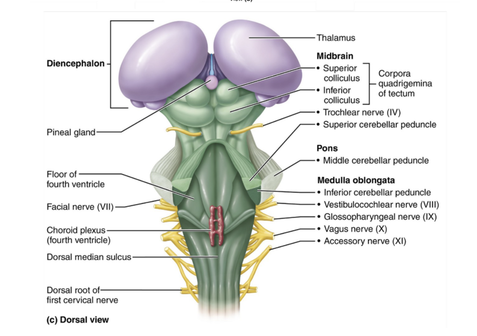
how many pairs of cranial nerves are directly associated w/ nuclei in the brain stem
10/12 of all the pairs
what structures form the two prominent ventral bulges of the midbrain containing the pyramidal motor tracts
cerebral peduncles

what functional roses are assigned to the periaqueductal gray matter in the midbrain
pain supression
fight/flight response
contains cranial nerve nuclei
in the midbrain corpora quadrigemina, how do the functions of the superior colliculi differ fro mthe inferior colliculi
superior; serves as visual reflex center
inferior; auditory relay center
degeneration of dopamine-releasing neurons in which midbrain causes parkinson’s disease
substantia nigra
what is the function of the red nucleus in the midbrain
relay nucleus for descending motor pathways that cause limb flexion
forms part of reticular formation
which cranial nerves originate from nuclei situated within the pons
trigeminal, abducens, and facial nerves

difference between longitudinal fibers and transverse fibers in the pons
longitudial; connect higher brain centers to the spinal cord
transverse; relay communication between motor cortex/cerebellum
what anatomical structures form the ventral longitudinal ridges on the medulla oblongata
pyramids (from the pyramidal motor tracts)
what functional crossing occurs at the crossing of the pyramids in the medulla oblongata
pyramidal motor tracts cross over to the to the opposite side of the CNS
ensures that each cerebral hemisphere controls voluntary movements of the contralateral side of the body
what information is relayed by the inferior olivary nuclei inside the olives of the medulla
they relay sensory stretch information from the muscles and joints to the cerebellum
which medullary nuclei relay ascending general sensory information from the spinal cord to the somatosensory cortex
nucleus gracilis and nucleus cuneatus
what wormlike midline structure connects the two cerebellar hemispheres
vermis
what are folia in the cerebellum
transversely oriented, slender gyri on the surface of the cerebellar cortex
what are the distinctive, branching treelike pattern of white matter inside
arbor vitae
which large specialized neuron orginate in the cerebellar cortex and convey output from the cerebellum
purkinje cells
what four steps describe the sequence of cerebellar motor processing
receives motor intent fro mthe cerebral cortex
receives body position momentum feedback from proprioceptors/sensory pathways
calculates optimal smooth muscle contraction
sends movement blueprint to motor cortices and brain stem
besides motor coordination, what nonmotor cognitive functions are influences by the cerebellum
thinking, language, emotion, and regulation of food intake
what is the primary function of the reticular activating system within the reticular formation
sends continous impulses to the cerebral cortex
maintains cortical alertness and filters out petitive/weak sensory stimuli