BIO 341 Unit 3 EU
 Call Kai
 Learn
 Practice Test
 Spaced Repetition
 Match
 Flashcards
 Knowt Play
1/47

There's no tags or description
Looks like no tags are added yet.
48 Terms
Generation of T-cell immune response
Infection can occur anywhere in the body
Myeloid dendritic cells roam body, capture antigens, and bring them to secondary lymphoid tissue to present to naive T cells
Infections in skin → draining lymph nodes
Infections in blood → spleen
Infections in mucosal tissue → mucosal secondary lymphoid tissues
Dendritic cell maturation
dendritic cells undergo a maturation process traveling from skin/periphery to lymph nodes
Immature dendritic cells in skin
very good at capture, uptake, and processing of antigens
Mature dendritic cells in lymph node
elaborate dendrites to aid in T cells activation
What happens to the rest of the T cells
pathogen specific T cells are 1 in 10^4-10^6 of all T cells
Naive T cells continue circulating
condensed chromatin
Little cytoplasm
Little RNA and protein synthesis
Once activated, takes a few days for T cell to proliferate and differentiate into effector T cell = delay time between infection and immune response
Homing of naive T cells to lymph nodes
circulating T cell enters the high endothelial venule in the lymph node
Binding of L-selectin to GlyCAM-1 and CD34 allows rolling interaction
LFA-1 is activated by chemokines bound to extracellular matrix
Activated LFA-1 binds tightly to ICAM-1
Diapedesis - lymphocyte leaves blood and enters lymph node

Once inside lymph node… (T cell)
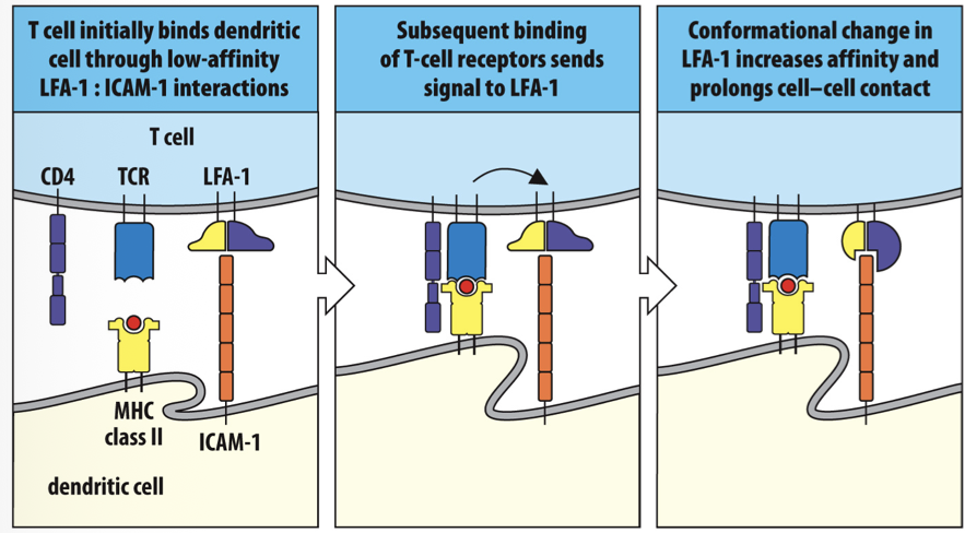
T cell initially binds dendritic cells roam body through low affinity LFA-1 and ICAM-1 interactions
Subsequent binding of T cell receptors send signal to LFA-1
Conformational change in LFA-1 increases affinity and prolongs cell-cell contact
Leaving the lymph node (T cell)
Most T cells won’t recognize antigen and will leave lymph by efferent vessel
S1P gradient directs cells toward efferent vessel
Naive T cells have S1P receptor on cell surface to follow gradient
Upon recognition of antigen, CD69 sequesters S1P receptor inside cell (T cells is stuck in lymph node)
Mature/effector T cell lose CD69, express S1P receptor again, and can leave lymph
Co-stimulatory molecules
T cell must recognize peptide:MHC complex but that does not lead to full activation
Activation requires co-stimulatory signal
CD28 receptor on T cells binds to B7 molecule on dendritic cell
Only dendritic cells, macrophages, and B cells have B7 (only expressed during infection)
Once T cells won’t recognize is activated, CD28 is replaced by CTLA4 (binds B7 20x stronger than CD28)
The SMAC
when naive T cell binds to dendritic cell, binding occurs in T ell synapse
Within the synapse are two areas
c-SMAC (central)
p-SMAC (peripheral)
ITAMs
Upon binding at T cell synapse, signaling beings by phosphorylation of ITAMs:
In the resting T cell the ITAMs of the CD3 complex are not phosprylated
Binding of MHC to the TCR leads to ITAM phosphorylation by Lck
Zap-70 binds phosphorylated zeta-chain ITAMs and is phosphorylated by Lck
effects of ZAP-70
T cell binds to dendritic cell → Lck phosphorylates ITAMs → ZAP-70 binds to phosphorylated ITAMs → PLC-gamma → PIP2→cleaved into DAG and IP3
→DAG
→PKC-theta → NFkB
Ras-MAPK → AP-1
→IP3
increases Calcium → Calcineurin → NFAT
3 transcription factors (NFkB, AP-1, and NFAT) made that change the pattern of gene expression → cell division, proliferation, and differentiation to mature/effector T cells
IL-2
IL-2 is in activated T-cells
cells constitutively express partial receptor
IL-2 expression leads to full receptor function and autocrine signaling
T-cell anergy
some T ells will respond to self-antigen by accident
Lack co-stimulatory molecule → T cell anergy
Anergy = unresponsive and cannot ever be revived
differentiation of CD4 T cells
CD4 T cells are helper T cells
recruit other cells to destroy pathogen
Several classes of CD4 T cells
Cytokines environment determines which T cells develop
Polarized T cell responses
activation and differentiation of T cells by cytokines → positive feedback mechanism
Polarized TH1 response = cell-mediated immunity
Polarized TH2 response - humoral immunity
Activation of CD8 T cells
CD8 T cells are cytotoxic so they need a much stronger signal to be activated (because they kill)
Dendritic cell usually activates CD4 T cells to secrete IL-2 and CD8 T cell → IL2 from the CD4 T cell drives proliferation and differentiation of CD8 T cells
Dendritic cells can also send the signal themselves to activate CD8 T cells to effector status → CD8 T cell makes IL2 driving its own proliferation and differentiation
post activation of T cells
Once T cells are activated they leave secondary lymphoid tissue and travel to infected tissues to do their work
leads to changes in many cell surface proteins
Relaxation of co-stimulators requirement
While activation of T cells required co-stimulators molecule, differentiated effector T cells do not have this requirement
Allows CD8 cells to bind to any cell with pathogen
Allows CD4 cells to bind more targets
cytokines and cytotoxins
effector T cell functions are mediated through a host of Tolkien’s and cytotoxins
cytotoxins - kill cells
Cytokine regulation of gene expression
effector T ells can disrupt target cells by altering gene expression through the release of cytokines
JAK-STAT pathway often activated in this pathway
CD8 effector cell function
once inside of a cell, pathogens become inaccessible to antibodies
CD8 T cells secrete lyric granules (vesicles) to kill target cell as well as pathogen
Once they have enough lyric granules again, CD8 T ells can kill multiple cells in succession
killing by apoptosis
CD8 T cells kill their target cells by inducing apoptosis
pokes holes in cell membrane
Activate nucleases to chop up DNA
Cell shrinks by shedding membrane-bound vesicles
Effector TH1 CD4 cells
Responsible for activating macrophages
binding of CD40 and IFN-gamma lead to macrophage activation
TH1 cell and infected macrophage come together → T cell beings to and activates macrophage → killing intravesicular bacteria (remember macrophages aren’t activated which is why they aren’t killing pathogen in leprosy)
Effector TFH CD4 cells
TFH cells are responsible activating naive B cells
After differentiation they move to B cell area of lymph node to complete their work
TFH cell recognizes peptide derived from B cell’s antigen → Naive B cell and TFH cell exchange signals that being the process of B cell activation
B cell activation
B cell activation requires cross-linking of BCR by the antigen
activates signaling pathway through ITAMs
Cross linking of B cell receptors by antigen → clustering of antigen receptors allows receptor-associated kinases to phosphorylate the ITAMs → Syk binds to doubly phosphorylated ITAMs and is activated on binding → changes in gene expression in nucleus
Co-receptors
Just like T ELLs, binding of B cells to antigen isn’t enough to fully activate
Must bind to co-receptor made up of 3 parts:
CR2 and CD21 = recognizes C3b fragments on pathogen
CD19 = signaling chain
CD81 = brings CD19 to the surface
BCR/co-receptor function
BCR and co receptor cooperate in B cell activation by a pathogen
BCR and co-receptor cooperate in B cell activation by a soluble antigen
→ both lead to changes in gene expression in nucleus
TFH cells activating B cells and TI antigens
Most B cells are activated by TFH cells in the secondary lymphoid tissues
Thymus independent antigens
Some B cells can be activated by certain antigens without T cell activation
No need for T cell
Follicular dendritic cells (FDC)
FDCs and macrophages present antigens to B cells
Express CR2 to capture antigens and present to B cells
Activation of B and T cells in lymph nodes
Naive B cells search for specific antigen displayed by FDC in the B cells area; naive T cells search for specific antigen presented by dendritic cells in the T cell area
Antigen activated T cells proliferate and differentiate; antigen activated B cells move to the boundary region
Antigen activated B cells present antigens Thymus independent to effector TFH cells, forming cognate interactions and cognate pairs
TFH secrete cytokines to aid B cells
Helper TFH cells conjugates with the B ell and beings to synthesize cytokines and CD40 ligand
The helper TFH cell reorients its cytoskeleton and secretory apparatus toward the B cell
Cytokines are secreted into the narrow space between the TFH cell and the B cell
clonal expansion
after conjugate pair forms from B and T cells, they must being clonal expansion
Primary focus occurs in medullary cords
lasts several days and produces B cells secreting IgM
Some B cells stay there and differentiate into plasma cells
The primary focus for expansion of antigen activated B cells in in the medullary cords → the secondary focus for expansion of antigen activated B cells is in the primary follicle → expansion of antigen activated B cells in the primary follicle creates the germinal center
Secondary focus
many B/T cell conjugates will leave medullary cords and move to primary follicle for expansion → germinal center
Somatic hypermutation and isotype switching occur here
Centroblast = large, metabolically active B cells that divide rapidly
cells have isotype switching occurring
Centrocytes = divide slowly, re-express cell-surface immunoglobulin that has been isotype switched
derivatives of centroblasts
Will die quickly if not bound to antigens
Selection of centrocytes
activated B cell→somatic hypermutation of immunoglobulin V regions in rapidly proliferating germinal center control blasts
→Germinal center centrocyte with low affinity surface immunoglobulin → BCR is not cross linked and centrcyte cannot present antigen to TFH cells → cell death
Or
→ germinal center centrocyte with high-affinity surface immunoglobulin → BCR is cross linked and antigen is presented to helper TFH cell → centrocyte receives help, survives and divides → centrocytes differentiate into plasma cells
cytokines from T helper cells determine B cell fate
Cytokines determine isotype switching (Ex: IL-4 induces IgG1)
If no binding to T cells through CD40, what will happen to B cells?
B cells will only secrete IgM (can’t isotype switch if they can’t bind to T cell)
Differentiation to plasma vs memory cells
centrocytes with identical BCR
Antigen selected centrocytes mature under the influence of an IL-10 secreting helper TFH cell → centrocytes differentiate into plasma cells → make antibodies that fight infection
Or
Antigen selected centrocytes mature under the influence of an IL-4 secreting helper TFH cell death Or→ centrocytes differentiate into memory B cells → the investment that prevents future infections from causing disease
Antibody effects functions - IgM, IgG, monomeric IgA
while IgM is the first antibody produced, it undergoes isotype switching
IgM, IgG, and monomeric IgA protect internal tissues
Most common circulating molecule is IgG
smaller than IgM and can reach more tissues
Relies on Fc receptor (FcRN) to be carried into tissues
Dimeric IgA
responsible for protecting mucosal surfaces
IgE
found in very small amounts in body
Not secreted as a soluble antibody, remains on cell surface
Cross linking of IgE antibody leads to release of histamine from mast cells
inflammatory mediator
Effects of inflammatory mediators
inflammatory mediators are released by mast ELLs, basophils, eosinophils
smooth muscle contractions in airways and gut to expel parasites
Increased permeability of blood vessels to help flush out parasites
Physical removal of parasites that are too big to be phagocytosed
In developed countries, we aren’t exposed to many parasites so mast cells are considered a detriment
some people make IgE that responds to innocuous substances like pollen
Antibodies from mothers
during pregnancy, from IgG from maternal circulation is transported across placenta and delivered to baby
At birth, baby has as high levels of IgG as mother with as wide a range of antigen specificities
Infants obtain IgA from breastmilk = passive transfer of immunity
goes to gut and binds to microbes; excreted through feces
Baby’s first antibodies
after birth, antibody level gradullary decreases until 6 months when baby begins to make its own antibodies
maternally derived IgG is catabolized and IgA is lost immediately in feces
IgG levels are lowest in children age 3-12 months
Problematic for premature babies who didn’t get much of mother’s IgG and take longer for immune system to be fully functioning
Also age where genetic deficiencies in immune system are found
Neutralizing antibodies
specific to an antigen and can prevent microbe from attaching to human epithelium
neutralizing toxins
many microbes release toxins that can be neutralized by IgG or IgA
Antibody activation of complement
Most antibodies don’t directly kill pathogens but rather recruit other cells/molecules to destroy pathogen
IgM, IgG3, and IgG1 can activate complement
Activation of complement by IgG
in order for IgG to activate the complement pathway, C1q must bind to at least 2 IgG molecules
are we sure we’re gonna do this (cell death)
IgG molecules bind to antigens on bacterial surface → C1q binds to two or more IgG molecules and initiates complement activation
Or
IgG molecules bind to soluble multivalent antigen → C1q binds to soluble immune complex and initiates complement activation
The role of RBC’s
RBCs aid in removal of immune complexes from the circulation through CR1;C3b binding
Small antigen:antibody complex forms in circulation and activates complement → immune complex is coated with covalently bound C3b → CR1 on an erythrocyte surface binds C3b-tagged immune complex → erythrocyte carries immune complex to the liver or spleen, where it it detached and taken up by a macrophage
Fc(gamma) receptors
complement its powerful because C3b coats pathogens permanently, delivering them for uptake and elimination
antibodies can bind just as well
Fc gamma receptors are specific for IgG and are expressed by macrophages and dendritic cells
aid in uptake of pathogen coated in IgG
Antibody dependent cell mediated cytotoxicity (ADCC)
we can use our knowledge of Fc receptors in therapy:
B cell lymphoma cells express CD20
Treat patients with anti-CD20 antibody (Rituximab)
Fc receptor on NK cell binds to Rituximab and signals NK cell to kill B cell lymphoma cell