MCB 245 cell types
 Call Kai
 Learn
Learn Practice Test
 Spaced Repetition
 Match
Match Flashcards
 Knowt Play
1/53

There's no tags or description
Looks like no tags are added yet.
Name | Mastery | Learn | Test | Matching | Spaced | Call with Kai | Chat |
|---|
No analytics yet
Send a link to your students to track their progress
54 Terms
Epithelial tissue structure
tightly packed cells with minimal extracellular matrix
Epithelial tissue functions
Covers body and organ surfaces, lines body cavities and organ cavities, forms glands
Types of Simple Epithelium
simple squamous, simple cuboidal, simple columnar, pseudostratified columnar
Types of Stratified Epithelium
squamous, cuboidal, columnar, transitional
Connective tissue structure
Contains cells, protein fibers, and ground substance
Connective tissue functions
Binds, supports, and protects other tissues and organs
Types of connective tissue proper
Loose (areolar, adipose, reticular)
Dense (regular, irregular, elastic)
Types of supporting connective tissue
Cartilage (hyaline cartilage, elastic cartilage, fibrocartilage, bone)
Types of fluid connective tissue
blood, lymph
Muscle tissue structure
Cells may be cylindrical, branching, or spindle-shaped; may contain contractile proteins (myofilaments)
Muscle tissue functions
Moves the skeleton, organ walls, or body structures; contractile, conductive, and excitable
Types of muscle tissue
Skeletal, Cardiac, Smooth
Nervous tissue structure
Contains neurons and glial cells
Nervous tissue functions
Neurons transmit nerve impulses and process information; glial cells support, protect, and nourish neurons
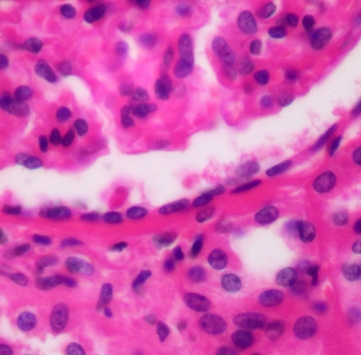
Simple Squamous Epithelium

Simple cuboidal epithelium

Simple columnar epithelium

Pseudostratified ciliated columnar epithelium

Stratified squamous epithelium

Stratified cuboidal epithelium

Stratified columnar epithelium

Transitional epithelium
Simple squamous function + location
Thinnest possible barrier to allow for rapid diffusion and filtration; secretion in serous membranes
Located in air sacs in lungs (alveoli), lining of lumen of blood and lymph vessels, serous membranes of body cavities
Simple cuboidal function + location
Absorption + secretion; forms secretory tissue of most glands and small ducts
Located in kidney tubules, thyroid gland follicles, surface of ovary, secretory regions of ducts and most glands
Non-ciliated simple columnar function + location
Absorption + secretion; secretion of mucin
Located in inner lining of most of digestive tract (stomach, small + large intestine)
Ciliated simple columnar function + location
Secretion of mucin and movement of mucus along apical surface of epithelium by cilia; oocyte movement through uterine tube
Located in the lining of larger bronchioles (air passageways) of lung and the uterine tubes
Non-ciliated pseudostratified columnar function + location
Protection
Located in lining of part of male urethra and epididymis
Ciliated pseudostratified columnar function + location
Protection; secretion of mucin and movement of mucus along apical surface of epithelium by cilia
Located in lining of larger airways, nasal cavity, pharynx, larynx, trachea, and bronchi
Keratinized stratified squamous function + location
Protection of underlying tissue from abrasion
Located in epidemies of skin
Non-keratinized stratified squamous function + location
Protection of underlying tissue from abrasion
Located in lining of oral cavity, pharynx, larynx, esophagus, lining of vagina, and anus
Stratified cuboidal function + location
Protection + secretion
Located in ducts of most exocrine glands and ovarian follicles
Stratified columnar function + location
Protection + secretion
Located in large ducts of salivary glands, conjunctiva covering eye, membranous part of male urethra
Transitional epithelium function + location
Accommodates urine volume changes (by distending or relaxing)
Located in urinary bladder, ureters, and part of urethra

Areolar (loose, connective proper)

Adipose (loose, connective proper)

Reticular (loose, connective proper)

Dense regular (dense, connective proper)

Dense irregular (dense, connective proper)

Elastic (dense, connective proper)

Hyaline cartilage (cartilage, supporting connective)

Fibrocartilage (cartilage, supporting connective)

Elastic cartilage (cartilage, supporting connective)

Compact bone (solid matrix, supporting connective)

Spongy bone (solid matrix, supporting connective)

Blood (fluid connective)
Areolar connective tissue
Abundant, viscous ground substance; few collagen and elastic fibers; scattered fibroblasts; many blood vessels
Protects tissues and organs; binds skin and some epithelia to deeper tissue
Located in papillary layer of the dermis, subcutaneous layer, surrounds organs, nerve cells, some muscle cells, blood vessels
Adipose connective tissue
Closely packed adipocytes; nucleolus pushed to edge of cell by large fat droplet; contains many blood vessels
Stores energy, insulates, cushions, protects
Located in subcutaneous layer, surrounds and covers some organs
Reticular connective tissue
Viscous ground substance; meshwork of reticular fibers, leukocytes, and some fibroblasts
Provides stroma (supportive framework) to lymphatic organs
Located in spleen, lymph nodes, and red bone marrow
Dense regular connective tissue
Densley packed, parallel arrays of collage fibers, fibroblasts squeezed between layers of fibers, scarce ground substance; limited blood supply
Attatches bone to bone and muscle to bone, resists stress applied
Located in tendons and ligaments
Dense irregular connective tissue
Collagen fibers randomly arranged and clumped together. fibroblasts in spaces among fibers, more ground substance than in dense regular, extensive blood supply
Withstands stresses applied in all directions, durable
Located in dermis, periosteum covering bone and cartilage, epineurium covering nerves, epimysium covering skeletal muscle, organ capsules
Elastic connective tissue
Elastic fibers, fibroblasts occupy some spaces between fibers
Allows for stretching and recoil
Located in walls of elastic arteries (aorta), trachea, vocal cords
Hyaline cartilage
Glassy-appearing matrix, irregularly arranged chondrocytes in lacunae
Provides support, forms most of fetal skeleton
Located in tip of nose, trachea, larynx, costal cartilage, epiphyseal (growth) plates and articular ends of long bones, fetal skeleton
Fibrocartilage
Really visible, numerous, parallel collagen fibers with limited ground substance, large chondrocytes in lacunae
Resists compression, acts as shock absorber
Located in intervertebral discs, pubic symphysis, menisci of knee joints
Elastic cartilage
Abundant elastic fibers that form weblike mesh, closely packed chondrocytes in lacunae
Maintain shape while permitting extensive flexibility
Located in external ear, epiglottis of larynx