M5: Chest Tubes, Airways, and Respiratory Procedures
 Call Kai
 Learn
 Practice Test
 Spaced Repetition
 Match
 Flashcards
 Knowt Play
1/51

There's no tags or description
Looks like no tags are added yet.
52 Terms
inhalation
diaphragm contracts, chest expands, negative P pulls air IN
exhalation
diaphragm relaxes, P pushes air OUT
abnormal lung sound examples
rhonchi (mucus in large airways), rales (fluid in alveoli), wheezes (narrowed airways), stridor (upper airway obstruction), pleural rub (inflamed pleura)
normal respiratory rates
adults → 12-20 breaths/min
children → 14-22 breaths/min
infants (<12mo.) → 30-60 breaths/min
What controls breathing?
medulla and pons sense CO2 (NOT O2)
high CO2 → fast, deep breathing
low CO2 → slow, shallow breathing
(CO2 drives breathing; rising CO2 lowers pH signaling brain to breathe faster)
external respiration
at the alveoli and pulmonary capillaries
O2 moves into blood, CO2 moves out of blood
internal respiration
at the systemic capillaries and body tissues
O2 moves into cells, CO2 moves from cells to blood
ventilation vs. respiration
ventilation → mechanics of breathing (airway, lungs)
respiration → gas exchange (alveoli, cells)
good ventilation, poor respiration → fluid in lungs
good respiration, poor ventilation → hypoventilation
perfusion
blood flow
pulmonary arteries carry blood to the lungs, veins return it to the heart, O2 binds Hb at alveolar-capillary membrane
hypoxia
low tissue oxygen
caused by poor ventilation/perfusion (V/Q mismatch)
pulmonary embolism
clot blocks lung blood flow, medical emergency
s/s → sudden dyspnea, chest pain, tachycardia, coughing blood, anxiety, “impending doom”
diagnosis → CTPA, D-dimer, V/Q scan, DVT u/s
treatment → anticoagulants, thrombolytics, IVC filter, O2, pain control
complications → lung tissue death, pulmonary HTN, cardiac arrest
goal of chest tube
remove air, blood, or fluid and restore negative pressure
chest tube indications
pneumothorax (air in pleural space collapses part/all of lung)
hemothorax (blood in pleural space from trauma/surgery)
pleural effusion/empyema (fluid/pus compresses lung and limits expansion)
after thoracic surgery (drains air/blood and restores pleural pressure)
3 chambers in a chest tube
collection chamber, water seal chamber, suction control chamber
collection chamber
function → collects drainage from pleural space
watch for → mark date/time/level each shift, sudden bright red surge means hemorrhage, notify MD if >100-200 mL/hr or bloody drainage
water seal chamber
function → 1-way valve that keeps air from re-entering the chest
watch for → tidaling w/ breathing is normal, continuous bubbling means air leak
suction control chamber
function → regulates the suction applied, often ~-20cm H2O per order, prevents excess suction on pleural cavity
watch for → gentle, steady bubbling is expected in a wet system
wet suction
water level in suction chamber sets suction (ex: 20cm)
gentle bubbling shows suction is working
check water level → evaporation can lower it
dry suction
dial sets suction level
no bubbling in suction chamber → indicator shows suction is on
quieter and easier to set up
things to keep at bedside for chest tube patient
vaseline gauze (Xeroform), occlusive dressing, new drainage system, sterile water
initial chest tube setup at bedside
hand hygiene, verify order (suction or water seal)
fill water seal to 2cm w/ sterile water
connect tubing securely, keep unit below chest
set dial (ex: -20) then raise wall suction until indicator shows
check tubing, apply occlusive dressing, assess tidaling
assessing a patient with a chest tube
respiratory status → rate/effort, SpO2, breath sounds, tracheal position, pain
drainage → amount, color, consistency, mark level and time each shift
tube/system → taped connections, no kinks or dependent loops, unit upright and below chest
site/skin → dressing dry/intact, check for crepitus (subcutaneous emphysema)
chest tube nursing considerations
collection chamber upright, below insertion site
tape to floor so it can’t tip
mark level per order, DO NOT EMPTY
report drainage >100mL/hr
note drainage → purulent vs. bright red
water seal tidals w/ breathing, continuous bubbling = air leak
suction set to -20mmHg
NEVER CLAMP A CHEST TUBE W/O MD ORDER
common chest tube problems
continuous bubbling in water seal → check connections/tubing for leak, assess pt, notify provider if persists
tubing disconnected → reconnect promptly using aseptic technique, assess pt
unit cracked or tipped over → set it upright or replace it, assess breathing
tube pulled out of the chest → cover site w/ sterile occlusive dressing per policy, call for help, assess for distress
no tidaling → check for kinks/loops/clots, may mean the lung has re-expanded, assess and report
potential chest tube complications

tension pneumothorax
signs → sudden dyspnea, tracheal deviation away from affected side, absent breath sounds, HOTN
actions → call for help, give oxygen, keep tube unclamped, prepare for decompression
hemorrhage
signs → sudden bright red surge or over 100 mL/hr, tachycardia, HOTN
actions → notify provider now, check v/s, secure IV access, prepare for volume
when to call provider emergently
sudden increase in drainage (>150-200mL/hr, bright red, clots), respiratory distress (tracheal deviation, hypoxia, spreading subcutaneous emphysema), continuous air leak (w/ no clear cause), device failure (or accidental tube removal)
assisting with chest tube removal
premedicate for pain, pt exhales or does the Valsalva maneuver as the tube is pulled, apply an airtight occlusive dressing, chest X-ray if warranted, watch for respiratory distress
chest tube removal preparation
verify order, lung re-expanded, minimal drainage, no air leak
explain the procedure in age-appropriate terms
premedicate 30-60min before
gather supplies → petroleum gauze, tape, sterile gloves, PPE
position semi/high Fowler’s
baseline → RR, SpO2, lung sounds, HR, pain, drainage
monitoring after a chest tube removal
assess breathing → q15-30min for 1-2hr, RR, effort, sounds, SpO2, pain
inspect dressing → drainage, bleeding, air leak
watch for pneumothorax → decreased breath sounds, tachypnea, cyanosis
chest x-ray → if ordered
deep breathing, coughing, IS use q2h
document → time, tolerance, dressing, findings, notifications
oropharyngeal airway
aka OPA, unconscious clients only (no gag reflex), size measured corner of mouth to angle of jaw (4-12cm), remove when gag reflex returns, watch for lip/tooth/tongue trauma
nasopharyngeal airway
aka NPA, conscious/semiconscious clients, size measured nose tip to earlobe (5-9cm), lubricate first, avoid w/ facial trauma or skull fracture, alternate nares q8-24hrs
endotracheal tube care
confirm placement (capnography, X-ray, breath sounds), secure tube, document cm mark at lip, cuff pressure 20-30 cm H2O checked q8-12hrs, oral care q2-4hrs, HOB elevated to prevent VAP, suction only as needed w/ sterile technique
**watch for unplanned extubation, obstruction, and right mainstem intubation
tracheostomy
surgical opening through the skin into the trachea in which a tracheal tube is inserted to keep the airway/trachea open, ensures O2/CO2 exchange
tracheostomy indications
prolonged mechanical ventilation, upper airway obstruction, inability to clear secretions, airway trauma/surgery, neuromuscular failure
parts of a tracheostomy tube
outer cannula → stays in the trachea
inner cannula → removed for cleaning or replaced
obturator → guides insertion of tube, KEPT AT BEDSIDE
cuff (if cuffed) → seals the airway, 20-30cm H2O
tracheostomy ties/dressings
ties sung → one finger fits underneath
two people change the ties
new trach → sterile gauze, kept dry and intact
cotton fibers can be aspirated or irritate the stoma so LINT-FREE DRESSINGS ONLY
monitor skin for breakdown/infection
tracheostomy nursing care
inner cannula → clean/replace at least q8-12hrs w/ sterile technique
stoma → clean/re-dress daily or when soiled, watch for infection/bleeding
ties → snug w/ one finger underneath, two people for ties change
cuff → keep 20-30cm H2O, check q8-12hrs
humidification → humidified oxygen or trach collar to thin secretions
swallow/speech → assess swallowing, involve speech therapy and communication aids
tracheostomy suctioning technique
prepare → sterile kit, suction at 100-150mmHg, catheter no more than half the tube diameter
preoxygenate → give oxygen before and between passes
insert → advance catheter w/o applying suction
withdraw → intermittent suction while withdrawing, 10s at most
reassess → SpO2, HR, breath sounds, rest before repeating
*STOP/REASSESS if SpO2 falls, HR drops or rhythm changes, client is in distress, 10 second limit is reached, suction only when needed not on a schedule
airway suctioning considerations
pre/post assessment, correct catheter size, pre-test suction, hyperoxygenate, maximum of 2 passes 10 seconds per pass w/ 30sec-1min between passes
what to keep at bedside in tracheostomy patient
obturator, suction equipment, oxygen, manual ventilation bag, spare trach tubes (1 same size, 1 size smaller)
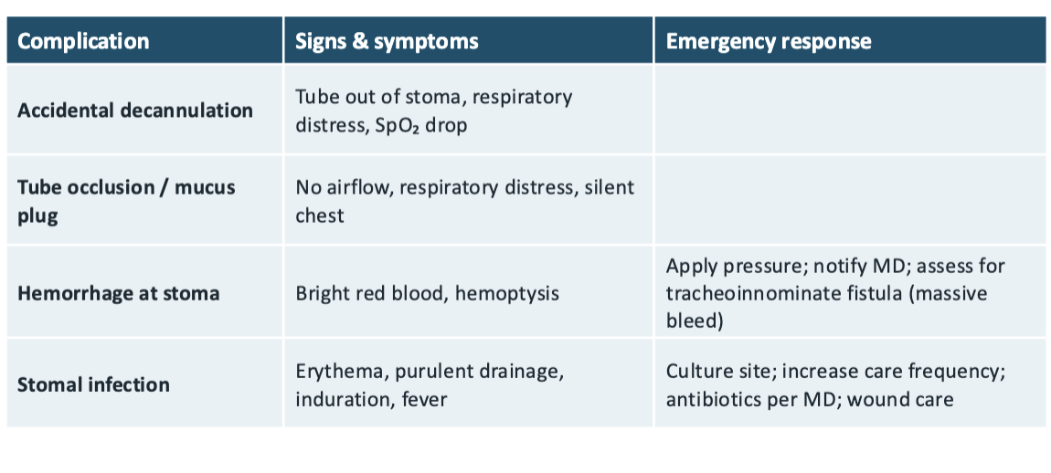
mucus plug/obstruction
signs → catheter will not pass, rising work of breathing, falling SpO2
actions → remove/replace the inner cannula, suction and give oxygen, call for help early
accidental decannulation
signs → tube out/displaced, distress, air movement at the stoma
actions → call for help, stay with patient, give oxygen to face and stoma, reinsert w/ obturator only if trained
trach complications and responses
before/after bronchoscopy
before → confirm consent, NPO per policy (often 6-8hrs), baseline v/s and breath sounds, review anticoagulants/allergies, explain what to expect
after → NPO until gag reflex returns, HOB elevated, monitor v/s and respiratory effort, mild sore throat and blood-tinged sputum are expected, REPORT bright red blood or increasing breathlessness
emergent findings after bronchoscopy
stridor/laryngospasm → high-pitched breath sounds or airway spasm
hemoptysis → coughing up bright red blood beyond streaks
pneumothorax → sudden chest pain, dyspnea, falling SpO2, decreased breath sounds
aspiration → coughing/choking w/ fluids
thoracentesis
needle removes pleural fluid to diagnose or relieve dyspnea
position → sitting upright, leaning forward over table
after → auscultate, check site, expect chest X-ray
monitor for pneumothorax and respiratory distress, document amount/character of fluid
thoracoscopy
scope passed through chest wall into pleural space
chest tube is often placed afterwards, monitor drainage/air leak/pain/infection, coach coughing/deep breathing/IS, report increased dyspnea or bleeding/fever
check insertion site, listen to patient’s lungs, check oxygen levels
A chest tube has continuous bubbling in the water seal chamber. What is your first action?
remove/clean/replace inner cannula, give patient oxygen if needed, call for help if low oxygen persists
A trach client’s SpO2 is falling and the suction catheter will not pass. What do you do?
gag reflex
A client one hour after bronchoscopy asks for water. What must you check first?