Kines 350 Exam 3
 Call Kai
 Learn
 Practice Test
 Spaced Repetition
 Match
 Flashcards
 Knowt Play
1/69

There's no tags or description
Looks like no tags are added yet.
70 Terms
What is the circulatory system?
works with cardiopulmonary or cardiorespiratory system
Cardiorespiratory:
transport O2 into muscles/tissues for cellular work
removal of CO2 from body
regulation of body temp
What are the two major adjustments of blood flow during exercise?
increase in cardiac output
redistribution of blood flow from inactive organs to active muscle
What are the parts of the circulatory system?
Heart: creates pressure to pump blood
Arteries and arterioles: carry blood away from heart
Capillaries: exchange of nutrients, O2 and CO2 within tissues
Veins and venules: carry blood toward heart

Know the flow of blood flow in the heart
Superior and Inferior Vena Cava brings deoxygenated blood to heart
Right Atrium
Right AV valve
Right Ventricle
Pulmonary Artery
Goes to lungs to oxygenate
(Pulmonary circuit)
Pulmonary Arteries
Left Atrium
Left AV valve
Left ventricle
Aorta
(Systemic circuit)
Layers of Cardiac muscle
Myocardium is heart muscle
heart wall includes: Epicardium, Myocardium (thickest), Endocardium
What is Myocardial Infarction (MI)
blockage in coronary blood flow results in cell damage/death
Coronary blood arteries supply heart with O2 needed for ATP producution for muscle contraction
Important: exercise training protects against heart damage
Which coronal artery is called the “widow maker”
Left Main coronary Artery- called widow maker because most men would have blockages there and it would stop blood flow to left ventricle
True or False does exercise training protect the heat from myocardial damage, such as heart attacks?
True: regular exercise is cardioprotective, it reduces the amount of myocardial damage, improves survival of heart attacks, and improves function of ATP-sensitive potassium channels
Compare heart muscle to skeletal muscle
Skeletal and heart muscle are striated, have z-discs, and aerobic
Skeletal: multinucleated, no junctional complexes, voluntary
Cardiac: single nucleated, have intercalated discs (help electrical impulses flow easily from fiber to fiber), involuntary
What is systole, diastole, what happens during rest and during exercise?
Systole- contraction phase, ejects blood
Diastole- relaxation phase, filling with blood
At rest: diastole time longer than systolic time (HR 75 bpm)
During exercise: duration of both systole and diastole are shorter (180 bpm)
What are the differences in pressure during the cardiac cycle? Heart sounds?
Diastole:
pressure in ventricle is low, filling blood from atria, AV valves open releasing pressure into ventricles
Systole:
pressure in ventricles rises, blood is ejected in pulmonary and systemic circuit
Heart Sounds:
1st sound: closing AV valves
2nd sounds: closing of aortic and pulmonary valves
Arterial Blood Pressure, Systolic, Diastolic, MAP
expressed as systolic/diastolic (180/20 mmHG)
Systolic Pressure: pressure generated by ventricular contraction
Diastolic Pressure: pressure in arteries during cardiac relaxation
MAP: mean arterial pressure- average pressure in arteries
DBP + 0.33 (Systolic BP-Diastolic BP)
What is Hypertension, aka High Blood pressure
Blood pressure that is 130/30 mmHG at rest
Primary Hypertension causes are unknown (genetic, dietary, aging)
Secondary Hypertension results from other diseases such as kidney disease
Risk Factors:
Left ventricular hypertrophy- works harder b/c pressure is high
Atherosclerosis and heart attack =- buildup of cholesterol
Kidney damage- vascular damage, damage other groups
Stroke- blood vessels bursts in brain (hemorrhagic stroke)

Label 1, 2, 6, 3 and 7
SA node
AV node
AV bundle
Right and Left bundle branches
Purkinje Fibers

What does an ECG do? Label the ECG chart
records heart’s electrical activity
P wave: atrial depolarization
QRS complex: ventricular depolarization and atrial repolarization
T wave: ventricular repolarization
What does ST segment depression suggest?
Myocardial ischemia: ventricles do not go through diastole long enough to fill up for Cardiac Output
What is Cardiac Output?
the amount of blood pumped out by the heart each minute
determines by Q= HRxSV
depends on training and gender
training lowers cardiac output @rest, improves heart disease
training allows for a larger SV to be able to pump out more volume of blood/min

How does the nervous system regulate heart rate? What is the difference between trained and untrained people?
Parasympathetic Nervous System:
Slows HR by inhibiting SA and AV node
Sympathetic Nervous System:
Increases HR by stimulating SA and AV node
Trained people increase or have a dominance of parasympathetic tone while simultaneously increasing sympathetic activity threshold, therefore more fit people get winded slower

What part of the brain controls the cardiovascular system?
The Medulla Oblongata via Aortic Baroreceptors and Carotid Baroreceptors (mechanoreceptors) which help determine stretch of blood vessel walls
What are Beta-blockers?
Beta-adrenergic blockers are drugs that can compete with epinephrine and norepinephrine (which ↑ HR) for beta receptors in the heart (↓ HR)
lower myocardial oxygen demand
lowers HR and contractility
Prescribed for people with Coronary Heart Disease
lowers for submax exercise and max exercise
What is Heart Rate variability?
the time between heart beats varies, the Standard deviation between each R-R interval
When our HRV is ↑ (the distance of R-R intervals vary) it means we have a healthy HR, parasympathetic is in control
Low HRV is a predictor of cardiovascular morbidity and mortality ↑ stress, ↑HRV, sympathetic is in control

How is Stroke Volume regulated?
End Diastolic Volume (EDV)
volume of blood in ventricles at the end diastole (preload)
Average Aortic blood pressure
pressure the heart must pump against to eject blood (afterload)
determines how much blood will be pumped out/overcome arterial pressure
Strength if the ventricular contraction (contractility)
enhanced by circulating epinephrine and norepinephrine
direct sympathetic stimulation

Explain End Diastolic Volume
“Frank-starling mechanism”- the more EDV, the bigger stretch of muscle at cross-bridges resulting in larger SV
Dependent on venous return
the more blood returned to the heart the more the heart can fill and pump out
Skeletal muscle pump: muscle contractions to force blood in the extremities to heart
Respiratory pump: changes in thoracic pressure to pull blood toward heart

Review
Got it!

What happens to the Hematocrit if someone is Anemic? At high altitudes?
Hematocrit is our RBCs in blood
If someone is anemic, hematocrit % decreases and hemoglobin decreases
At high altitudes, when we are dehydrated, increase of hematocrit % and increase of hemoglobin activity
What is the direct relationship of pressure and resistance for blood flow?
change of pressure (MAP- right atrial pressure)/ resistance= blood flow
high resistance= little blood flow (vasoconstriction)
low resistance- more blood flow (vasodilation)

What are the factors that determine the resistance in our blood vessels?
Length of vessel
Viscosity of blood
Radius of vessel (greatest influence)
Sources of Vascular resistance are:
MAP decreases throughout system circulation
Largest BP drop occurs across arteries
True or False the changes of arterial mixed venous (arterial O2 volume- venous O2 volume) remain the same during exercise
False, as exercise increases, O2 is demanded by skeletal muscle, so venous O2 levels are less than arterial.
How is O2 delivery accomplished?
Increased Cardiac Output (increase of HR and SV)
HR: adults about 220 bpm-age
SV: increase then plateaus to about 40-60% of VO2max
no plateau for trained individuals, keeps increasing due to improved ventricular filling and ↑ EDV and SV at ↑ HR
Redistribution of blood flow
What is the arteriovenous difference (a-VO2 difference)?
the amount of O2 that is taken up from 100 mL blood
increases during exercise due to higher O2 uptake in tissues
Fick equation: relationship between CO and a-VO2 difference, and VO2
VO2=Q x a-VO2 difference

Go over each graph about cardiovascular variables during exercise
systolic BP and MAP increases, while diastolic doesn’t change that much
SV increases and then plateaus in untrained individuals, they must increase HR
As VO2max increases, CO also increases, in untrained individuals their SV platerus so their HR must keep increasing
Heart rate continues to increase
Arteriovenous difference increases as VO2max increases reaching above steady state
What are some impacts of body position that affect Stroke Volume?
Effect on gravity on venous return
for example: when someone faints, lie them on their side since blood won’t pool near our legs
Upright exercise results in increased SV: due to large EDV, increased venous return
Supine exercise (swimming) results in small increase in SV

Where does blood get redistributed to during exercise?
During rest our GI and integumentary system mostly use blood
During exercise our Heart needs a larger CO, so blood is coming in and out faster and our skeletal muscles need O2 to be delivered for ATP production
How does local blood flow get regulated during exercise?
Using autoregulation (local control: tissues send messages to blood vessel then to the brain to vasodilate or vasoconstrict)
blood flow will increase to meet metabolic demands of tissue
The SNS vasoconstricts to organs that do not need blood flow at the moment (about 20-30% reduced)
What do Vasoactive factors do?
They are produced in endothelium or arterioles
promotes smooth muscle relaxation
results in vasodilation and increased blood flow

What happens to systole and diastole in response to exercise?
The changes in Heart Rate and Blood pressure depend on type, intensity, environmental condition and duration of exercise
Pressure in systole increases during exercise, pumping at a higher SV and HR
Pressure in diastole doesn’t change much to allow for filling
True or False: emotional influence effects BP and HR
True, if we are more emotionally stresses, our BP and HR increases pre-exercise

What happens to our HR, SV and CO during the transition from rest to exercise?
Our CO is factored by our HR and SV
onset of exercise: rapid increase of HR, SV, and CO
plateaus in submaximal exercise (oxygen demand=oxygen consumption)
during recovery from exercise: decrease in HR, SV, and CO toward resting levels (EPOC)
depends in duration and intensity of exercise, and training state of subject
high intensity calls for more rapid and slow component of EPOC slowing down HR/CO recovery
duration of exercise calls for more slow component of EPOC, which prolongs recovery of cardiovascuation
trained recover much faster
What happens to BP, HR , CO during incremental (steadily increasing) exercise? What is double-product?
As work rate increases= HR and CO increase linearly, reaches plateau @VO2max
Blood Pressure- MAP increases linearly
Systolic= increases
Diastolic= remains constant
Double product (RPP)- workload and oxygen demand by myocardium to do work as HR increases and systolic BP increases

True or False at the same oxygen uptake, arms works less since they are less heavy than legs.
False: arms are working against gravity, which means the heart must work harder to pump blood to upper extremities, greater RPP for arm than leg
HR increases due to high SNS stimulation
BP increases due to vasoconstriction

What happens to recovery of HR and BP during Intermittent (HITT) exercise?
Recovery of HR and BP depend on fitness level, temperature and humidity, and duration of intensity of exercise
Diagram: as our work increases from rest, our HR increases, as we go back to rest our HR does not go fully back down to rest so it will fluctuate, as seen.

What happens to Cardiovascular changes during prolonged exercise?
CO is maintained
SV gradually will decrease due to dehydration (sweating) and reduced plasma volume which gets taken up by muscles so (less blood in general)
increase of hematocrit % (RBC)
less EDV and therefore less SV
What is cardiovascular drift?
the drift when SV gradually plateaus and HR has to increase to maintain CO.
Why is there sudden cardiac deaths during exercise?
while sudden cardiac deaths are uncommon, it can be causes by many diff factors.
In children and adolescents
Genetic anomalies of coronary arteries (stress tests can help us detect)
cardiomyopathy
myocarditis
In adults
coronary artery disease
cardiomyopathy

What is the Central Command Theory?
It is the afferent nervous system that regulates cardiovascular adjustments for exercise via feedback
Located in higher brain centers
Fine-tuned feedback from
heart mechanoreceptors (like muscle spindles and golgi tendon organs) detect stretch reflex
muscle chemoreceptors (K+ and lactic acid)
muscle mechanoreceptors: sensitive to force and speed of movement
baroreceptors: sensitive to change in arterial blood pressure

Go over diagram and make sure you understand what happens during exercise
Yep!
What is the percentage of all gass partial pressures that make up the total pressure of gas mixture in the air
O2= 20.93%
CO2=0.03%
N2=79.04% (this is fully exhaled in and out, our body does not use Nitrogen)

Why is blood partial pressure lower in the venules/veins than in arteries/arterioles?
In the systemic circuit, the partial pressure in the arteries must be larger than the partial pressure in the tissue or target area, so that diffusion can occur and O2 can be uptaken for working muscles
The veins that uptake CO2, a byproduct of metabolic systems, so the PO2 in veins is less than the PO2 in arteries due to the consumption in tissues

What is Fick’s Law of Diffusion?
it is the rate of gas transfer (V gas) proportional to the tissue area ( A), diffusion coefficient (D), and the difference between partial pressure of gasses on each side of tissue (p1-p2) and inversely proportional to the thickness of tissue (T)
Vgas=(A/T) x D x (p1-p2)
What happens to the blood flow around the lungs at rest and during exercise?
At rest= the blood pools towards the base of lungs standing due to gravity
During exercise= blood flow increases in top of lung (apex)
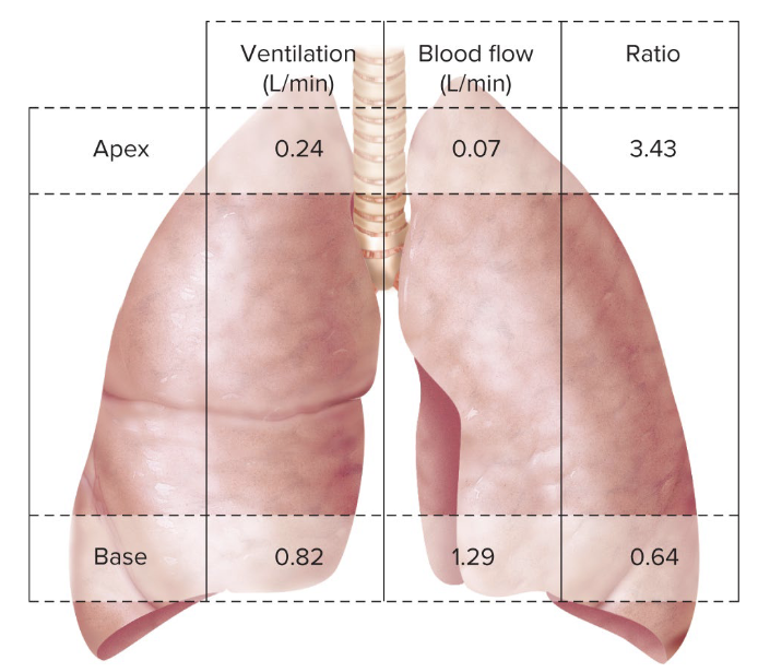
What is the Pulmonary ventilation/perfusion ratio? What does this ratio mean at rest and during exercise?
Pulmonary Ventilation/Perfusion ratio helps us determine how much air is flow and blood flow is going to the lungs, how well the air in the lungs can be available for blood to bind to O2 and drop of CO2
APEX of lungs (standing at rest)- V/Q ratio <0.1 underperfused (blood tries to work against gravity)
BASE of lungs (standing at rest) V/Q ratio >0.1 overperfused (blood is mostly at the base due to gravity)
During exercise- light-to-moderate improves V/Q ratio and heavy intense might make this ratio inequal

How is O2 transported in the blood? What does this mean between sex differences during exercise? What determines the amount of O2 that can be transported?
Hemoglobins in blood bind to O2 molecules- 4 O2 for each 1 Hb (about 99% is transported by Hb)
Males carry more Hb volume in blood therefore can carry more O2 per volume of blood, which reflects on their physical deliverance of O2 to working muscle for better performance
Hb concentration determines how much O2 can be transported

What does the Oxyhemoglobin curve help us determine?
It shows us how much saturation of O2 is in our blood from the PO2 at the veins and at the arteries.
Humans are around 97% saturated, meaning we carry most of our O2 by Hb
In Veins at rest we have less O2 saturation
In Arteries we have full (~100%) O2 saturation

What 3 major factors can contribute to a “rightward” shift of the Oxyhemoglobin curve?
pH of the blood-a decreases pH (meaning more acidic) lowers Hb-O2 affinity
favors offloading O2 to tissues
Body temperature- body temp increasing lowers Hb-O2 affinity
2-3 DPG (byproduct of RBC glycolysis)
during possible altitude exposure

What is the difference between Myoglobin and Hemoglobin?
Myoglobin is inside the muscle cells and has a higher affinity to bind to O2
It can store O2 inside muscle cells for onset exercise until respiratory system can deliver
Myoglobin binds to O2 at a lower PO2 from the Diffusion Gradient

How is CO2 transported out of our body from muscle cells?
Dissolves in plasma (10%)
Bound to Hb (20%)
Bicarbonate (70%)
Pulmonary ventilation removes H+ ions from blood by the HCO3- reaction to form Bicarbonate buffer which maintains our acid-base balance in our blood
Increased ventilation, CO2 exhalation (reduced PCO2 and pH increases)
Decreased ventilation, buildup of CO2 (increases PCO2 and pH decreases)

What happens during ventilation at exercise? What about PO2 and PCO2
Ventilation will rapidly increase at onset of exercise, followed by slower rise as we reach steady state
PO2 will slightly decrease at the onset of exercise do to muscle tissues needing O2 for aerobic metabolism and alveolar ventilation must catch up
PCO2 will slightly increase as muscles begin to make CO2 as a byproduct of metabolism and it will enter bloodstream
Does environment play a role in ventilation during prolonged exercise?
Yes, in hot and humid environments Ve drifts upward for thermoregulation, as body heat increases, SV and venous return decreases

What are some differences between untrained and trained individuals with O2 saturation, arterial PO2, arterial pH and ventilation as VO2max increases?
O2 saturations:
untrained: not changed as exercise intensity increases
trained: decreases because as muscles are doing more work O2 is being offloading
Arterial PO2:
untrained: happens around the same ventilation threshold (50%-75% VO2max)
trained: as work increases for elite athletes CO increases and loading of O2 can’t keep up so the pressure drops
Arterial pH
untrained: happens around same ventilation threshold (50%-75% VO2max)
trained: happens around same ventilation threshold (60%-85% VO2max)
Ventilation
untrained: ventilation rapidly increases at a lower intensity (50%-75% VO2max)
trained: ventilation rapidly increases at a slightly higher intensity (60%-85% VO2max)
lactate threshold corresponds to ventilatory threshold
Are there sex differences for breathing during exercise?
Yes, women have smaller airways than men so they have a higher resistance to airflow and limits to maximum ventilatory capacity, resulting in a greater work to breathe during exercise
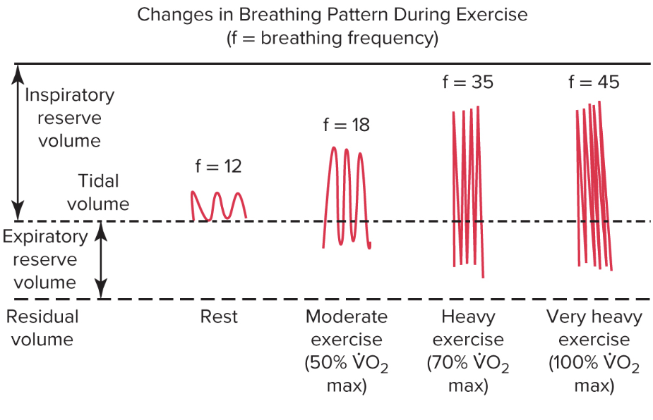
How does inspiratory, expiratory, and tidal wave differ as exercise intensity increases?
At rest, our tidal volume is small and we mostly are inspiratory
As we increase exercise our inspiratory volume increases and our expiratory decreases
At very intense exercise tidal volume levels out and we begin to breathe more rapidly
What is an abdominal “stitch” related to exercise?
It is an exercise-related transient abdominal pain (ETAP)
it can occur during many sports activities
irritation of parietal peritoneum (lining of abdominal cavity)
to reduce the risk of ETAP, avoid large amounts of food and water prior ti exercise
How is inspiration and expiration controlled?
Remember: skeletal muscles are innervated by alpha motor neurons, which are somatic and voluntary, however breathing is both voluntary and involuntary, this is because motor control centers in the medulla control this
Inspiration: preBotzinger complex in the medulla oblongata that controls inspiration via spinal cord motor neurons
Exhalation: retrotrapezoid nucleus/parafacial respiratory group controls exhalation
input by chemoreceptors and mechanoreceptors
Tell me the specifics of input to respiratory control center
Humoral (blood-borne) chemoreceptors-
Central chemoreceptors- located in the medulla, they are sensitive to PCO2 and H+, (as PCO2 increases, H+ concentration increases for acid-base balance), increase ventilation
Peripheral chemoreceptors- they are aortic and carotid bodies, sensitive to PCO2, PO2, H+ and K+ in blood
Neural input chemoreceptors and mechanoreceptors - coming from motor cortex and skeletal muscle receptors such as mechanoreceptors, muscle spindles, golgi tendon organs
As arterial PCO2 increases what happens to ventilation?
As arterial PCO2 increases, our ventilation increases to expel arterial CO2 out as exhalation our PO2 stays relatively the same
As arterial PO2 keeps dropping what happens in response to Ventilation?
As arterial PO2 drops, ventilation decreases, until PO2 reaches a hypoxic threshold in which ventilation quickly responds if O2 keeps dropping after hypoxic threshold
How does the ventilatory control center control breathing during submax exercise and heavy exercise?
Submax exercise: primary drive: higher brain centers (central command)
fine tuned by humoral chemoreceptors and neural feedback from muscle
Heavy exercise: a linear rise in Ve occurs due to
increasing blood H+ (from lactic acid) stimulates carotid bodies
increasing K+, body temp, and blood catecholamines may also stimulate breathing
True or false, ventilation is lower during exercise following endurance training, trained individuals have a better pH balance
True, at the same VO2max from untrained and trained individuals corresponds to the workload (trained person at 70% of VO2max breathes harder since their muscles are doing more work)
at the same workloads breathing is more increased at untrained individuals
True or False: training changes the structure of lungs, like other adaptations
False: training does not alter lung structure
True or False: Pulmonary ventilation/gas exchange is NOT a limitation in healthy individuals during low-to-moderate and high intensive exercise
True: the pulmonary system delivers oxygen at the same level
however respiratory muscle fatigue does occur during high intensity and gas exchange does limit exercise performance in some elite endurance athletes (more venous constriction and offloading O2 in muscle tissue)