Ciliate and Apicomplexan Lab
 Call Kai
 Learn
Learn Practice Test
 Spaced Repetition
 Match
Match Flashcards
 Knowt Play
1/45

Earn XP
Description and Tags
9/28/26
Name | Mastery | Learn | Test | Matching | Spaced | Call with Kai | Chat |
|---|
No analytics yet
Send a link to your students to track their progress
46 Terms

what does the image show?
Balantidium coli in trophozoite stage

what are the characteristics of Balantidium coli in trophozoite stage?
oval-shaped, large curved macronucleus
small micronucleus
cyststome
peristomal cilia
food vacuoles
what can Balantidium coli cause?
can cause intestinal lesions that result in a disease similar to amoebic dysentery (although this is rare)
Balantidial dysentery
where can Balantidium coli in trophozoite stage be found?
mainly in loose stools
infections are established when infective cyst is ingested with fecally-contaminated food/water
what hosts can Balantidium coli be found?
also infects pigs
human infections with Balantidium coli are especially common in pig-rearing areas
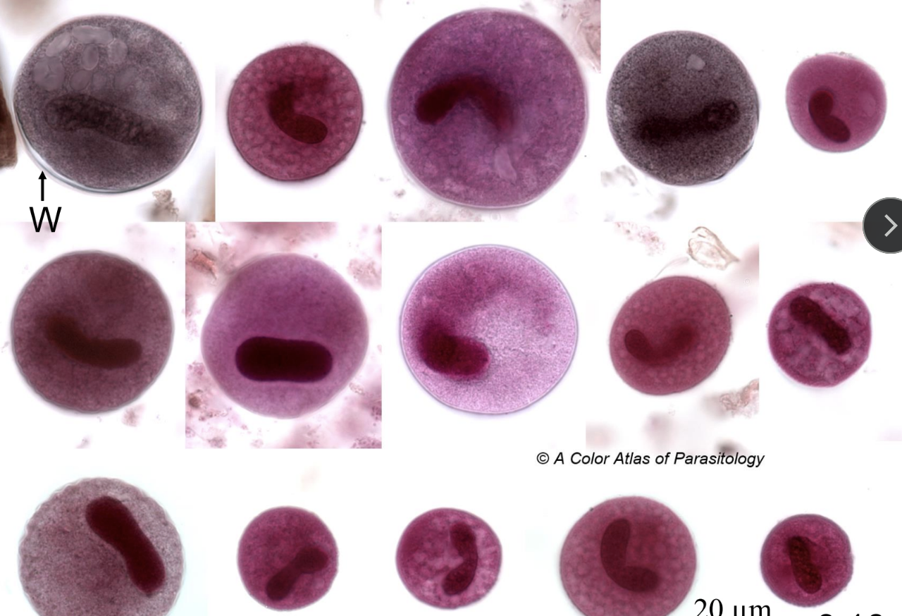
what does the image show?
Balantidium coli in cyst stage

what are the characteristics of Balantidium coli in cyst stage?
smooth, round shape
large curved macronucleus
cyst wall
why is it important to note the cyst wall of Balantidium coli in cyst stage?
cyst wall becomes separated from the cell in some specimens due to heat fixation of the bacteria onto the slide
this causes the cytoplasm to recede and make the cyst wall more obvious
what is the infective stage for of Balantidium coli?
cyst stage
found mainly in formed stools

what does the image show?
Balantidium coli in trophozoite stage
in a colon section (lower magnification)

how does Balantidium coli in trophozoite stage affect the colon?
lesion in colon is caused by the parasite
MM = muscularis mucosae
trophozoites can also be found in lymphatic vessels in the submucosa
(indicates spread of infection)
what is muscularis mucosae?
thin layer of muscle in gastrointestinal tract
separated from submucosa (bottom layer)
what is the submucosa?
thin layer of dense irregular tissue that supports the mucosa and joins it to the muscle layer
can Balantidium coli spread to other sites in the tissue/body?
yes (can go from muscularis mucosae to lymphatic vessels in the submucosa)
BUT only rarely causes this pathology in humans
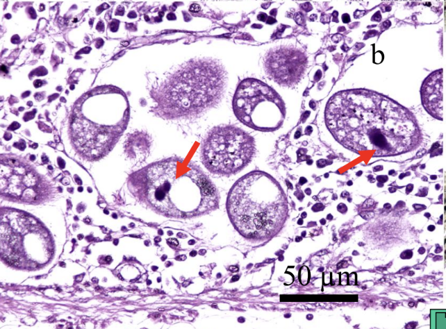
what does the image show?
Balantidium coli in trophozoite stage
in a colon section (higher magnification)
what is notable in the higher magnification of Balantidium coli in trophozoite stage in a colon section?
lesions in colon caused by trophozoites
note the macronucleus in some specimens

what does the image show?
Plasmodium falciparum in ring stage/early trophozoite
(in a blood smear)
what are the characteristics of Plasmodium falciparum in ring stage?
applique forms
double chromatin dots
multiple infections in some cells
high parasitemia
nonenlarged/nondecolorized infected cells
absence of Schuffner’s dots
what are applique forms (in Plasmodium falciparum ring stage)?
parasite is closely opposed to the edge of the erythrocyte
usually appears flattened against the membrane
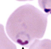
what are double chromatin dots (in Plasmodium falciparum ring stage)?
chromatin dot has two separate masses (double dot)
appearance resembles a signet ring (although only applies when dots are relatively close together)

what are multiple infections in some cells (in Plasmodium falciparum ring stage)?
when there are multiple infections in the red blood cell by Plasmodium
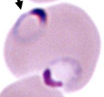
what is high parasitemia (in Plasmodium falciparum ring stage)?
high % of red blood cells are infected with parasites
for Malaria → 5% or more of RBC are infected (indicates severe malaria)
what is nonenlarged/nondecolorized infected cells (in Plasmodium falciparum ring stage)?
helps rule out specific malaria species like Plasmodium vivax and Plasmodium ovale
(which target immature red blood cells; which enlarges them and makes them lose color)
what is absence of Schuffner’s dots (in Plasmodium falciparum ring stage)?
Schuffner’s dots → tiny spots that appear in some forms of Malaria parasites
absense of them indicate infection of Plasmodium falciparum species
(Plasmodium vivax and Plasmodium ovale have the dots)
why is important to be able to distinguish Plasmodium falciparum (ring stage)?
organism kills more people annually than all other eukaryotic parasites combined
due to pathogenicity, failure to recognize P. falciparum infection in smears may have fatal consequences
what happens after ring stage in Plasmodium falciparum?
infected cells disappear from circulation by adhering to endothelial cells of blood vessels in the tissues
makes diagnosis difficult
can potentially lead to cerebral malaria
what is recrudescence in Plasmodium species?
merogony results in new infected erythrocytes (reappear in the peripheral blood at 48-hour intervals)
some individuals who are “cured” of infection may develop symptoms years later → due to resurgence of previously low, non-detectable levels of parasitemia
what is relapse compared to recrudescence (in Plasmodium species)?
recrudescence can occur in all species of Plasmodium
has different mechanism than relapse (which only occurs in Plasmodium vivax or Plasmodium ovale)

what does the image show?
Plasmodium falciparum in gametocyte
(in a blood smear)
what are the two pathways for merozoites (in Plasmodium falciparum)?
some meroziotes penetrate erythrocytes (RBCs) and differentiate into gametocytes
some merozoites undergo merogony → create more daughter cells
what are the special characteristics of gametocytes in Plasmodium falciparum?
(have unique appearance)
elongate
have nucleus (often obscured by hemozoin) less than ½ the length of the cell
what are the characteristics of microgametocytes in Plasmodium falciparum?
may be shorter and more blunt-ended
have a lighter blue cytoplasm
have a nucleus greater than ½ the length of the cell
surrounding erythrocyte is usually not visible
when do gametocytes of Plasmodium falciparum produce gametes?
do not produce gametes until they reach the midgut of a mosquito

what does the image show?
Plasmodium vivax in ring stage/early trophozoite
(in a blood smear)
what are the characteristics of Plasmodium vivax in trophozoite/ring stage?
enlarged
decolorized infected erythrocytes
prominent Schuffner’s dots
ameboid shape (shows pseuopod-like extensions)
hemozoin granules
why is the infected red blood cells enlarged and decolorized in Plasmodium vivax in trophozoite/ring stage?
become enlarged and decolorized due to infection of Malaria
only present in Plasmodium species like Plasmodium vivax and Plasmodium ovale
what are Schuffner’s dots in Plasmodium vivax?
fine pink/reddish granules in the cytoplasm of infected red blood cells

what is an ameboid shape of the trophozoite in Plasmodium vivax?
irregular, constantly changing form driven by the protrusion and retraction of temporary cytoplasmic extensions called pseudopods
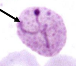
what are Hemozoin granules in Plasmodium vivax?
(may be relatively inconspicuous in trophozoites of this species)
insoluble, brownish-black crystals of toxic heme produced by malaria parasites when they break down host hemoglobin inside red blood cells
what do merozoites of Plasmodium vivax usually infect?
preferentially infect only young erythrocytes (reticulocytes)
how pathogenic is Plasmodium vivax?
parasitemia is relatively low
while malaria caused by Plasmodium vivax historically has been “benign” → severe and fatal infections with this species are now known to occur

what does the image show?
Plasmodium oocyst
(in tissue section)
what Phylum is Balantidium coli in?
Phylum Ciliphora
what Family is Balantidium coli in?
Family Balantidiidae
what Phylum is Plasmodium falciparum and Plasmodium vivax in?
Phylum Apicomplexa
what Family is Plasmodium falciparum and Plasmodium vivax in?
Family Plasmodiidae