Unit 7 - DNA Viruses
 Call Kai
 Learn
 Practice Test
 Spaced Repetition
 Match
 Flashcards
 Knowt Play
1/120

There's no tags or description
Looks like no tags are added yet.
121 Terms
Herpes viridae: genome/envelope, representative human diseases
dsDNA; enveloped
HSV, VZV, EBV, CMV, HHV-6/7/8
Adenoviridae: genome/envelope, representative human diseases
dsDNA; non-enveloped
respiratory, conjuctival, and GI disease
Papillomaviridae - genome/envelope, representative human diseases
dsDNA; non-enveloped
warts and HPV-associated cancers
Poxviridae - genome/envelope, representative human diseases
dsDNA; complex virion
smallpox, mpox, molluscum contagiosum
parvoviridae
ssDNA; non-enveloped
parvovirus B19/fifth disease
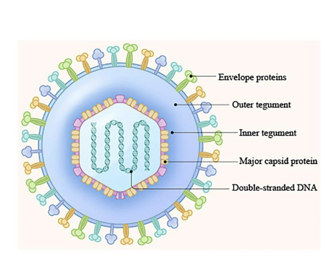
what is the structure of herpesviridae?
Four layers:
dsDNA genome
Icosahedral capsid
protein shell around dsDNA
Tegument
protein layer between capsid and envelope
Envelope
lipid membrane enables attachment and fusion
virus replicates inside nucleus of host cell
what are the 8 human herpesviruses (HHV)/ herpes viridae?
HSV-1/ HSV-2
VZV/ HHV-3
EBV/ HHV-4
CMV/ HHV-5
HHV-6/ HHV-7
HHV-8/ KSHV
-all can establish latency and reactivate later
HSV-1/ HSV-2 - major syndromes, latent reservoir
oral/genital lesions (cold sores), keratitis, encephalitis, neonatal herpes
latent reservoir in sensory neurons liek trigeminal nerve root ganglion or sacral root ganglia
VZV/ HHV-3 - major syndromes, latent reservoir
Varicella (chicken pox); shingles
latent reservoir in sensory nerve ganglia like sacral nerve root ganglia
EBV/ HHV-4 - major syndromes, latent reservoir
infectious mononucleosis; lymphoid/epithelial tumors
latent reservoir in B lymphocytes
CMV/ HHV-5 - major syndromes, latent reservoir
asymptomatic or mono-like infection; especially important for transplants
latent reservoir in leukocytes and endothelial cells
HHV-6/ HHV-7 - major syndromes, latent reservoir
roseola (sixth disease) in children
latent reservoir in T cells and other cells
HHV-8/ KSHV - major syndromes, latent reservoir
Kaposi sarcoma; primary effusion lymphoma
latent reservoir in B cells/ endothelial lineage
process of latency and reactivation
primary infection
viral replication and shedding occurs after exposure; symptoms may be absent or present
latent phase
viral genome persists within defined host cells with limited gene expression
reactivation
immune changes may allow renewed replication, shedding or symptomatic disease

how is herpes simplex virus (HSV) transmitted?
through direct contact with active vesicles or body fluids
cannot penetrate intact skin
travels through breaks in skin or mucous membranes in mouth or genital areas
asymptomatic shedding makes transmission possible with hard to notice symptoms
what are the two types of herpes simplex virus (HSV) and what do they primarily effect
HSV1
primarily oral, ocular, and CNS infections
~60-80% of Americans infected
HSV2
primarily genital and neonatal herpes
clinical manifestations of oral herpes (typically HSV1)
mucosal vesicles inside or around mouth
tingling pain precedes formation
vesicles break open, crust over, and heal
two types:
herpes labialis → cold sore
herpetic gingivostomatitis

clinical manifestations of genital herpes (typically HSV2)
vesicles appear around genitals, buttocks, or rectum
symptoms include fever, body aches, swollen lymph nodes, or headache
recurrent outbreaks due to latency
estimated 2-4x increased risk of HIV transmission
clinical manifestations of ocular herpes (typically HSV1)
swelling around eyelids
keratitis can result in ulceration
may lead to blindness
clinical manifestations of HSV - herpetic whitlow
infection of fingers
common among healthcare workers exposed to oral secretions
often observed in thumb-sucking children with HSV-1 oral infection
clinical manifestations of neonatal herpes (HSV2)
transmission from mother to fetus
highest risk (~50%) in mothers with unrecognized primary infection
85-90% of cases occur during birth
provide antiviral therapy during delivery to reduce risk
Clinical manifestations of HSV - HSV encephalitis
HSV-2 in neonates
HSV-1 in older children and adults
may result in fever, seizures, altered mental status, confusion
what is the preferred method of diagnosis of HSV
nucleic acid amplification test (NAAT) such as PCR
amplify viral DNA from lesion
PCR is recommended when HSV encephalitis is suspected
what are additional methods (other than NAAT) for diagnosis of HSV
Viral culture
MRC-5 cells used to grow virus
identification of viral strain with monoclonal Abs or molecular
Serology
positive results (type-specific IgG) cannot distinguish recent or past infection (due to reactivation of virus)
Direct fluorescent antibody (DFA)
swab of vesicle incubated with type-specific Ab
less sensitive
how is HSV treated?
Antiviral drugs → help reduce shedding and transmission during childbirth
nucleoside analogs
Acyclovir
Acyclovir monophosphate converted by host into acyclovir triphosphate (ACV-TP)
ACV-TP inhibits viral DNA replication
incorporates in synthesized DNA, but lacks 3’ -OH group resulting in premature chain termination
Valacyclovir
Famicyclovir
how can an HSV infection be prevented?
no vaccine
condoms as a barrier
reduce risk, but don’t eliminate it
gloves
healthcare workers to prevent herpetic whitlow
how is varicella-zoster virus (VZV) transmitted
through direct contact or inhalation of infectious vesicles
virions produced at high levels, making them very infectious
what are the two distinct diseases of varicella-zoster virus (VZV)
Varicella: chickenpox
Herpes-zoster: shingles
VZV - Varicella: chickenpox clinical manifestations in children vs. adults
Children: blister-like rash with intense itching and fever
vesicular lesions on face and trunk
secondary bacterial infections due to scratching
Adults, neonates, or pregnancy: more severe
pneumonia, premature labor, congenital malformations
VZV - herpes zoster: shingles clinical manifestations
after chickenpox, virus travels to sensory nerve endings in dorsal ganglia cells → latency
reactivation
15-30% → usually older individuals
postherpetic neuralgia
>1 year of pain
immune suppression
what is the typical and preferred diagnosis for varicella-zoster virus (VZV)
typically diagnosed based on clnical presentation
preferred: NAAT (PCR) from infected vesicle material are best diagnostic material
what are other methods (other than NAAT) to diagnose VZV
Viral culture
more difficult than HSV
Direct immunofluorescent assay (DFA)
vesicle swab or fluid
serology
IgM is not reliable due to reactivation
IgG detectable through ELISA (after pt. has formed Abs to virus)
ELISA is most common serology test
treatment of VZV
airborne isolation to stop inhalation of respiratory droplets
antivirals
acyclovir or valacyclovir → adults and immunocompromised individuals
prevention of VZV
Vaccine:
1995: live, attenuated varicella virus
12 months or older, adolescents, adults
problematic breakthrough cases
mild illness, contagious
recommended booster
2005: combined w/ MMR as MMRV (called Proquad)
2017: Shingrix as recombinant virus
recommended for person aged 50 years or older to combat shingles
2 doses 2-6 months apart
Epstein-Barr Virus (EBV) transmission
~40-100% of adults exposed
intimate contact with salivary secretions (kissing; sharing drinks, cups, utensils; children’s toys)
blood transfusions
bone marrow/solid organ transplants
sexual contact
perinatal contact
how does EBV infect cells of the body?
infects B lymphocytes and epithelial cells
complement receptor 2 (CR2/CD21) used for viral attachment and entry into B cells
after entry into cell, virus replicates
lyses host cell
releases infectious virions

Clinical manifestations of EBV
Children: typically asymptomatic to mild infection
Adolescents: infectious mononucleosis
classical triad: fever, cervical lymphadenopathy, sore throat (pharyngitis)
other symptoms: splenomegaly, hepatomegaly, periorbital edema
Cancers
Burkitt’s lymphoma
Nasopharyngeal carcinoma
Hodgkin lymphoma
EBV cancers - Burkitt’s lymphoma
malignant neoplasm of B cells in germinal center
90% of lymphomas in transplant patients at EBV+
overexpression of c-myc leads to increased cellular proliferation
often presents as tumors in jaw or face region
EBV cancers - nasopharyngeal carcinoma
nearly all cases are linked to EBV
no clear oncogene
hyperendemic in certain regions suggesting additional factors (China, Taiwan, Malaysia, Indonesia)
EBV cancers - Hodgkin lymphoma
~50% of cases linked to EBV
Reed-Sternberg cells present in lymph nodes
derived from B lymphocytes
multinucleated
sometimes looks like an “owl’s eye”
presence of cell is necessary for diagnosis

how is EBV diagnosed?
Classical symptoms
absolute lymphocytosis
50% of total leukocytes are lymphocytes
10-20% are reactive
mostly T cells responding to virus
elevated liver enzymes (ALT, AST)
seen in 50% of patients
Serology
infection of B cell causes polyclonal activation
B cells proliferate and secrete Abs
heterophile Abs
EBV-specific Abs

EBV diagnosis - heterophile Abs
Abs that may cross-react with Ags from a different species → Ags on animal erythrocytes
IgM class
good screening tool (Mono-spot)
~40% of pts. will have detectable amounts in 1st week; 90% by week 4
Paul Bunnel presumptive test
test for _____ _____
Pt. serum added to sheep RBCs → look for agglutination
pos. titer > 56
refer to Davidson differential test (confirmatory test)
what can cause false positives when testing for heterophile Abs in EBV diagnosis?
Forsmann Abs
produced after bacterial infections
found in 98% of the population
Serum sickness
Abs made after exposure to animal proteins in vaccines
not common anymore
use EBV-specific Abs to confirm suspected cases
EBV diagnosis - EBV-specific Abs
ELISA detection method
high sensitivity (95-99%)
detect Abs to Ags at different stages of infection
initial viral replication
Early Antigen (EA)-D: diffuse distribution in nucleus and cytoplasm
EA-R: restricted to cytoplasm only
late Ags appearing during lytic cycle
viral capsid Ags (VCAs)
latent Ags
Epstein-Barr Nuclear Ag (EBNA): 1, 2, 3A, 3B, 3C
Latent Membrane Protein (LMP): 1, 2A, B
EBV specific Ags - initial viral replication
early antigen (EA)-D: diffuse distribution in nucleus and cytoplasm
EA-R: restricted to cytoplasm only
EBV specific Ags - late antigens appearing during lytic cycle
Viral Capsid Antigens (VCAs)
EBV specific Ags - latent Ags
Epstein-Barr Nuclear Ag (EBNA): 1, 2, 3A, 3B, 3C
Latent Membrane protein (LMP): 1, 2A, B
EBV-specific Abs - what Abs indicate acute infectious mononucleosis (IM)
IgM anti-VCA
anti-EA-D
EBV-specific Abs - what Abs indicate convalescence
IgG anti-VCA
Anti-EBNA
Anti-EA-R
EBV-specific Abs - what Abs indicate past infection
IgG anti-VCA
Anti-EBNA
what do these panel results for EBV specific Abs indicate:
VCA IgM: neg
VCA IgG: neg
EA: neg
EBNA: neg
negative for EBV
what do these panel results for EBV specific Abs indicate:
VCA IgM: pos
VCA IgG: pos
EA: pos
EBNA: neg
acute EBV infection
what do these panel results for EBV specific Abs indicate:
VCA IgM: neg
VCA IgG: pos
EA: pos (neg)
EBNA: pos
convalescent EBV infection
what do these panel results for EBV specific Abs indicate:
VCA IgM: neg
VCA IgG: pos
EA: neg
EBNA: pos
past EBV infection
what do these panel results for EBV specific Abs indicate:
VCA IgM: neg
VCA IgG: pos
EA: pos
EBNA: pos (neg)
chronic EBV infection
how is EBV diagnosed?
NAAT (PCR)
viral load assays help monitor treatment
important for transplants and malignancies
viral culture
not practical in most cases
how is EBV treated?
self-limiting
rest plus medication for fever/pain relief
antivirals
for severe infection including encephalitis or hepatitis
acyclovir or valacyclovir
how is cytomegalovirus transmitted?
close, prolonged contact with infectious body secretions (intimate sexual contact, blood transfusions, solid organ transplants, perinatally)
virus can be isolated in saliva, urine, stool, vaginal, and cervical secretions, semen, breast milk, blood
50-80% of adults in US are CMV+ by age 40
30-50% of children are CMV+ by 5
how is cytomegalovirus (CMV) transmitted in the body
Infects a wide variety of cells
endothelial and epitelial cells
PMNs (neutrophils)
monocytes and macrophages
lymphocytes
persists in WBCs in latent stage
monocytes, DCs, myeloid progenitors
may reactivate later due to autoimmunity, stress, etc.
Cytomegalovirus (CMV) - congenital infections
most common congenital infection in US
1 in 200 babies born with it → most severe cases can result in mother losing baby
1 in 5 babies with congenital _____ will have long-term health problems
low birth weight
hepatosplenomegaly
Reactive lymphocytosis
petechiae in 50% of cases
hearing loss/deafness
lower risk is mom is already seropositive at time of conception
CMV clinical manifestations - primary infection
asymptomatic in immunocompetent host
may present as self-limiting, mild IM-like illness
sore throat lymphadenopathy, fatigue
major concern for immunocompromised individuals
organ transplant recipients
HIV+ patients
in both populations, infection can result in hepatitis, pneumonia, colitis, encephalitis, retinitis
increased rate of graft rejection or graft vs. host
CMV clinical manifestations - infection after transplant
due to large percentage of CMV+ patients, blood products are screened for ____
primary infection may happen if ____- patient transfused/transplanted with ____+ blood product
transplant pts may go through reactivation of their own infection
____+ patient is purposefully immunocompromised before transplant → virus reactivates
what is the preferred method of diagnosis for CMV?
NAAT (PCR)
congenital infections can be screened using amniotic fluid before birth or saliva after birth
transplants can be screened using plasma or whole blood
both donor and recipient
after transplant, quantitative PCR can help track if infection happens
what are other methods to diagnose CMV (other than NAAT)
serology via ELISA
IgM will not indicate recent infection due to reactivation
IgG is more helpful
4 fold increase in titer can suggest recent infection
otherwise, its likely a past infection
histology
biopsy tissue and stain
viral culture
MRC-5 cells
historical method, not frequently used
treatment and prevention of CMV
screening of blood products of transplants
leukoreduce products
filter WBCs to remove before transfusion/transplantation
leukoreduced products are considered safe for most patients
CMV immune globulin
IgG Abs against CMV
in combination with antivirals helps to reduce CMV death/disease
Barriers to infection
condoms
handwashing → especially after handling children or toys
antiviral drugs
nucleoside analogs (valganciclovir and ganciclovir)
cidofovir and foscarnet → target DNA polymerase in drug resistant CMV
Human Herpesvirus 6 (HHV-6)
transmitted via respiratory droplets
two species:
6A and 6B
infects children early in life (6-18 months)
causes Roseola
sixth disease - soft pink rash on torso and extends to face and extremities
five other diseases cause rash during childhood: measles, scarlet fever, Rubella, parvovirus B19, additional one that doesn’t exist
common complication is febrile seizures
diagnosis with PCR and viral load testing
Human Herpesvirus 7 (HHV-7)
>90% of adults are infected
shed in saliva of ~80% of healthy adults
infects T cells (via CD4) as well as other host cells
downregulates CD4 expression, which can interfere with HIV infection
primarily asymptomatic
may cause Roseola
clinically identical to HHV-6
rarely causes severe infection
testing rarely required
Human Herpesvirus 8 (HHV-8)
<1% of general population is a carrier → most US cases are found in HIV+ individuals
linked to cancerous condition called Kaposi sarcoma
cancer cells from the lymphatic endothelium are found on the skin or mucous membranes
tumor lesions appear purple, red, or brown patches
commonly seen on feet, legs, and face
may be seen in mouth, GI tract, lungs, and lymph nodes
diagnosis is made by biopsy of lesion
PCR performed on tissue
treatment is typically antiretroviral therapy for HIV along with chemotherapy
what family of viruses are able to encode its own machinery for genome transcription
poxviridae
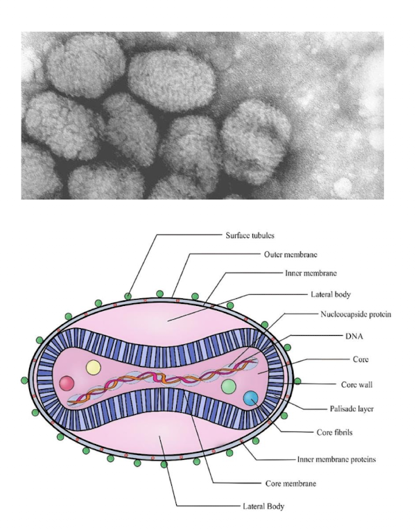
features of the poxviridae family of viruses
large dsDNA virus, roughly the same size as Chlamydia
“Brick-like” shape with inner membrane and outer envelope
no conventional icosahedral capsid
cytoplasmic replication → encodes its own machinery for genome transcription
what are the species of poxviridae viruses?
83 total
Variola virus (smallpox)
cowpox virus
Mpox virus
vaccinia virus
molluscum contagiosum virus
what are general clinical manifestations of the poxviridae family of viruses?
causes fluid-filled vesicles (pustules) to form on the skin
damage and craters formed called poxes
Variola virus (smallpox) transmission
exclusive to humans
no animal reservoir or vector
transmitted through:
respiratory droplets
direct contact with vesicles
indirect contact with contaminated items
no asymptomatic carriers
highly infectious
~30% mortality
clinical manifestations of variola virus (smallpox)
incubation period: 1-3 weeks
prodromal: 2-4 days
fever, headache, body aches
may be infectious at this stage
early rash: ~4 days
oral lesions - small red spots on tongue and mouth
change into sores, break open, virus spreads to all parts of the body
pustular rash and scabbing: ~5-7 days later
rash usually more concentrated on face and extremities
~6 days: pustules form crust and scab over
scabs fall off within 2-3 weeks
no longer contagious
may leave pox scars
how is variola virus (smallpox) diagnosed
mainly through signs and symptoms
immediately contact public health authorities before specimen collection or transport (it has been functionally eradicated)
Confirmation via NAAT (PCR)
collect specimens for each disease stage
Prodromal: nasopharyngeal swabs, whole blood
Early rash: tonsillar swab, lesion biopsy, whole blood
pustules: lesion fluid, whole blood
scabs: lesion scab, whole blood
treatment and prevention of smallpox (variola virus)
Vaccine
first vaccine developed by Edward Jenner in 1796
Milkmaids exposed to cowpox (Vaccinia virus) were immune to smallpox
Jenner inoculated 8 year old neighbor with fluid from cowpox sore from milkmaid
exposed neighbor to smallpox → immune to infection
1939: Vaccinia virus in smallpox vaccine was not cowpox, but likely horsepox
provide immunity against small pox
still used today
small pox has been declared eradicated since 1980
last US case 1949, last global case 1977
ACAM2000
Live vaccinia virus
Bifurcated needle with two prong
pustule should appear within 1 week
JYNNEOS
live-attenuated vaccinia virus
2 doses administered via subcutaneous injection
why did global smallpox eradication succeed?
favorable biology
humans were the only natural reservoir; no silent animal reservoir maintained variola transmission
recognizable and traceable
characteristic rash and transmission mainly after illness begins supported detection, isolation, and contact tracing
effective public health tools
potent, stable vaccinia vaccine; bifurcated-needle delivery, global surveillance and ring vaccination around cases
Mpox (Monkeypox) transmission
may spread to anyone through close, personal, skin-to-skin contact including direct contact with rash, especially sexual contact or through saliva or upper respiratory secretions
low risk from indirect contact through fomites
clinical presentation of Mpox
symptoms start within 3 week of exposure
after rash, rash develops within 1-4 days
Mpox risk and disease severity
less severe and lower mortality compared to smallpox
Molluscum contagiosum transmission
skin-to-skin contact and fomites
common in children and also occurs with sexual contact
Molluscum contagiosum clinical manifestations
firm, dome-shaped papules with central umbilication
benign, but may appear anywhere on body
may become itchy, sore, or swollen
typically resolves without scarring
diagnosis of molluscum contagiosum
usually clinical (signs/symptoms)
biopsy/histology can reveal intracytoplasmic Henderson-Patterson inclusion bodies in atypical cases
Parvovirus B19 (fifth disease) - virus features
non-enveloped, ssDNA virus
smallest DNA virus (22-26 nm)
typically associated with veterinary pathogens in dogs and cats
what are clinical manifestations of parvovirus B19 (fifth disease)
erythema infectiosum (fifth disease)
develops classic “slapped-cheek” facial rash
can be followed by a lacy body rash in children
adults:
arthralgia or arthritis can be more prominent than rash
transmission of parvovirus B19
respiratory droplets
blood and blood products
vertical transmission
pathogenesis of parvovirus B19
infects erythroid precursors
may temporarily suppress erythropoiesis
P Ag as RBC receptor for entry
Individuals with sickle cell disease or chronic hemolytic anemias are high risk → transient aplastic crisis (RBC cell production isn’t keeping up with need of the body)
severe fetal anemia
<5% of pregnant women
may lead to miscarriage or stillbirth (hydrops fetalis)
Erythema infectiosum (fifth disease)
most infections occur during childhood (80% adults seropositive by 65, 20% of cases asymptomatic)
incubation period: 14-18 days
prodromal phase includes fever, headache, runny nose
adults may have painful, swollen joints
classic “slapped cheek” rash appears
secondary rash may appear later on chest, rash, trunk, etc.
adults may only present with secondary rash
Parvovirus B19 - laboratory diagnosis
serology
B19-specific IgM can support recent infection
IgG usually indicates past exposure
interpret with symptom timing
Molecular testing
B19 DNA PCR (whole blood, amniotic fluid, tissue/biopsy) is especially useful in aplastic crisis, persistent infection, or immunocompromised patients
pregnancy
maternal testing and fetal evaluation depend on timing and clinical risk; coordinate with obstetric specialists
what are the non-herpes DNA viruses?
Adenovirus
HPV
Variola
Mpox
Molluscum
Parvovirus B19
Adenovirus - shedding/transmission and treatment/prevention
respiratory, ocular, or fecal shedding; prolonged after illness
usually supportive treatment; hand hygiene/ disinfection; type 4/7 vaccine only select military
HPV - shedding/transmission and treatment/prevention
transmission through skin/mucosal contact, often silent infection for months/years
treat lesions/dysplasia; vaccination and cervical screening
Variola - shedding/transmission and treatment/prevention
transmission after symptoms; infectious until scabs separate
eradicated; vaccination/public-health response if suspected
Mpox -shedding/transmission and treatment/prevention
close contact with lesions/material until all scabs shed and new skin forms
supportive treatment; vaccines for eligible groups, specialist for severe disease
Molluscum - shedding/transmission and treatment/prevention
transmission through skin contact, fomites, autoinoculation while lesions present
cover lesions, avoid scratching; often resolves without therapy
parvovirus B19 - shedding/transmission and treatment/prevention
supportive care; special management for aplastic crisis, pregnancy, or chronic anemia
where do most DNA viruses replicate?
in the nucleus
poxviruses are the exception
Herpesviruses establish _____; PCR positivity or seropositivity requires clinical interpretation
latency
PCR is central for identifying DNA viruses, but specimen and timing determine ____
usefulness
skin lesion, CSF, plasma, cervical specimen and neonatal urine answer different questions
prevention is ______-specific
virus
VZV, HPV and orthopox vaccines exist; HSV vaccination is not currently available
Molecular testing - clinical presentation: fresh oral/genital vesicles
what test/specimen should be used and what are key caveats
HSV NAAT from lesion
caveat: Type HSV-1 vs HSV-2