Dog fish- Shark muscles
 Call Kai
 Learn
 Practice Test
 Spaced Repetition
 Match
 Flashcards
 Knowt Play
1/39

There's no tags or description
Looks like no tags are added yet.
40 Terms
axial muscles
-trunk muscles
-epibranchial muscles
-hypobranchial muscles
-branchiomeric muscles
epibranchial muscles term
muscles located above (dorsal) the pharynx
hypobranchial muscles term
muscles located below (ventral) the pharynx
branchiomeric muscles term
muscles of the visceral arches
appendicular muscles
-pectoral fin abductors (located dorsally)
-pectoral fin adductors (located ventrally)
adduct
-to pull towards
-add to the midline

abduct
-to pull away
-to move away from the midline

trunk terms
-horizontal skeletogenous septum
-myomere
-myoseptum (singular)
-epaxial muscles
-hypaxial muscles
-linea alba

horizontal skeletogenous septum
membrane of connective tissue
which divides the dorsal and
ventral myomeres (epaxial and
hypaxial musculature)
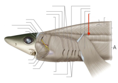
myomere
-bands of muscle along the sides
used for locomotion
-contained Z-shaped myomeres

myoseptum (singular)
-plural= myoseptae
-connective tissue sheets that
serve to separate the myomeres

epaxial muscles
located on the dorsal surface of the dogfish

hypaxial muscle
located on the ventral surface of the dogfish

linea alba
connective tissue that separates the
left and right hypaxial muscles mid-ventrally

origin
-fixed attachment
-attachment of a muscle that remains relatively fixed during muscular contraction
insertion
-moves with contraction
-The attachment of a muscle tendon to a moveable bone or the end opposite the origin
epibranchial muscles
-includes epaxials and hypaxials
-dorsal to the cucullaris are these
dorsal longitudinal bundles of muscle
-represent the anterior continuation
of the epaxial musculature of the head
-located anteriorly to the back of chondrocranium
-Action: elevate the cranium during prey capture
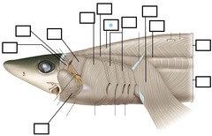
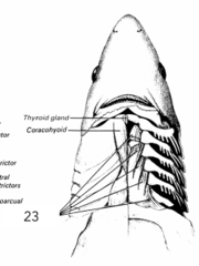
hypobranchial muscles
-hypaxial muscles located ventral to the gills
-coracomandibular
-corahyoid
-coracoarcuals
-corabranchials

coracomandibular
-the long, midventral, and nearly
cylindrical muscle exposed on
reflection of the intermandibularis
-Action: opens mouth; depresses
the lower jaw;
expand gill pouches

coracohyoid
-lies deep to the coracomandibular
-Action: opens the mouth; depresses
the lower jaw;
expand gill pouches

coracoarcuals
-nearly triangular, paired structure
-lie medially between the ventral
superficial branchial constrictors and
anterior to the hypaxial musculature
-Action: opens the mouth;
depresses the lower jaw;
expand gill pouches

coracobranchials
-a series of 5 muscles that fan out from
the coracoid bar, coracoarcuals,
and the walls of the pericardial cavity
to the ceratobranchial and basilbranchial cartilages
-Action: opens the mouth;
depresses the lower jaw;
expand gill pouches
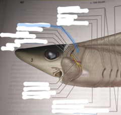
branchiomeric muscles
-preorbitalis
-levator palatoquadrati
-spiracularis
-levator hyomandibulae
-adductor mandibulae
-cucullaris
-superficial constrictors

superficial constrictor muscles
-intermandibularis
-interhyoideus
-dorsal hyoid constrictor
-ventral hyoid constrictor
-dorsal branchial constrictor
-ventral branchial constrictor

preorbitalis
-Action: closes the mouth
(adducts the lower jaw-
elevates the Meckel's cartilage);
protrude upper jaw

adductor mandibulae
-large muscle that is ventral to the
levator hyomandibulae muscle and
posterior to the angle of the mouth
-Action: closes the mouth;
(adducts the lower jaw-
elevates the Meckel's cartilage)
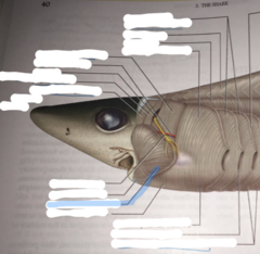
levator palatoquadrati
-lies anterior to the spiracle
-Action: elevates the upper jaw (palatoquadrate)

spiracularis
-on the anterior wall of the spiracular valve
-Action: elevates the upper jaw
(palatoquadrate); closes the spiracular valve

spiracle
used to take in water and ventilate the gills

levator hyomandibulae
-lies posterior to the spiracle
-Action: elevates hyomandibular cartilage
cucullaris
-triangle (isosceles triangle) that lies
dorsal to the constrictors
-Action: elevates the pectoral girdle

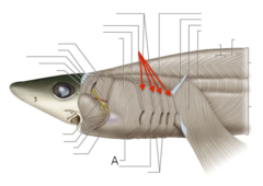
superficial constrictors
-make up a block of muscles that
surrounds the pharyngeal slits
-they are subdivided into dorsal and ventral portions
-separated by vertical connective tissue
partitions termed raphes, which
look like white lines on the muscle surface
-Action: compress the gill pouches

raphe
connective tissue that separates
the superficial constrictors

superficial constrictors list
-intermandibularis
-interhyoideus
-dorsal hyoid constrictor
-ventral hyoid constrictor
-dorsal branchial constrictor
-ventral branchial constrictor

intermandibularis
-lies in between the Meckel's cartilages
-Action: elevates the floor of the mouth,
reducing the volume of the oral cavity

interhyoideus
-a second, thin, and less extensive muscular sheet
that lies deep to the intermandibularis
-Action: elevates the floor of the mouth,
reducing the volume of the oral cavity

dorsal hyoid constrictor
-1st constrictors, lie in between the
adductor mandibulae and the first
pharyngeal slit
-raphes extending dorsally and
ventrally from the first slit separate
the hyoid constrictors from
the first of 4 superficial constrictors
-Action: compress gill pouches

ventral hyoid constrictors
-bottom part of large gill muscle
closest to the head
-1st constrictors, lie in between the
adductor mandibulae and the first
pharyngeal slit
-raphes extending dorsally and
ventrally from the first slit separate
the hyoid constrictors from the first
of 4 superficial constrictors
-Action: compress gill pouches

dorsal superficial branchial constrictor
-constrictors are similar in form and extend
between the raphes associated with each slit
-the branchial constrictors, which compress
the branchial region, extend medially
-superficially, they overlap one another
so that only their outermost portions are visible
-Action: compress the gill pouches
ventral superficial branchial constrictor
-constrictors are similar in form and
extend between the raphes associated
with each slit
-The branchial constrictors, which
compress the branchial region, extend
medially
-Superficially, they overlap one another
so that only their outermost portions are
visible
-Action: compress the gill pouches
