SPAUD 343: Final Exam
 Call Kai
 Learn
 Practice Test
 Spaced Repetition
 Match
 Flashcards
 Knowt Play
1/167

There's no tags or description
Looks like no tags are added yet.
168 Terms
peripheral auditory system
made up of outer, middle, and inner ear
central auditory system
system that’s made up of everything past the vestibulocochlear nerve (CNVIII)
outer ear
made up of pinna and external acoustic meatus
middle ear
made up of tympanic membrane, ossicles, eustachian tube, and tympanic cavity
inner ear
made up of cochlea, semicircular canals, and vestibulocochlear nerve
cochlea

cochlear duct
endolymph-filled membranous tube inside the bony cochlea

vestibule
contains utricle and saccule

semicircular canals

basilar membrane
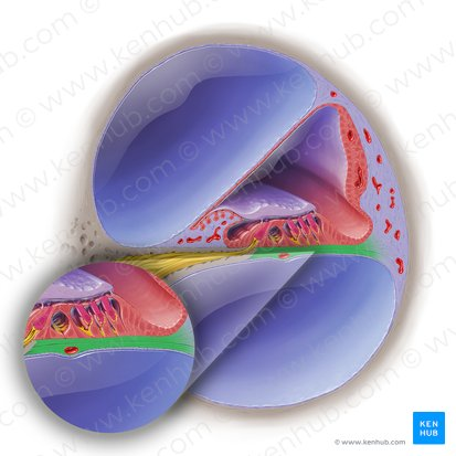
scala vestibuli
perilymph filled

scala tympani
perilymph filled
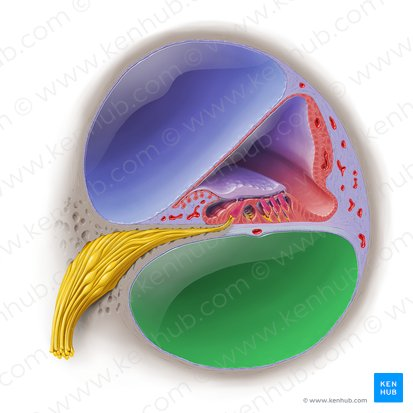
scala media
endolymph filled

tonotopic organization
sound organization on cochlea
low frequencies peak at apex, high frequencies peak at base

neural impulse
mechanical to electric (electrochemical) energy
like action potential; hair cells bend, tiplinks open, K comes in top, Ca come in battom
Ca causes neurotransmitter release
central auditory pathway
cochlear nerve
cochlear nuclei
superior olivary complex
lateral lemniscus
inferior colliculus
medial geniculate body
auditory cortex
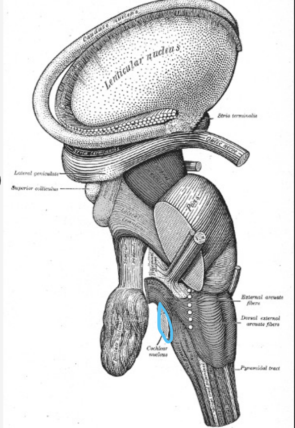
cochlear nuclei
part of central auditory pathway
in the brainstem, where pons and medulla meet
divided into dorsal and ventral cochlear nuclei
is also tonotopically arranged
goes to left and right superior olivary complex
superior olivary complex
part of central auditory pathway
is ipsilateral and contralateral → gets info from left & right ear → binaural hearing
helps with direction of sound
has medial and lateral divisions; medial helps with low-freq. hearing and lateral helps with high-freq. hearing

stapedius reflex
loud sounds cause this
stiffens stapedius muscle and keeps ME bones from moving too much and causing damage
lateral lemniscus
part of central auditory pathway
tract of 6 pathways (axons) → all auditory info
SOC projects 4 tracts thru this to the inferior colliculus
CNC projects 2 tracts directly to this
bilateral representation is stronger from the opposite ear (important bc helps if one side is damaged)
yellow on image

inferior colliculus
part of central auditory pathway
left and right divisions, connected
maintains tonotopic organization that originated in the cochlea
regulates the startle reflex

startle reflex
reaction to loud unexpected sound
regulated in inferior colliculus
type of reflex
hyperacusis
too easily startled by sounds
rare condition
not just being jumpy, almost painful, can come from aging
location of cochlear nucleus complex
in the brainstem where the pons and medulla meet
superior olivary complex
involved in sound localization and stapedius reflex
binaural hearing
getting hearing info from both ears
superior olivary complex
first spot responsible for binaural hearing
medial SOC
low frequency hearing
binaural hearing
lateral SOC
function is higher frequency hearing
functions of SOC
sound localization and stapedius reflex
six
how many tracts does the lateral lemniscus have?
4 and 2
in the lateral lemniscus, tracts are from the superior olivary comples and tracts are from the cochlear nucleus
opposite
in the lateral lemniscus, the bilateral representation is stronger in which ear?
inferior colliculus
final bilateral portion in the central auditory pathway
inferior colliculus
regulates the startle reflex
inferior colliculus
damage to this can result in hyperacusis
thalamus
where the medial geniculate body is located
medial geniculate body
acts as a relay station
relays auditory tracts to the auditory parts of the cerebral cortex, ipsilaterally
ventral division of MGB
function of this is basic sound info: frequency, intensity, binaural info
dorsal division of MGB
function of this is to focus attention: establish/maintain our attention to source of sound
auditory cortex
main central auditory pathway ends at the primary this
41 and 42
broadmann’s map location of primary auditory cortex
wernicke’s area
area important for attaching meaning to hear speech
arcuate fasciculus
connects broca’s to wernicke’s area
helps with syntax
broca’s area
responsible for understanding syntactic construction
sensorineural
90% of hearing loss is this kind
sibilants
those with sensorineural hearing loss have difficulty with this group of sounds
sensorineural hearing loss
damage to the vestibulocochlear nerve or inner ear (usually organ of corti’s hair cells) causes this kind of hearing loss
auditory processing disorder
difficulty processing and interpreting auditory language symbols
pure word deafness
another name for auditory agnosia
pure word deafness
also called auditory agnosia
you can speak, read, and write, but can’t understand language
brainstem
most of the structures of the vestibular system are located here
vestibular system
detects, codes, and process movement
helps us sit upright, walk, and perform other spatial environment functions
works with our proprioception and sense of vision
peripheral vestibular system
components of this system are the semicircular canals and vestibule
central vestibular system
components of this system are CN VIII and brain structures
semicircular canals
three fluid-filled canals that correspond to our 3D world
anterior/superior canal
controls coronal plane (tilt head R/L)
posterior canal
controls sagittal plane (nod head up and down)
horizontal/lateral canal
controls transverse plane (shake head no)
ampulla
big bumps on the semicircular canal

crista ampullaris
sensory organ of rotation (semicircular canals)
cupula
hair cell membrane (gelatinous, mass-filled, embedded with stereocilia) of semicircular canals
macula
sensory organ of vestibule
otolithic membrane
hair cell membrane of vestibule
macula
helps decipher head position (no movement needed)
hair cells of crista ampullaris and macula
where the vestibular branch of the vestibulocochlear nerve connects
vestibulocochlear reflex
automatic eye movements that stabilize images in the retina during head and body motion
reticular formation
connection with the vestibular nuclear complex involved in motion sickness and vomiting
labrynthitis
infection of the inner ear that affects the vestibular nerve, leads to vertigo, nausea, and vomiting
vestibular (acoustic) schwannoma
tumor on the vestibular branch of CN VIII
slow growing
conceptual level
first step of sensory motor speech
conceptual level
level of coming up with what you want to say (cognition)
linguistic and motor planning
two parts of linguistic planning level
conceptual level areas
prefrontal cortex, limbic system (whole cerebral cortex) are involved in this
linguistic planning
concerned with language content, form, and use
semantics and meaning
function and area of the “content” of language (broca’s and wernicke’s area)
pragmatics
function and area of “use” of language (right hemisphere)
syntax and morphology
function and area of “form” of language (broca’s area)
motor planning
plans and arrangements of phonemes
motor planning/sequencing
overall spatial and temporal (timing) goals that include knowing the phonemes and having a rough target
premotor cortex and supplementary motor area
areas involved in the linguistic and motor planning level
linguistic and motor planning and motor programming
two levels involved in sequencing
apraxia and dysarthria
problems with motor programming could be these
planning and programming
problems with are more conceptual, problems with are more about coding to muscles
motor programming level
deals with transitions between phonemes
motor programming level
deals with precise movements of articulators with their targets, timing, and force
cerebellum, basal ganglia, and supplementary motor area
motor programming uses information from these
apraxia
disorder of motor planning and programming
apraxia
characterized by groping for articulatory placement, no muscle weakness, sequencing deficits, distorted substitutions, errors in articulatory placement, and inconsistency in errors
apraxia
difficulty coordinating movements, difficulty with prosody (intonation, stress), speech planning, rhythm, consonant clusters
aphasia
difficulty coming up with words, recognizing words, reading/writing, speaking/listening
apraxia
damage to supplementary area, insular cortex, broca’s area, basal ganglia result in this
perisylvian cortex
brain area around sylvian fissure
autism and epilepsy
childhood apraxia of speech sometimes occur with these
childhood apraxia of speech
distortions, substitutions, phoneme sequencing problems
area of conceptual level
whole cerebral cortex
prefrontal cortex
limbic system
damage to conceptual level
cognitive thinking problems (dementia)
area of linguistic planning
broca’s area
wernicke’s area
right hemisphere
damage of linguistic planning
aphasia
area of motor planning
premotor cortex, supplementary motor cortex