Anatomy II - Eye and Orbit
 Call Kai
 Learn
 Practice Test
 Spaced Repetition
 Match
 Flashcards
 Knowt Play
1/90

There's no tags or description
Looks like no tags are added yet.
91 Terms
lateral canthus
outer corner of eye

opthlamic artery
blood supply to orbit
supra-orbital artery
opthlamic artery gives rise to
superior orbital foramen
supraorbital artery passes out of orbit via
lateral rectus and superior oblique
what muscles arent affected in oculomotor palsy
medial canthus
inner corner of eye

Palpebra inferioris
lower eyelid

Palpebra superioris
upper eyelid
lacrimal caruncle
sebaceous glands

sclera

limbus

cornea

pupil

iris

cornea
the iris is deep to
pupil
opening in iris
cornea
pupil is deep to
axons of retinal ganglion cells
optic nerve carries
Fovea
area of greatest visual acuity and center of visual field
retina

optic disc
Region at the back of the eye where the optic nerve meets the retina. It is the blind spot of the eye because it contains only nerve fibers, no rods or cones, and is thus insensitive to light.
aqueous humor
located anterior to lens, on either side of iris
vitreous humor
located posterior to lens
palpebral part of orbicularis oculi
muscle that mediates blinking
muscle of facial expression
facial nerve (CN VII)
motor innervation of palpebral part of orbicularis oculi
sensory
is the cornea sensory or motor
Ophthalmic Division of the Trigeminal Nerve (CN V1)
sensory innervation of cornea
Corneal Reflex
Touching the Cornea at the Limbus with a benign stimulus such as a wisp of cotton elicits a blink response
CN V1 and VII
corneal/blink reflex tests which nerves as a unit
frontal bone
roof of orbit
zygomatic bone
lateral wall of orbit
maxilla and sphenoid bone
floor of orbit
ethmoid bone
medial wall of orbit
floor and medial wall
where is the orbit bone the thinnest
Orbital Fracture
Following blunt trauma to the orbital margin, the concussion may cause either of these bones to fracture, while the orbital margin may remain intact.
Causes bleeding into orbit
diplopia
double vision
superior oribital fissure
separates the greater and lesser wings of sphenoid bone
inferior orbital fissure
CN V2 arises out of the
subarchnoid space
what surrounds CN II
elevated intracranial pressure
optic disc margins blurred suggest
Papilloedema
swelling of the optic disc due to increased intracranial pressure
frontal nerve
CN V1
supraorbital nerve is a branch of __________ which is a branch of ______
oculomotor nerve (CN III)
levator palpebra superioris muscle is supplied by
levator palpebrae superioris

levator palpebrae superioris
superior tarsus muscle
eyelid evelators
sympathetic
the superior tarsus muscle is under ________ control
tarsal glands
cilary glands
secrete oil to prevent lacrimal fluid from overflowing eyelids
chalazion
obstructed tarsal glands resulting in inflammatory nodule facing inwards towards eye
hordeolum
obstructed Ciliary Gland resulting in inflammatory nodule facing outward
Palpebral part of orbicularis oculi
eyelid closer
eyeball primary position

adduction and abduction

elevation

depression

extorsion and intorsion

elevation and adduction
actions of superior rectus muscle
oculomotor nerve (CN III)
superior, inferior, medial rectus muscle innervation
depression and adduction
action of inferior rectus muscle
abduction
lateral rectus muscle action
abducens nerve (CN VI)
lateral rectus muscle innervation
adduction
action of medial rectus muscle
depression, abduction, intorsion
superior oblique muscle action
trochlear nerve (CN IV)
superior oblique muscle innervation
Oculomotor nerve (CN III)
inferior oblique muscle innervation
elevation, abduction, extorsion
action of inferior oblique muscle
1. abduct
2. elevate while abducted
3. depress while abducted
4. adduct
5. elevate while adducted
6. depress while adducted
clinical testing of extra-ocular muscles
lateral rectus and abducens nerve (CN VI)
Clinical testing of extra-ocular muscles abducting tests
test superior rectus and oculomotor nerve (CN III)
Clinical testing of extra-ocular muscles elevate while abducted tests what nerve
inferior rectus and oculomotor nerve (CN III)
Clinical testing of extra-ocular muscles while depressed and abduct tests
medial rectus and oculomotor nerve (CN III)
Clinical testing of extra-ocular muscles while adducting test
inferior oblique and oculomotor nerve (CN III)
Clinical testing of extra-ocular muscles while elevating and adducting tests
superior oblique and CN IV
Clinical testing of extra-ocular muscles depress while adducted tests
internal carotid artery
the ophthalmic artery is a branch of the
short ciliary nerves
Carry post-ganglionic parasympathic axons
ciliary ganglion
contain cell bodies of postganglionic parasympathetic neurons
oculomotor nerve (CN III)
innervation of preganglionic parasympathetic axons
ciliary
contraction removes tension on lens and causes it to thicken for near vision
Sphincter Pupillae
contraction causes pupil to constrict, reducing light entering the eye
far vision
ciliary muscle relaxed
suspensory ligaments tensed
near vision
ciliary muscles contracted
suspensory ligament relaxed
long ciliary nerve
nasociliary nerve
carry postganglion sympathetic axons
CN V1
nasociliary nerve is a branch of
Dilator pupillae
sympathetic target of eye
causes pupil to dilate, increasing light entering to eye
ciliary muscles
sphincter pupillae muscle
Parasympathetic Targets
horner's syndrome
loss of sympathetic supply
horner's syndrome
Pupillary constriction (Sphincter Pupillae, supplied by parasympathetic division, is unopposed)
Mild Ptosis of the Upper Eyelid (denervation of Superior Tarsus Muscle)
internal carotid artery
cranial nerves III, IV, V1, V2, and VI
cavernous sinus contains
danger zone
area bordered by upper lip, lateral aspect of nose, and medial corner of eye superior to supraorbital ridge
oculomotor nerve palsy
1.Eye oriented down and out (All muscles weakened except Lateral Rectus and Superior Oblique Muscles, which are abductors and, in the case of the Superior Oblique, a depressor)
2.Pupil dilated (Dilator Pupillae, supplied by the sympathetic division, is unopposed)
3.Ptosis (weakness of Levator Palpebrae Superioris Muscle)

abducens nerve palsy
lateral rectus muscle weakened and eye goes inward

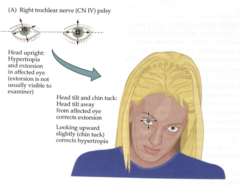
trochlear nerve palsy
superior oblique muscle is weakened
1. eye directed upward with intorsion deficit
will often tilt his/her chin down to correct