Pathophysiology of GI tract: Diseases of the GI tract, liver and exocrine pancreas
 Call Kai
 Learn
 Practice Test
 Spaced Repetition
 Match
 Flashcards
 Knowt Play
1/52

There's no tags or description
Looks like no tags are added yet.
53 Terms
Pathophysiology of the gastrointestinal
• The breakdown of normal motor, secretory, barrier, or immune functions across its segments, leading to distinct clinical disorders.
Cells of the Stomach
G Cells, Gastrin:
• Initiated by food entering the stomach
• Stimulate parietal cells and chief cells
Parietal Cells, Intrinsic Factor:
• Needed for the absorption of vitamin B12
Parietal Cells, HCl:
• Activate pepsinogen to create an optimal pH of 2 and denature proteins
Chief Cells, Pepsinogen:
• Not activated until stomach acid has a pH of 6
Secretion in the Stomach

Neural and Hormonal Control of the GI Tract
Neural Control of the GI Tract
Parasympathetic Nervous System:
Vagus Nerve
Increased gastric motility
Increased gastric acid secretions
Sympathetic Nervous System:
Inhibits gastrointestinal activity
Hormonal Control of the GI Tract
•Gastrin
Increases gastric motility
Promotes stomach emptying
• Secretin
Decreases gastric acid secretions
• Cholecystokinin
Inhibits gastric emptying
Diseases of Mouth
Mouth is the entry point of various pathogens and
microorganisms.
• Thrush (Oral Candidiasis) - Acute fungal infection, caused by the yeast Candida albicans, which occurs when the commensal microbe grows in white patches on the tongue and oral mucosa.
• Caused by – Candida albicans (Yeast)
• Symptoms – Itching and rash at the infected site.
Thrush (Oral Candidiasis)
• Risks - In adults it causes opportunistic infection mainly in those whose immunity is lowered by, e.g., steroids, antibiotics or cytotoxic drugs.
In children it occurs most commonly in bottle-fed babies.
• Diagnosis – Physical examination of the lesions in mouth;
• Blood test – Candida specific antibody test is used to detected candidiasis – IgM, IgA & IgG.
• Treatment – Antifungals (Clotrimazole, Fluconazole, etc.), Antiseptics, etc.
Gingivitis
• This is inflammation of the gums.
• May be Acute or Chronic.
• It occurs in response to accumulation of bacterial plaque around the teeth.
• It gradually destroys the tissues supporting the teeth.
Symptoms:
Swollen gums
bleeding gums
Risks:
Poor oral hygiene
smoking or chewing tobacco
older age, etc.
Dental Caries
• cause destruction of the calcified tissues of the teeth.
• untreated - microorganisms may invade the pulp, causing inflammation and infection.
• Bacteria - produce acids that demineralize the enamel.
• Dextran, a sticky polysaccharide produced from sucrose, causes the bacteria to stick to the teeth.
• Masses of bacterial cells, dextran, and other debris adhering to teeth constitute dental plaque.
• Filling or root canal treatment helps to treat the decay of pulp caused due to the infection.

Diseases of Pharynx
Tonsilitis
• Inflammation of palatine tonsils, palatine arches the walls of the pharynx.
it is caused by Viruses and Streptococcus pyogenes.
There may be production of abscess at the site of infection.
The infection, if untreated may spread to the neck.
Repeated infection may lead to chronic inflammation, fibrosis and permanent enlargement.
Surgical removal of the tonsils (Tonsillectomy) can help to reduce the pain and inflammation.
NSAIDs, analgesics and antibiotics can be used

Diphtheria
• Causative agent - Corynebacterium diphtheriae
• This is a bacterial infection of the pharynx – extends to the nasopharynx and trachea, caused by a bacteria in which thick fibrous membrane forms over the area and may obstruct the airway.
• The microbe produces powerful exotoxins that may severely damage cardiac and skeletal muscle, the liver, kidneys and adrenal glands.
• It may be treated by using antitoxin to diphtheria toxin.
• Vaccination with diphtheria toxoid is the best way to prevent its occurrence.
Diseases of Salivary Glands
Mumps
• Caused by - Mumps virus
• acute inflammatory condition of the salivary glands, especially the parotids.
• virus spread by inhalation of infected droplets.
• Viruses multiply elsewhere in the body before spreading to the salivary glands.
Complications affect:
the brain, causing meningitis or meningo-encephalitis
the testes, causing orchitis (testicular inflammation)
Pancreatitis
heart problems,
miscarriage
Risks:
Unvaccinated individuals > high risk
children between 2-12 years
Symptoms:
Pain while swallowing
Fever
Loss of appetite
Tumor of Salivary Glands
Salivary adenoma
• Benign tumor of the parotid gland > most common
• A second tumor may develop in the same gland several years - malignant type.
Carcinoma
• Malignant tumor > affect parotid glands.
• Some forms have a tendency to infiltrate nerves in the surrounding tissues, causing severe pain.
Symptoms:
Swelling near the jaw or in neck or mouth
difficulty swallowing
numbness in part of the face
Causes:
Genetic mutations (predominantly)
smoking & tobacco chewing
Diseases of Esophagus
Gastro-esophageal reflux disease (GERD)
• Reflux of gastric juice and acids from the stomach into the esophagus > due to a fault in the lower esophageal sphincter to close and prevent the gastric contents from entering into the esophagus.
• Results in inflammation of the lower esophagus.
• Reflux persists > fibrous tissues form at the site of inflammation.

GERD - Reflux of gastric contents is associated with
• increase in the intra-abdominal pressure, e.g. in pregnancy, constipation and obesity.
• low levels of secretion gastrin > reduced sphincter action at the lower end of the esophagus.
• the presence of hiatus hernia.
Symptoms:
heartburn
nausea
regurgitation
discomfort in upper abdomen
Barrett’s esophagus
• consequence of Reflux Esophagitis.
• stratified squamous epithelium of lower
esophagus is replaced by columnar epithelium
(columnar metaplasia).
• not treated on time > progress to Carcinoma of
Esophagus then Adeno-carcinoma.
Symptoms:
Difficulty in swallowing food
Chest pain
heartburn
regurgitation
Risk:
Family history
Gender (Men are more likely to develop it)
Smoking
GERD
Obesity
Achalasia
• Neuromuscular dysfunction due to which the cardiac sphincter fails to relax during swallowing > progressive dysphagia and dilatation of the esophagus.
• It occurs when the intra-mural neurons in the wall of the esophagus are damaged.
Symptoms:
heartburn
regurgitation of food
belching
chest pain
vomiting
Causes:
Loss of nerve cells from esophagus
Risk:
Allgrove syndrome
Herpes infection
Measles
Autoimmune diseases
Carcinoma of Oesophagus
• Unknown (idiopathic)
Contributory factors:
Diet and personal habits:
Heavy smoking
Alcohol consumption
Esophageal disorders:
Esophagitis (especially Barrett’s esophagus in adenocarcinoma)
Achalasia
Hiatus hernia
Diverticula
Other factors:
Family history: tylosis (keratosis palmariset plantaris)
Genetic factors (coeliac disease, epidermolysis bullosa, tylosis)
HPV infection
Diseases of Stomach
Congenital and Acquired Condition
Pyloric Stenosis
• narrowing of the pyloric opening (male children) as a congenital defect (infantile pyloric stenosis).
• rarely observed in adults > due to acquired inflammatory fibrosis or by invasion of tumors
• In both the types, there is hypertrophy as well as hyperplasia of the circular layer of muscularis in the pyloric sphincter accompanied by mild degree of fibrosis.
Symptoms:
Vomiting after feeding/eating
Stomach contractions
Dehydration
Changes in bowel movement
Risks:
Gender (More seen in males)
Premature birth
Family history
Smoking during pregnancy
Complications:
Jaundice (rare)
Stomach irritation
Inflammatory Conditions
Gastritis:
• the lining of the stomach becomes inflamed
cause: upper abdominal discomfort like indigestion or dyspepsia.
• may progress to peptic ulcer or gastric cancer.
Types:
Acute Gastritis - a transient acute inflammatory involvement of the stomach, mainly mucosa, and is usually a response to irritant drugs (NSAIDs like Aspirin, etc.), highly spiced food, excessive alcohol consumption, malnutrition, smoking, bacterial infections (Helicobacter pylori, diphtheria, salmonellosis, pneumonia, staphylococcal food poisoning), emotional factors (like shock, anger), trauma & surgery.
Chronic Gastritis - a milder but longer-lasting condition. associated with Helicobacter pylori but is sometimes due to autoimmune disease or chemical injury.
Autoimmune gastritis - presence of circulating antibodies and is sometimes associated with other autoimmune diseases such as Hashimoto’s thyroiditis and Addison’s disease. May lead pernicious anemia and increased risk of stomach cancer.
Peptic Ulcer Disease (PUD)
• degeneration and necrosis of gastrointestinal mucosa exposed to acid-peptic secretions.
• They are open sores that develop on the inner lining of the stomach and duodenum.
• occur in any part of the GIT which are exposed to HCl and Pepsin, but majority occur in Stomach and Duodenum.
• Acute Peptic Ulcer > result of severe stress (physiological or psychological). Multiple and small mucosal erosions.
• Chronic Peptic Ulcers > Helicobacter pylori infection is found associated in 90% of duodenal and 70% of stomach ulcer cases.
Symptoms:
Burning stomach pain
Bloating
Intolerance to fatty food
Heartburn
Risks:
Smoking
Alcohol consumption
Consuming spicy food
Stress
Complications:
Hemorrhage
Perforation
Anemia
Gastric outflow obstruction
Gastric cancer

Diseases of Small and Large Intestine
• Congenital Abnormality

Meckel’s Diverticulum
• an outpouching containing all the layers of the intestinal wall in their normal orientation. common in males.
• Causes – occurs as a result of incomplete absorption of the vitelline duct by the fetus during the 7th week of pregnancy.
• Symptoms – GI bleeding, Abdominal pain, Obstruction of the bowels, diverticulitis (swelling of the intestinal wall).
• Other congenital conditions:
Intestinal atresia (congenital absence of intestinal lumen)
Intestinal stenosis (congenital narrowing of the intestinal lumen)
Intestinal malrotation (failure of normal rotation of midgut)

Hernia
• Protrusion of an organ or part of an organ through a weak point or aperture in the surrounding structures.
• External hernia - protrusion of the bowel through a defect or weakness in the peritoneum.
• Internal hernia - herniation that does not present on the external surface
formation of Hernia:
Local weakness
Increased intra- abdominal pressure
• Strangulated hernia - blood flow in the hernial sac is obstructed; results in infarction or gangrene of the affected loop of intestine.

Sites of Hernias
Inguinal hernia – Inguinal canal; occurs more commonly in males.
Femoral hernia - femoral canal through which the femoral artery passes.
Umbilical hernia – umbilicus, where the umbilical blood vessels from the placenta enter the fetus before birth.
Incisional hernia - repeated stretching of fibrous (scar) tissue formed after previous abdominal surgery.
Hiatus hernia - protrusion of a part of the fundus of the stomach through the esophageal opening in the diaphragm.
Causes:
Congenital
Aging
Damage from injury
Pregnancy
Previous surgery
Obesity
Symptoms:
Bulge or Lump in the affected area (which disappears on lying down)
Pain
Heartburn
Difficulty in swallowing
Chest pain (in case of Hiatus hernia)
Risks:
Family history
Pregnancy
Smoking
Premature birth
Cystic fibrosis
Inflammatory Bowel Disease (IBD)
• disorders that involve chronic inflammation of the digestive tract.
2 conditions:
• A. Crohn’s disease
• B. Ulcerative colitis
A. Crohn’s disease
• inflammation of the lining of the digestive tract, often involving deeper layers of the digestive tract.
• There’s chronic patchy inflammation with oedema – causing partial obstruction of the lumen – skip lesions.
Complications:
Fistula formation
Malabsorption,
Development of malignancy
Bowel obstruction
Ulcerative colitis
• Involves inflammation and ulcers along the lining of the colon and rectum.
• Begins in the rectum, and extends upwards into the sigmoid colon, descending colon, transverse colon, and sometimes may involve the entire colon.
Complications:
Fulminant colitis (Toxic megacolon)
Perforated colon
Perianal fistula
Colon cancer
Severe dehydration
Symptoms:
Diarrhea
Blood in stools
Reduced appetite
Weight loss
Risks:
Smoking
Familial history
Excessive use of NSAIDs
Causes:
Mostly Idiopathic; may be due to immune system
malfunction or hereditary
Enteric Fever (Typhoid & Paratyphoid)
• Causative agents – Salmonella typhi (Typhoid) and S. para-typhi A or B (Paratyphoid).
• Mode of spread - Through typhoid carriers, Fecal-oral transmission route.
• Symptoms – High fever, Stomach pain, Constipation or Diarrhea, Rashes.
• Risks – Close contact with an infected person, Drinking contaminated water, Travelling to affected areas.
• Complications- Myocarditis, Endocarditis, Pneumonia, Pancreatitis.
• Prevention – Washing hands properly, Avoid drinking untreated water, Avoid raw fruits and vegetables, Consumption of hot food, Getting vaccinated.
Dysentery
• condition in which diarrhea is accompanied with abdominal cramps and passage of mucus in the stools.
Types:
Bacillary dysentery:
• Causative agent - Shigella species
• Vector - common housefly
• Superficial transverse ulcerations of mucosa of the bowel wall occur in the region of lymphoid follicles. The mucosa overlying the lymphoid follicles become necrosed and the surrounding mucosa shows congestion and oedema.
• Complications - haemorrhage, perforation, stenosis.
Amoebic dysentery:
• Causative agent - Entamoeba histolytica
• Vector - common housefly, rodents, cockroaches
• The disease primarily affects the large intestine. The parasites produce the characteristic flask-shaped ulcers.
• Complications - amoebic liver abscess, amoebic hepatitis, perforation, haemorrhage.
• Symptoms – Loose water stools, Blood in stools, Fever, Mucus in stools, Bloating, etc.
• Risks – Contaminated food, Poor hand washing, Swimming in contaminated water - lakes

Appendicitis
• Acute inflammation of the appendix
• common in people between 10-30yrs.
• Causes – Blockage in the lining of appendix, Obstructive (Faecolith, Calculi, Tumor, etc.), High protein intake diet, Bacterial infections.
• Symptoms – Sudden pain in the right side of the lower abdomen, Nausea and Vomiting, Loss of appetite, Constipation or Diarrhoea, Bloating, Flatulence.
• Complications – Ruptured appendix, Appendix abscess
The Liver
Functions
• Storage of nutrients
• Maintains blood glucose
• Produces bile, plasma proteins, blood clotting factors, cholesterol, and lipoproteins
• Takes part in metabolic processes
Storage Processes
• Glycogenesis - Glucose is converted to glycogen
• When glycogen supply is low - Gluconeogenesis (Proteins and fats are converted to glycogen)
• Glycogenolysis (Glycogen is converted to glucose) Maintain blood glucose level
Diseases of Liver
• Jaundice - yellow pigmentation of the skin or sclerae of the eye by bilirubin > a result of elevated levels of bilirubin in the blood (hyperbilirubinemia) (serum bilirubin exceeds 2 mg/dl).
Types:
• 1. Pre-hepatic jaundice - result of increased hemolysis of red blood cells that results in production of excess bilirubin > excess bilirubin is unconjugated (unconjugated bilirubin is fat soluble) it cannot be excreted in the urine, therefore remains normal in colour.
• Neonatal haemolytic jaundice - high rate of hemolysis coupled with a shortage of conjugating enzymes in the hepatocytes of the still immature liver in the neonates.
Jaundice (cont.)
• Intra-hepatic jaundice - result of damage to the liver itself by Viral hepatitis, Toxic substances (drugs, Amoebiasis, Cirrhosis, etc.)
• Excess bilirubin accumulates in the liver.
• Because it is mainly in the conjugated form, it is water-soluble and excreted in the urine making urine dark in colour.
• Post-hepatic jaundice - obstruction to the flow of bile in the biliary tract due to Gallstones in the common bile duct, Tumor of the head of the pancreas, Fibrosis of the bile ducts, following cholangitis or injury by the passage of gallstones.
• In this situation excess bilirubin is also conjugated and is therefore excreted in the urine.
• Effects of raised serum bilirubin: Pruritus (itching), Pale feces due to absence of stercobilin, Dark urine due to the presence of increased amounts of bilirubin
Gilbert’s Syndrome
• It is a familial, genetically-determined, autosomal disease of the liver.
• characterized by mild, benign, unconjugated hyperbilirubinemia which is not due to hemolysis.
• due to reduced conjugation of bilirubin or impaired hepatic uptake of bilirubin.
• Symptoms – Occasional yellowing tinge of the skin and whites of the eyes due to slightly elevated levels of bilirubin as seen in Jaundice
• Risks – Gender (Males are at a high risk), Both the parents carry the abnormal gene for the disorder.
• Complications – Low levels of the enzyme (UDP-glucuronosyl transferase) causing Gilbert’s syndrome may increase the side effects of other drugs (like Irinotecan, etc.) that are metabolized by the same enzyme.
Hepatitis– inflammation of the liver
Mild
• Impaired hepatocyte function
Severe
• Impaired hepatocyte function
• Necrosis and obstruction of blood and bile flow
Cause
• Idiopathic – fatty liver
• Infection – viral or non-viral
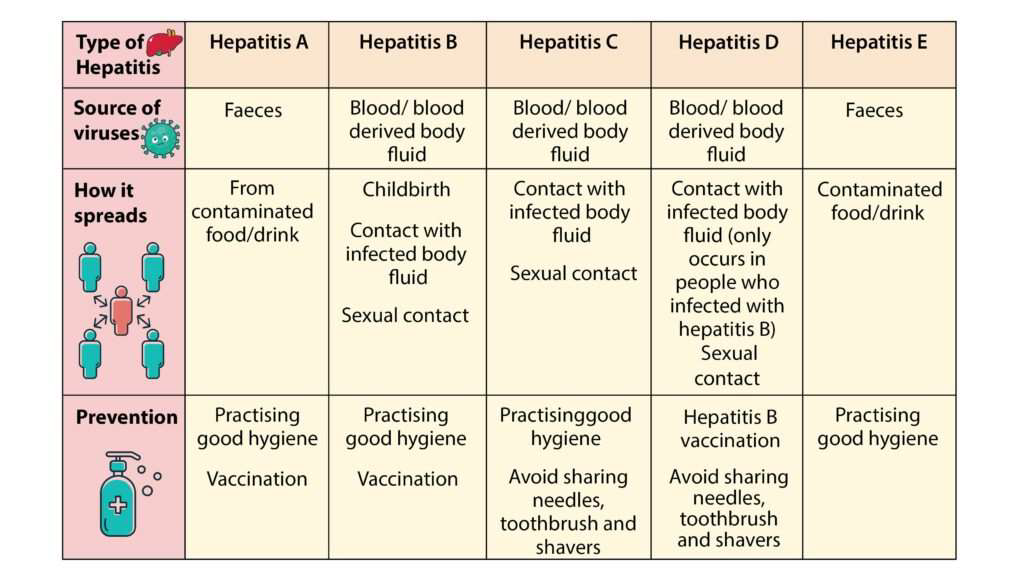
Viral Hepatitis
• infection and inflammation of the liver by hepatotropic
viruses.
• 5 main varieties of these viruses causing distinct types of viral hepatitis:
Hepatitis A virus (HAV)
Hepatitis B virus (HBV)
Hepatitis C virus (HCV)
Hepatitis delta virus (HDV)
Hepatitis E virus (HEV)
• Only HBV is a DNA virus, all other human hepatitis viruses are RNA viruses.
Hepatitis A
• affects mainly children, causes a mild illness, often asymptomatic - Infection is spread by the fecal– oral route, e.g. via contaminated hands, food, water and fomites.
• Usually a benign, self-limiting disease and has an incubation period of 15-45 days.
• Mostly affect 5-14 years; adults are often infected by spread from children.
• Symptoms – Vomiting, Low-grade fever, Clay-coloured bowel movement, Dark urine, Yellowing of eyes, Intense itching.
• Risks – HIV positive, Clotting factor disorders, Physical contact with infected person.
• Complications – Loss of liver function, etc.
• Prevention – Hepatitis A Vaccine.
Hepatitis B (serum hepatitis)
• transmitted parenterally, e.g in recipients of blood and blood products, patients treated by renal dialysis and hospital workers exposed to blood.
• The virus has a longer incubation period - 30-180 days.
• The infection causes more severe form of illness e.g acute hepatitis B, chronic hepatitis, progression to cirrhosis.
• It leads to the development of hepatocellular carcinoma.
• Symptoms – Abdominal pain, Dark urine, Fever, Loss of appetite, Weakness, Jaundice.
• Risks – Unprotected sex, Sharing of needles, Infant born to an infected mother.
• Complications –Cirrhosis, Liver cancer, Liver failure.
• Prevention – Hepatitis B vaccine.
Hepatitis C
• Acquired by blood transfusions, blood products, hemodialysis, parenteral drug abuse and accidental cuts and needle-pricks in health workers & unprotected sex, especially with the presence of blood.
• Acute HCV hepatitis is milder than HBV hepatitis, but HCV has a higher rate of progression to chronic hepatitis than HBV.
• asymptomatic infection, diagnosed later when cirrhosis or chronic liver failure becomes evident.
• Considered more important cause of chronic liver disease worldwide than HBV.
• Symptoms – Dark urine, Fluid buildup in abdomen, Swelling of legs, Bruising and bleeding easily, Jaundice, Poor appetite.
• Risks – HIV, Piercing or tattoo, Infant born to an infected mother.
• Complications – Cirrhosis, Liver cancer, Liver failure.
• Prevention – practice safe sex
Hepatitis D
• A defective virus for which HBV is the helper; hepatitis D develops when there is concomitant hepatitis B infection.
• Co-infection – HDV infection and hepatitis B occur simultaneously.
• Superinfection – HDV infects a chronic HBsAg carrier.
• Symptoms – Abdominal pain, Loss of appetite, Vomiting, Dark urine, Fatigue.
• Risks – Unprotected sex, Infant born to an infected mother, HVB infected individuals.
• Complications – Cirrhosis, Liver cancer, Liver diseases.
• Prevention – Get vaccinated, Practice safe sex.
Diseases of Liver Viral Hepatitis

Diseases of Liver Viral Hepatitis
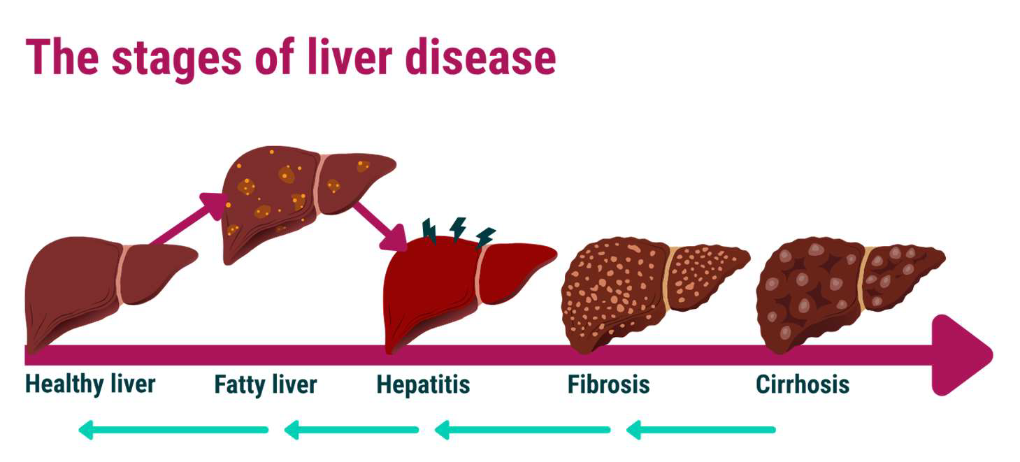
Cirrhosis
• Progressive destruction of the liver
Stage 1
• Fatty liver. Asymptomatic and reversible.
Stage 2
• Alcoholic hepatitis & Irreversible.
Stage 3
• End stage cirrhosis.
• Liver failure occurs when 80- 90% of the liver is destroyed.
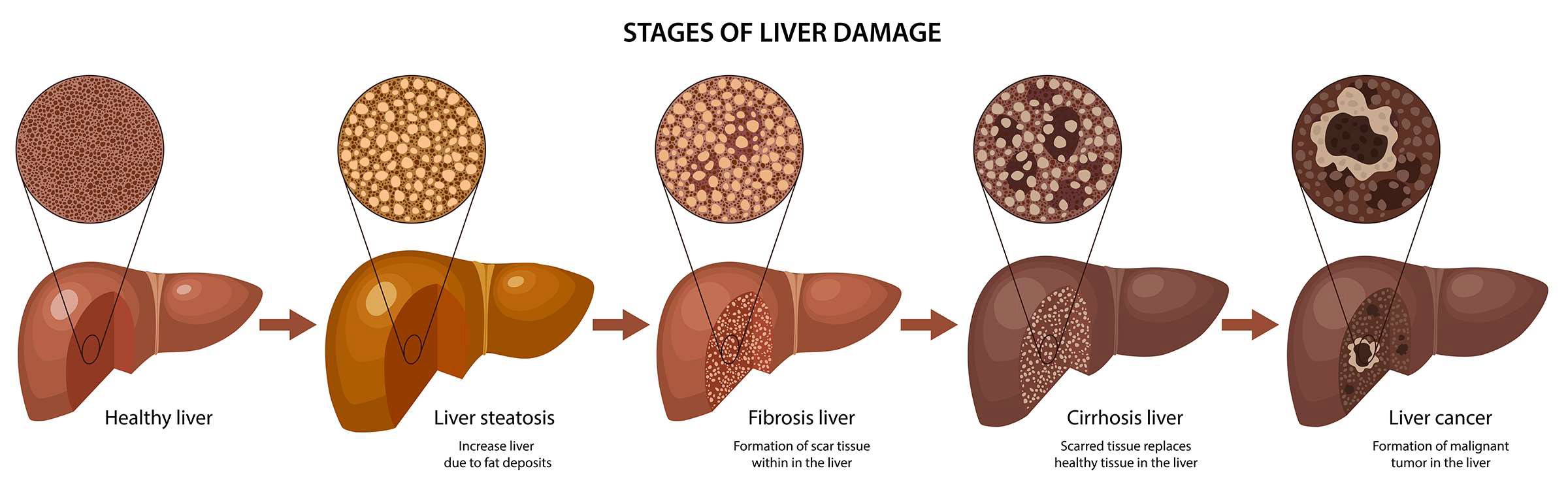
Liver Cancer
• Initial signs are mild
• Diagnosis occurs with advanced stages
• Hepatocellular Carcinoma
• Most common primary tumor of the liver
• Metastatic Liver Cancer
• Arises from areas served by the hepatic vein
Diseases of the Biliary Tract
• Gallstones (Cholelithiasis)
• Consist of deposits of the constituents of bile, most commonly cholesterol.
• Many small stones or one or more large stones may form but they do not necessarily produce symptoms.
• Causes – Too much cholesterol in bile, Too much bilirubin in bile, obstruction of bile duct.
• Symptoms – Sudden and rapidly intensifying pain in upper right portion of the abdomen, Pain in right shoulder, Back pain between shoulder blades.
• Risks – Gender (Females are more prone), Age above 40, Low fiber diet, Familial history, Diabetes, Liver disease.
• Complications – Cholecystitis - blockage of common bile duct, blockage of pancreatic duct, gallbladder cancer.
• Prevention – Don’t skip meals, Consume high fiber food, Drink enough water
Pancreas
• Exocrine Organ: secretes digestive enzymes and electrolytes
• Endocrine Organ: secretes insulin and glucagon
Important Molecules -
Trypsin, chymotrypsin, carboxypeptidase (Break proteins)
Ribonuclease (Break nucleic acids)
Pancreatic Amylase (Break starch)
Lipase (Break lipids)
Diseases of Pancreas
• Pancreatitis - Inflammation of the pancreas.
• Proteolytic enzymes produced by the pancreas are secreted in inactive forms, which are not activate until they reach the intestine – this protects the pancreas from digestion by its own enzymes.
• If these precursor enzymes are activated while still in the pancreas, pancreatitis results.
Acute pancreatitis
• sudden onset and lasts for a few days.
• severity depends on the amount of pancreatic tissue involved.
• The condition occurs in adults between the age of 40 and 70 years and is commoner in females than in males.
• There is elevation of serum amylase level within the first 24 hours and elevated serum lipase level after 3 to 4 days (specific for pancreatic disease).
Pancreatitis (cont.)
Chronic pancreatitis
• Either due to repeated attacks of acute pancreatitis or may arise gradually without evidence of pancreatic disease.
• It is more common in men - associated with fibrosis and distortion of the main pancreatic duct.
• Protein material secreted by the acinar cells blocks the tiny acinar ducts leading to the formation of encapsulated cysts, which are a feature of acute and chronic pancreatitis.
• Causes – Infection of pancreas, Injury to the abdomen, Obesity, Pancreatic cancer, Gallstones, Alcohol consumption.
• Symptoms – Upper abdominal pain, Rapid pulse, Tenderness in the abdomen, Abdominal pain that worsens after eating.
• Risks – Excessive alcohol consumption, Obesity, Family history of pancreatitis, Smoking.
• Complications – Pseudocyst, Kidney failure, Diabetes, Malnutrition, Pancreatic cancer,