PSIO 201 Lab Unit IV
 Call Kai
 Learn
 Practice Test
 Spaced Repetition
 Match
 Flashcards
 Knowt Play
1/140

There's no tags or description
Looks like no tags are added yet.
141 Terms
CNS
brain and spinal cord
PNS
nerves and ganglia

function of sensory (afferent) neurons
conduct signals from receptors to the CNS

function of interneurons
conduct signals between neurons

function of motor (efferent) neurons
conduct signals to target organ
which nervous system detects stimuli and transmits info from receptors to the CNS?
the sensory nervous system
two breakdowns of the sensory nervous system includes
somatic sensory and visceral sensory

somatic sensory
consciously perceived from receptors (eyes, ears, and skin)
visceral sensory
sensory input that is not consciously perceived from receptors of blood vessels and internal organs (heart)
what nervous system initiates and transmits info from the CNS to the effectors?
motor nervous system
what are the two breakdowns of the motor nervous system?
somatic motor and autonomic motor
somatic motor
motor output that is consciously or voluntarily controlled; effector is skeletal muscle
autonomic motor
motor output that is not consciously or is involuntarily controlled; effectors are cardiac and smooth muscle and glands.
dendrites
branch like structures that receive signals from other neurons and carry them to the cell body. allows for neuron to collect info from many sources
soma (cell body)
holds nucleus and essential organelles, maintains cell health and integrates signals from dendrites.
axon hillock
where the soma meets the axon, known as the trigger zone. generates and AP if incoming signals reach threshold
axon
long fiber that carriers AP’s away from cell body to communicate with neurons, muscles, or glands. the length allows communication to travel long distances
myelin sheath
fatty insulating layer that surrounds part of the axon in myelinated neurons, functions to boost speed and efficiency of signal transmission.
nodes of ranvier
small gaps between myelin segments, allows for the signal to jump from node to node, thus speeding up the transmission in myelinated neurons.
axon terminals
branching ends of an axon that lead to small bulbs called synaptic end bulbs which contain neurotransmitters.
synaptic end bulb
bulb that releases neurotransmitters into the gap, or synaptic cleft, between neurons. neurotransmitters carry the signal to the next cell and complete the communication.
autonomic nervous system has two divisions, what are they?
parasympathetic and sympathetic divisions.
sympathetic autonomic nervous system
Prepares the body for intense physical activity by activating the “fight or flight” response.
parasympathetic autonomic nervous system
Promotes rest and recovery, helping to conserve energy through “rest and digest” processes.
neuroglia
cells that support, protect, and furnish nutrients to neurons, and augment the speed of action potentials. divided between the CNS and PNS each type with unique roles.
name the neuroglia in the CNS
astrocytes
oligodendrocytes
microglia
ependymal cells
name the neuroglia in the PNS
schwann cells
satellite cells
function of astrocytes (CNS)
star shaped cells most abundant in CNS. anchor neurons to blood vessels, regulate brain’s extracellular environment, form blood-brain barrier. (limits substances from entering the brain from the blood.)
function of oligodendrocytes (CNS)
the myelin sheath around CNS axons, speeds up nerve transmission
function of microglia (CNS)
small, mobile cells that serve as immune cells. monitor for damage or infection and essential for immune defense.
function of ependymal cells (CNS)
cells that produce, monitor, and circulate CSF, their cilia help CSF flowing through ventricles and spinal cord.
function of schwann cells (PNS)
myelin sheath for PNS, allows for rapid action potential transmission.
function of satellite cells
surround and support neuron cell bodies located in ganglia. help maintain optimal environment for nerve function.
__________ form myelin sheaths around the axons of neurons in the CNS, whereas __________ form myelin sheaths in the PNS.
oligodendrocytes; schwann cells

what are the 4 principal regions of the brain?
brainstem
cerebellum
diencephalon
cerebrum
3 specific regions of the brainstem?
medulla oblongata, pons, and midbrain

name this principal region:
diencephalon

name this principal region:
cerebellum

name this principal region:
cerebrum
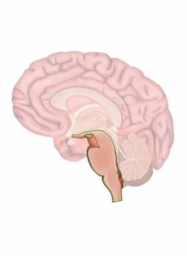
name this principal region:
brainstem

name this specific region of the brainstem:
midbrain w/ superior colliculi and inferior colliculi

name this specific region of the brainstem:
medulla oblongata

name this specific region of the brainstem:
pons

name the function of the medulla oblongata
respiratory and cardiovascular control
name the function of the pons
contains some respitatory centers

name the function of the midbrain
vision, hearing, and motor control
name the function of the superior and inferior colliculi
corpora quadrigemina contains reflex centers
superior = signals from retina and visual cortex
inferior = central relay and auditory processing

name the function of the cerebellum
smooths movements, regulates posture
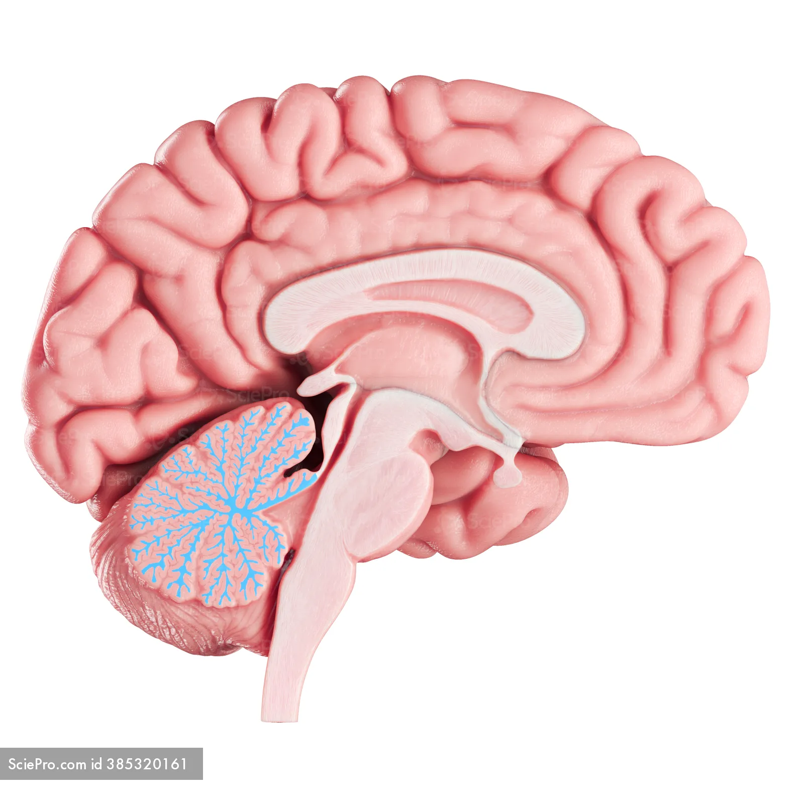
name this specific region of the cerebellum
arbor vitae

name this specific region of the cerebellum
vermis
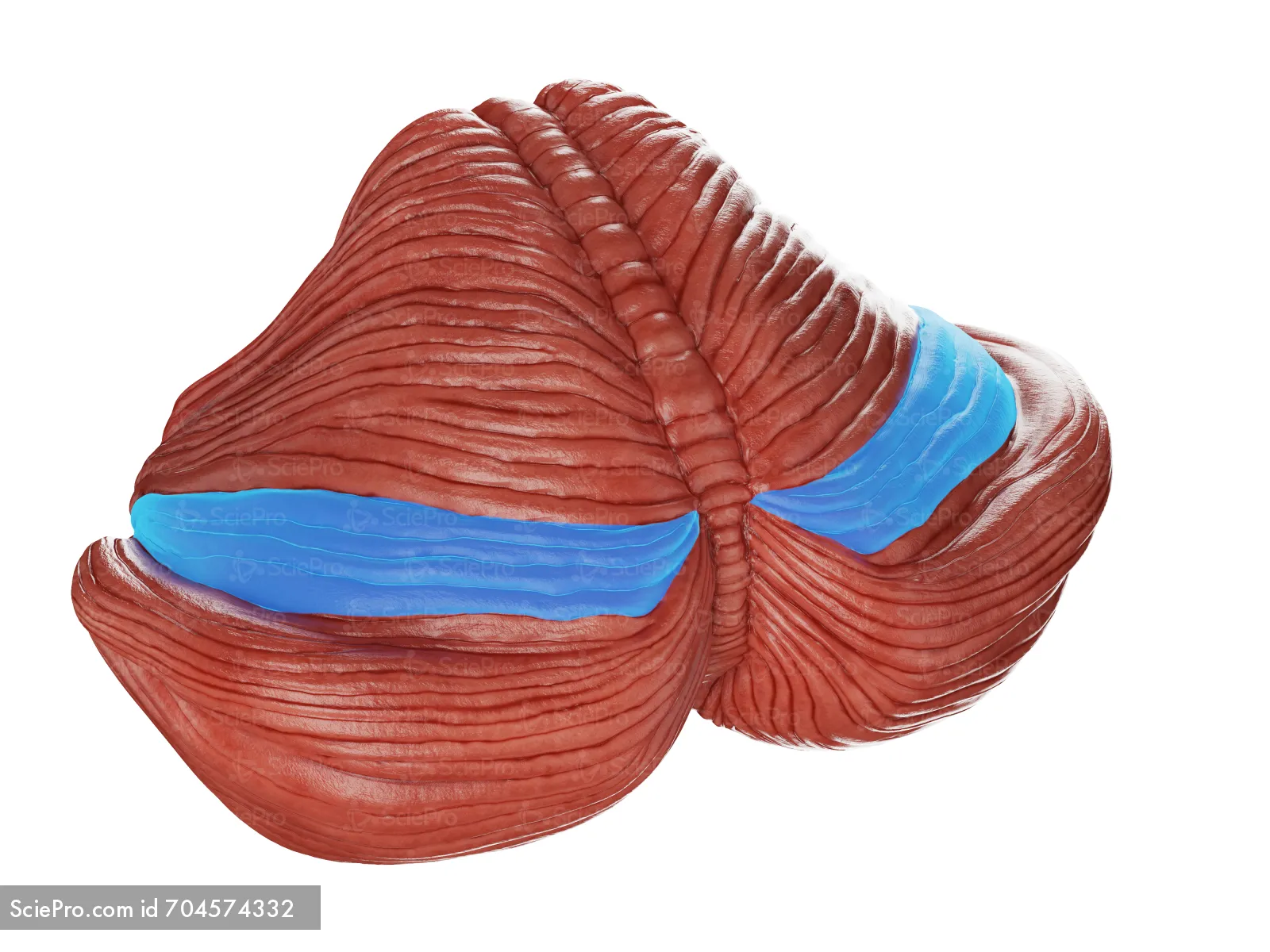
name this specific region of the cerebellum
folia
list the 3 specific regions of the cerebellum
folia, vermis, arbor vitae
list the 3 specific regions of diencephalon
epithalamus, thalamus, and hypothalamus

name this specific region of the diencephalon
epithalamus

name this specific region of the diencephalon
thalamus

name this specific region of the diencephalon
hypothalamus
what is the function of the epithalamus?
includes the pineal gland that produces and secretes melatonin.
what is the function of the hypothalamus?
manage homeostasis. 4 specific structures too.
list the 4 specific structures of the hypothalamus
mammillary bodies
optic chiasm
infundibulum
pituitary gland

name this specific structure of the hypothalamus:
mammillary bodies

name this specific structure of the hypothalamus: (pink)
optic chiasm
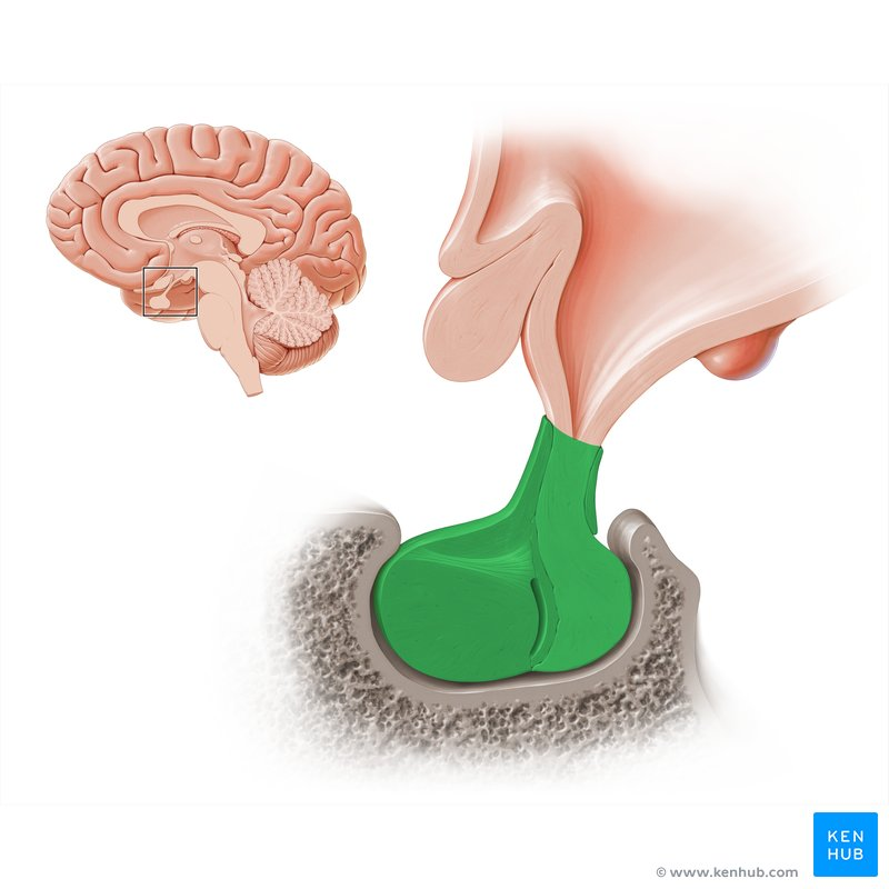
name this specific structure of the hypothalamus (connecting piece):
infundibulum

name this specific structure of the hypothalamus:
pituitary gland
what is the function of the mammillary bodies?
involved in reflexes like swallowing and in processing smell and taste
what is the function of the optic chiasm?
allows for visual information from each eye to be processed and support visual perception.

name this specific region of the cerebrum (A)
cerebral cortex (grey matter)

name this specific region of the cerebrum (B)
cerebral white matter
name the function of white matter in the cerebrum
to communicate as fast as possible due to its myelinated axons on the inside of the cerebrum
name the function of gray matter in cerebrum
not as fast signaling because of unmyelinated axons
what is the function of the intermediate mass of the thalamus?
connects the two halves of the thalamus
what is the function of the thalamus?
relays information to the cerebral cortex

name this structure in the thalamus (purple)
pineal gland
list the 3 specific regions of the cerebrum
corpus callosum
fornix
choroid plexus

name this structure of the cerebrum
corpus callosum
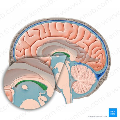
name this structure of the cerebrum
choroid plexus

name this structure of the cerebrum
fornix
what is the function of the fornix in the cerebrum?
carries info that helps form memories
what is the function of the corpus callosum in the cerebrum?
connects left and right brain hemispheres
what is the function of the choroid plexus in the cerebrum?
blood → CSF

what is this surface feature of the cerebral cortex?
central sulcus

what is this surface feature of the cerebral cortex?
longitudinal fissure

what is this surface feature of the cerebral cortex?
precentral gyrus
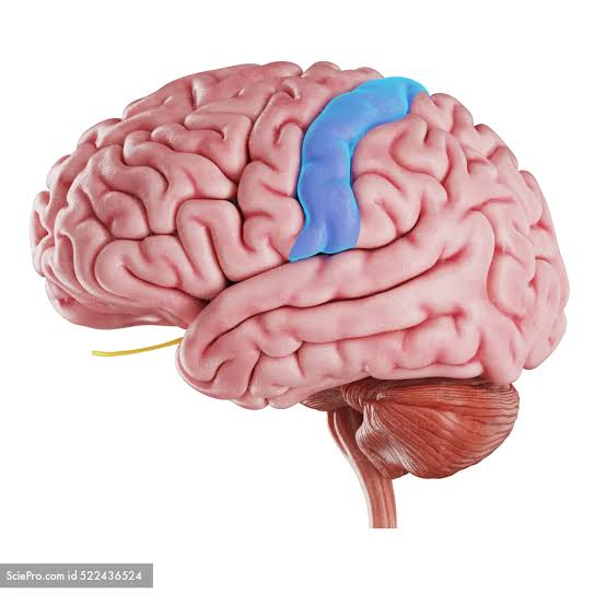
what is this surface feature of the cerebral cortex?
postcentral gyrus

what is this surface feature of the cerebral cortex?
lateral sulcus

what is this surface feature of the cerebral cortex?
transverse fissure

what is this surface feature of the cerebral cortex?
parieto-occipital sulcus

gyrus
elevation or fold in cerebral cortex that increases surface area for neurons

sulcus
shallow groove between elevations
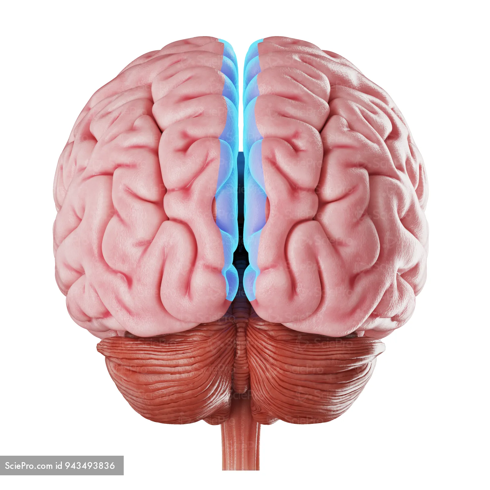
fissure
deep groove separating the cerebrum from cerebellum
what three types of functional areas are there in the cerebral cortex?
sensory cortices
motor cortices
association areas

name this sensory cortices:
primary somatosensory cortex

name this sensory cortices:
primary auditory cortex
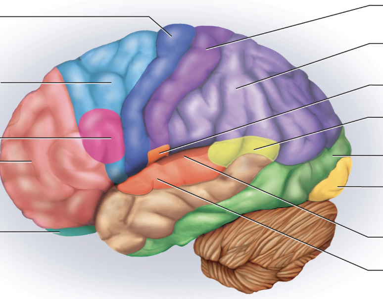
name this sensory cortices: (small orange spot)
primary gustatory cortex

name this sensory cortices:
primary visual cortex

name this sensory cortices:
primary olfactory cortex

name this association area:
somatosensory association area

name this association area: (light blue)
motor association area
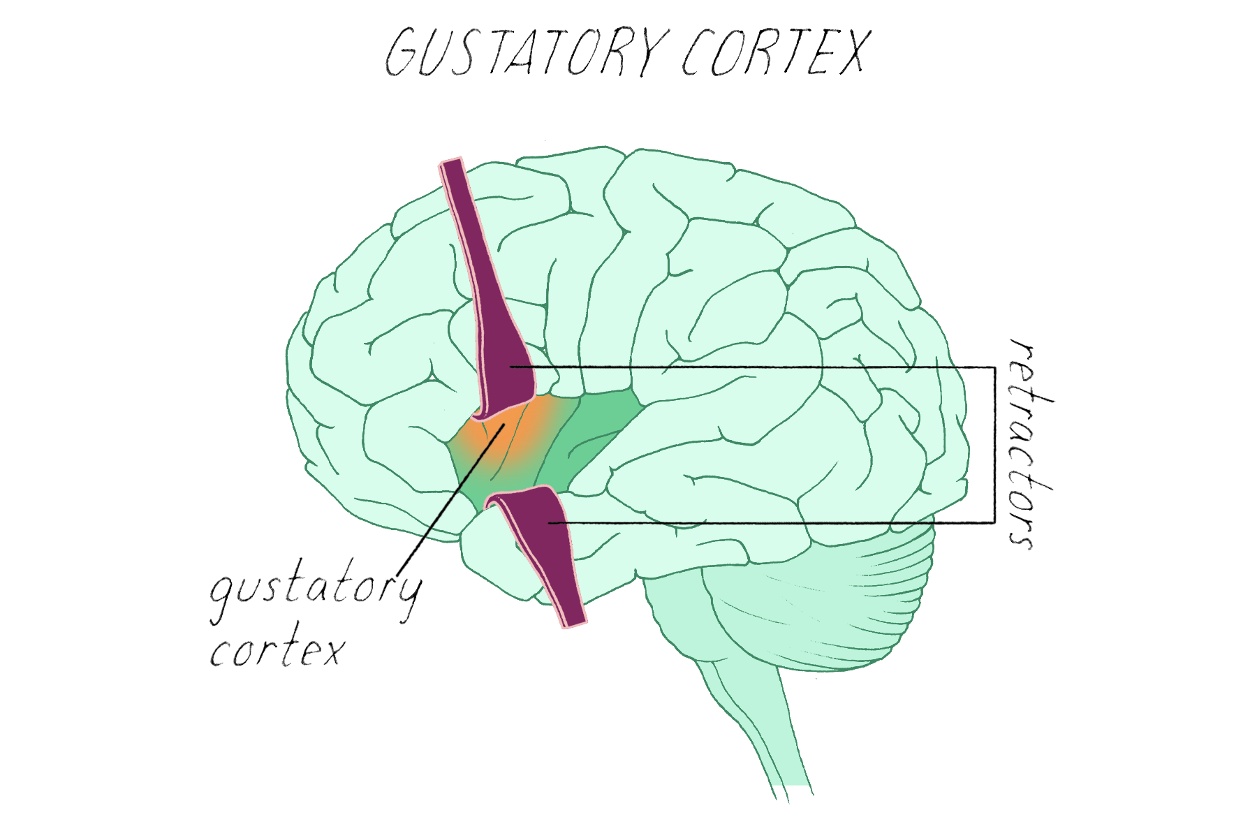
name this association area:
gustatory association area
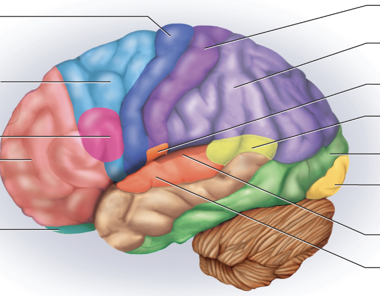
name this association area: (green above the yellow)
visual association area

name this association area: (teal)
olfactory association area