histology week 9 (ch. 12 & 13 GI Pts 1 and 2)
 Call Kai
 Learn
 Practice Test
 Spaced Repetition
 Match
 Flashcards
 Knowt Play
1/102

There's no tags or description
Looks like no tags are added yet.
103 Terms

What are A, B, C, D, and E?
A: muscularis mucosae
B: lamina propria
C: lumen
D: epithelium
E: adventitia

What are a, b, c, d, e, and f?
A: muscularis propria outer longitudinal layer
B: muscularis propria inner circular layer
C: myenteric plexus
D: submucosa
E: lymphoid aggregate
F: submucosal plexus

What are A and B?
a: surface epithelium functional surface
B: lamina propria

What is in the black outline?
Muscularis mucosae (deepest layer of mucosa)

What are A and B?
A: Muscularis mucosa
B: submucosa

What are A, B, and C?
A: epithelium
B: lamina propria
C: muscularis mucosa

What is this showing?
Esophageal submucosal glands

What are A and B?
Inner circular layer of muscularis externa
Outer longitudinal layer of muscularis externa

What are A and B?
A: outer longitudinal layer of esophagus
B: inner circular layer of esophagus

What are A, b, and c?
A: skeletal muscle (somatic)
B: skeletal and smooth muscle
C: smooth muscle (visceral)

What are A, B, C, and D?
A: trachea
B: esophagus
C: adventia
D: serosa

What are A and B?
A: adventitia
B: serosa

What are A and B?
A: esophagus
B: stomach

What structure and tissue type are on the left and right?
Left: stratified squamous epithelium of the esophagus
Right: simple columnar epithelium of the stomach

What are a, B, C, D, and E?
A: cardia
B: fundus
C: body
D: antrum
E: pylorus

What is this?
Surface epithelium of gastric mucosa

What is this?
Rugae gastric folds

What is this?
Rugae gastric folds

Which gland type is this?
Simple tubular

Which gland type is this?
Simple branched tubular

Which gland type is this?
Simple coiled tubular

Which gland type is this?
Simple acinar
Which gland type is this?
Simple branched acinar


Which gland type is this?
Compound tubular

Which gland type is this?
Compound acinar

Which gland type is this?
Compound tubuloacinar

What is each section of the gastric gland?
A: gastric pit or foveola
B: isthmus
C: neck
D: base of gland
E: gastric gland

What cells are in each section of the gastric gland?
A: surface mucous cells
B: parietal cells
C: neck mucous cells, stem cells, parietal cells
D: peptide cells, parietal cells, neck mucous cells, neuroendocrine cells

Which gastric region is this?
What are A and B?
Cardiac region
A: stratified squamous epithelium
B: simple columnar epithelium

What gastric sections is this?
What are a, b, c, and d?
A: gastric pit
B: lamina propria
C: pits
D: duct

What cell type is circled?
Parietal cells

What cell type is this?
What is in the black border?
Parietal cell
Tubular vesicular cytoplasm

What cell type is this?
What is in the black border
Parietal cell
Intracellular canaliculi

What cell thpe is in the blue border?
Chief cell

Which cell type is in the blue?
Which cell type is in the black?
Blue: Parietal cells
Black: chief cells
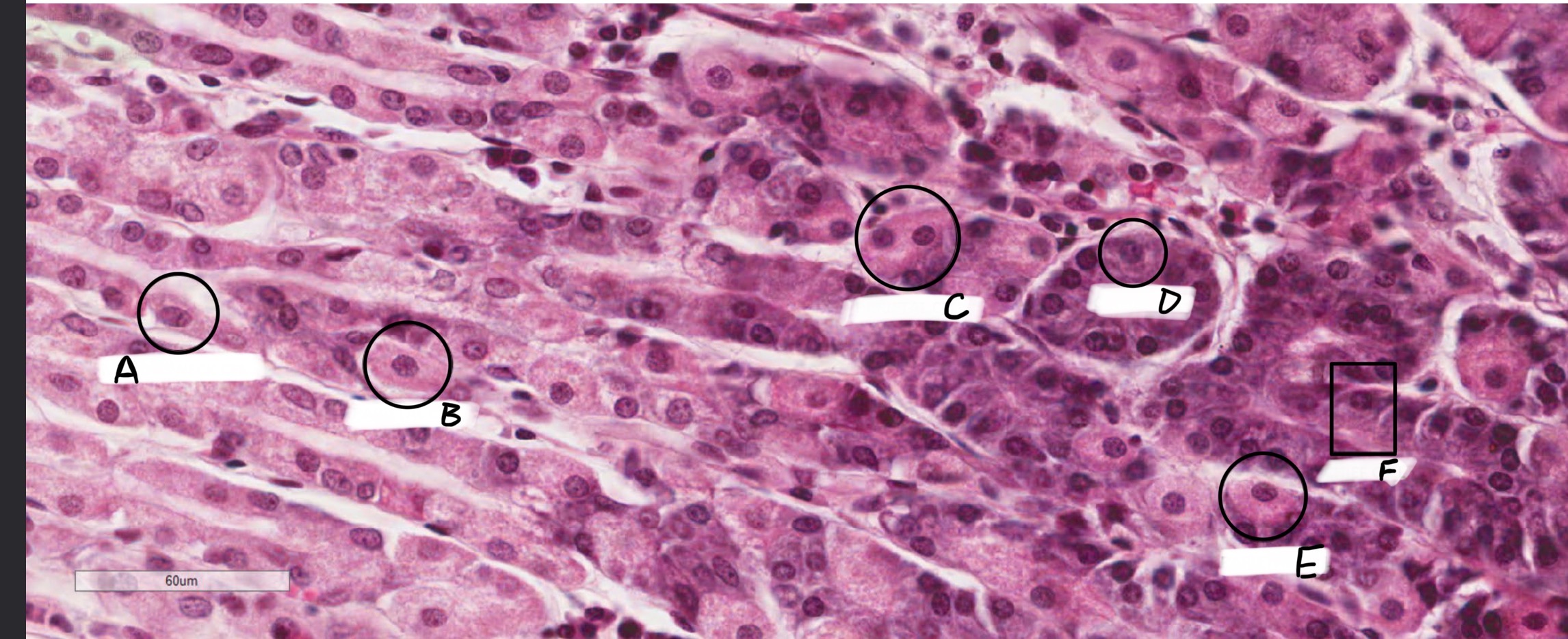
What are a, b, c, d, e, and f?
A; mucous neck cell
B: parietal cell
C: prietal cell
D: chief cell
E: parietal cell
F: chief cell

What are A, B, and C?
A: poorly stained surface epithelia cells
B: concentrated clusters of parietal cells
C: enteroendocrine cells, chief cells, and parietal cells

What is this?
Enteroendocrine cell

What are the layers of the muscularis externa?
1: inner oblique
2: middle circular
3: outer longitudinal

What are A, B, and C?
A: inner oblique
B: middle circular
C: outer longitudinal

What are A, b, and c?
A: inner oblique
B: middle circular
C: outer longitudinal

What are A, B, and C?
A: inner oblique
B: middle circular
C: outer longitudinal
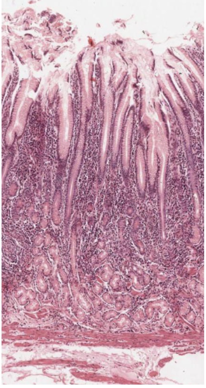
Which section of the stomach is this?
Pylorus

What are A, B, and C?
A: duodenum
B: pyloric sphincter
C: stomach (pylorus)

What are A, B, and C?
A: surface epithelium
B: lamina propria
C: muscularis mucosae

What is this?
Submucosa

What are A and B?
B: outer longitudinal layer of muscularis externa
A: inner circular layer of muscularis externa

What is this?
Outer longitudinal layer of muscularis externa

What is this?
Inner circular layer if muscularis externa

What is in the brackets?
Serosa/adventitia

What are the segments of the small intestine?
A: duodenum
B: jejunum
C: ileum

What are A, B, and C?
A: duodenum
B: pyloric sphincter
C: stomach pylorus

What is this?
Corrugations in the small intestine

What are A and B in the small intestine?
A: plicae circularis
B: villi

What is this?
Stomach pylorus

What is this?
Pyloric sphincter

What is this?
Duodenum

Villi
What are the cells in blue?
What are the dark longer cells?
Goblet cells
Enterocytes

What structure is this?
Microvilli
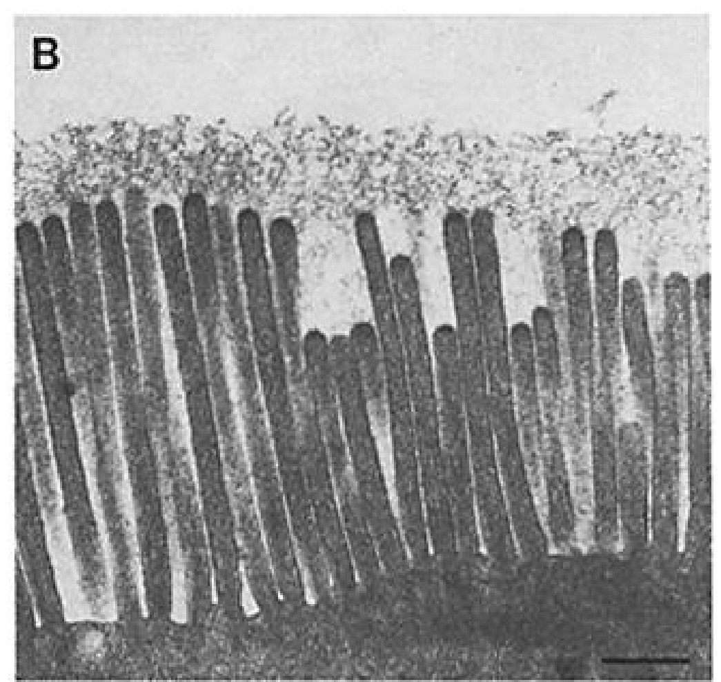
What structure is this?
Microvilli

What is the thin layer by the arrow?
Glycocalyx

What is this?
Brush border (glycocalyx + microvilli)

What are a, b, c, d, and e?
A: basement membrane
B: intestinal gland
C: artery
D: lacteal
E: vein

What are the arrows pointing to?
lacteals

What is this?
What type of gland is it?
Crypts of Lieberkuhn
Simple branched tubular

What is this?
Paneth Cell

What is this?
What are the arrows pointing to?
paneth cell
Acidophilic apex granules

What is this?
Paneth cells

What is the arrow pointing to?
Enteroendocrine cell

What is this cell?
Enteroendocrine cell

What are A and B?
A: enterocyte
B: goblet cell

What are A and B?
A: enteroendocrine cell
B: paneth cell

What cell type is circled?
Enteroendocrine cell

Duodenum
What are A, b, c, d, and e?
A: villi
B: mucosa, intestinal glands
C: submucosa, brunner’s glands
D: submucosa
E: muscularis externa

What are A and B?
What is in blue?
A: mucosa
B: brunner’s glands
Blue: muscularis mucosae

What is circled?
Brunner’s glands in duodenum

What is this?
What is the arrow highlighting?
Jejunum
No brunner’s glands

What is this?
Jejunum

What is this?
Jejunum

What are a and B?
A: Myenteric plexus
B: submucosal plexus

What are A, B, C, ans D?
A: villi
B: plicae circularis
C: submucosa
D: muscularis externa

What are A, B, and c?
A: inner circular layer
B: muenteric plexus
C: outer longitudinal layer

What is circled?
Myenteric plexus

What are A, B, C, D, and E?
A: mucosal glands
B: muscularis mucosae
C: submucosae
D: muscularis externa
E: submucosal plexus

What is this?
Submucosal plexus

What is this?
Gut associated lymphoid tissue GALT
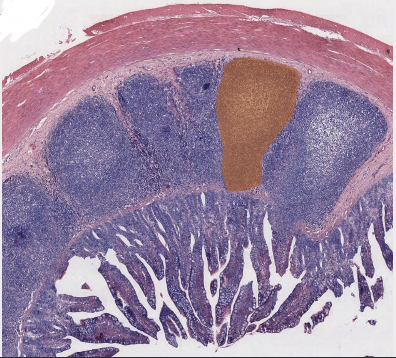
What is highlighted?
Peyer’s patch

What is this?
Peyer’s patch

What is this?
Microfold cell

What are a, b, and c?
A: M cell
B: lympg\hocyte
C: macrophages

What are a, b, c, d, e, f, g, and h?
A: appendix
B: cecum
C: ileocecal valve
D: ileum
E: ascending colon
F: taenia coli
G: right colic (hepatic) flexure
H: transverse colon

What are I, j, k, l, m, n, and o?
I: left colic (splenic) flexure
J: haustra
K: descending colon
L: sigmoid flexure
M: sigmoid colon
N: rectum
O: anjs

What is this?
What tissue type is this?
Tenia coli
Logitudinal smooth muscle

Muscularis externa
What are a, b, and c?
A: myenteric plexus ‘
B: outer longitudinal taenia coli
C: inner circular layer

What is this?
What is the white in the middle?
Appendix
Fat in the submucosa

What is this?
Appendix in longitudinal section

What is this?
Reminants of fat cells and large vessels in submucosa of appendix

What are A, B, C, and D?
A: anorectal junction ‘
B: rectim
C: external anal sphincter
D: internal anal sphincter

What are A, B, C, D, E, and F?
A: internal sphincter
B: intestinal (colic) glands
C: rectoanal junction
D: rectum (simple epithelium)
E: anal canal (stratified nonkeratinized epithelium)
F: submucosa (rich veins)

What are A and B/
A: skeletal muscle
B: smooth muscle