Spinal Cord
 Call Kai
 Learn
 Practice Test
 Spaced Repetition
 Match
 Flashcards
 Knowt Play
1/87

There's no tags or description
Looks like no tags are added yet.
88 Terms
Normal Lines on the spine
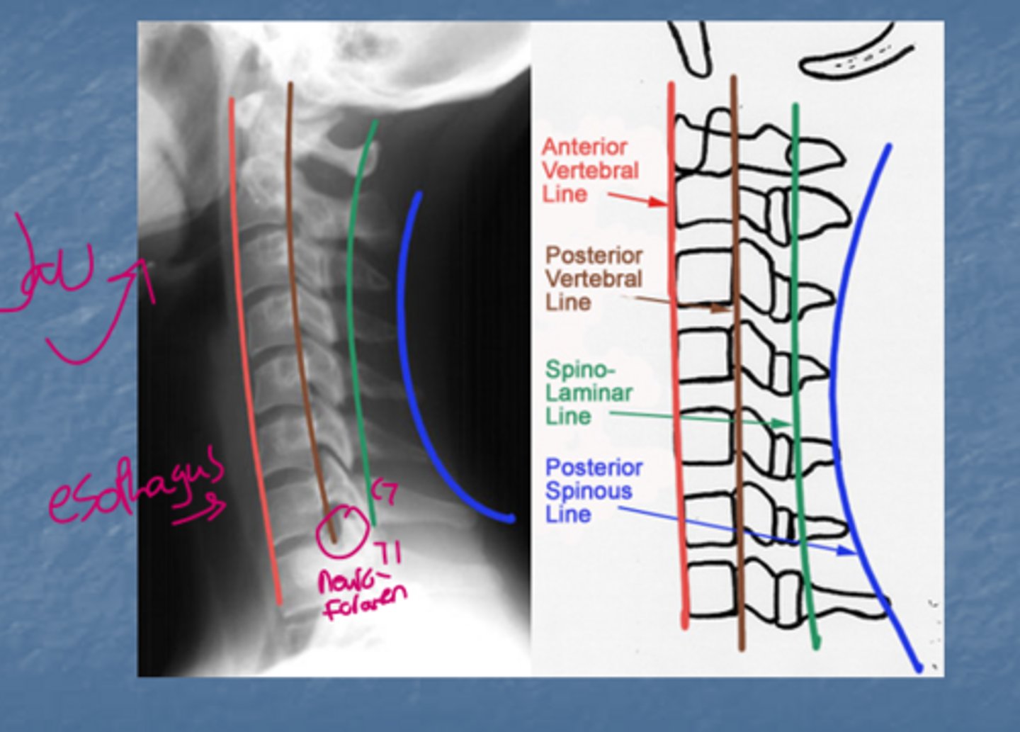
Spino-laminar junction
-Junction between the spinous processes and laminae.
-We dont want widening of this junction which could indicate injury
C1 and C2 are also called what, respectively?
C1-atlas (atlas held up the earth in greek mythology)
C2-axis (has an odontoid process that allows C1 to spin on its "axis")
antlanto-axial line
Joint between C1 and C2. Its a pivot joint that allows rotation for the head. When we are looking at the width of this line we don't want it greater than 3 mm.
What views of the spine do we get?
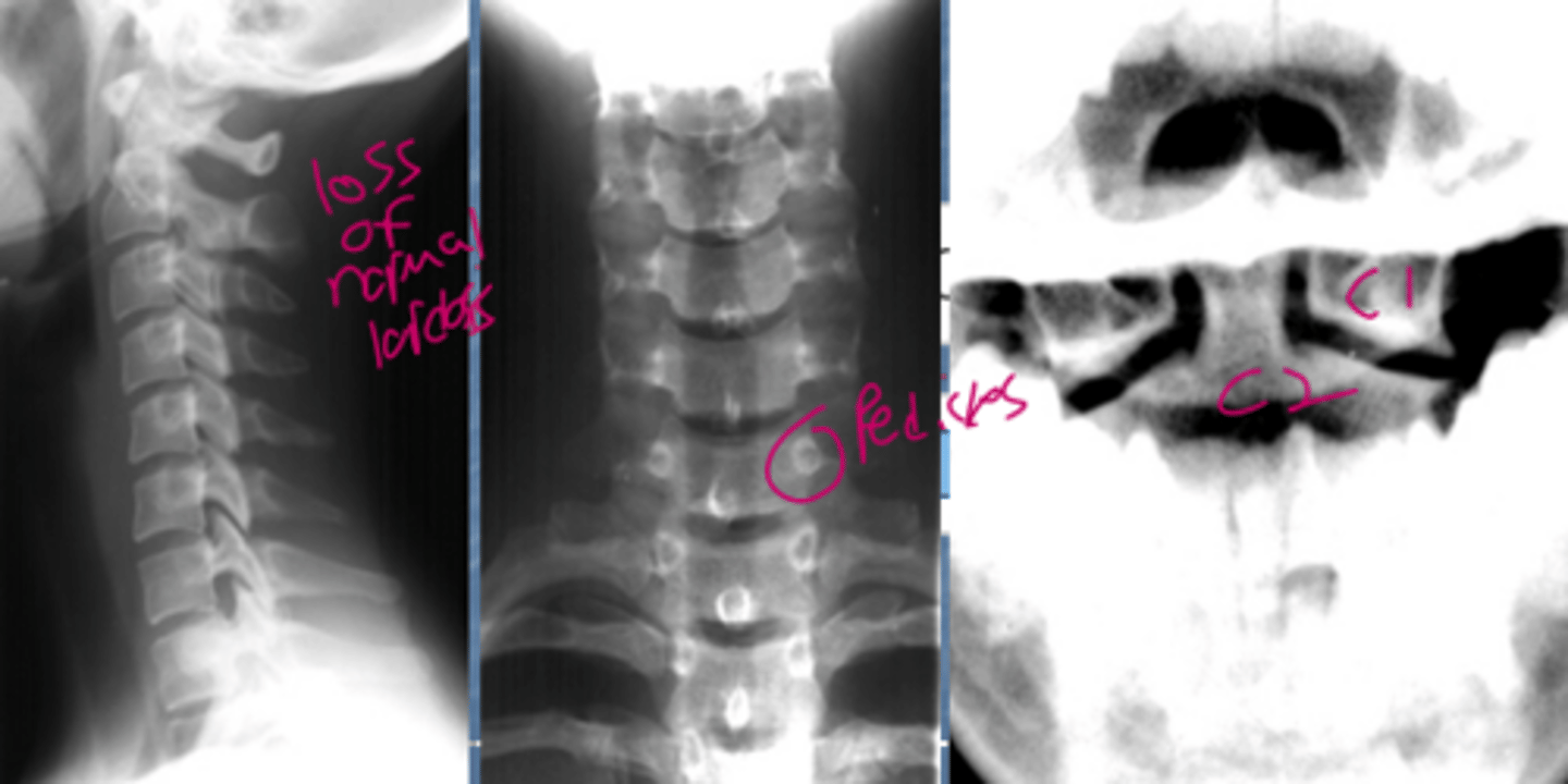
Lateral, frontal, open mouth
What does the oblique view allow us to see?
Neural foramen
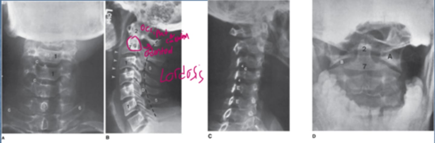
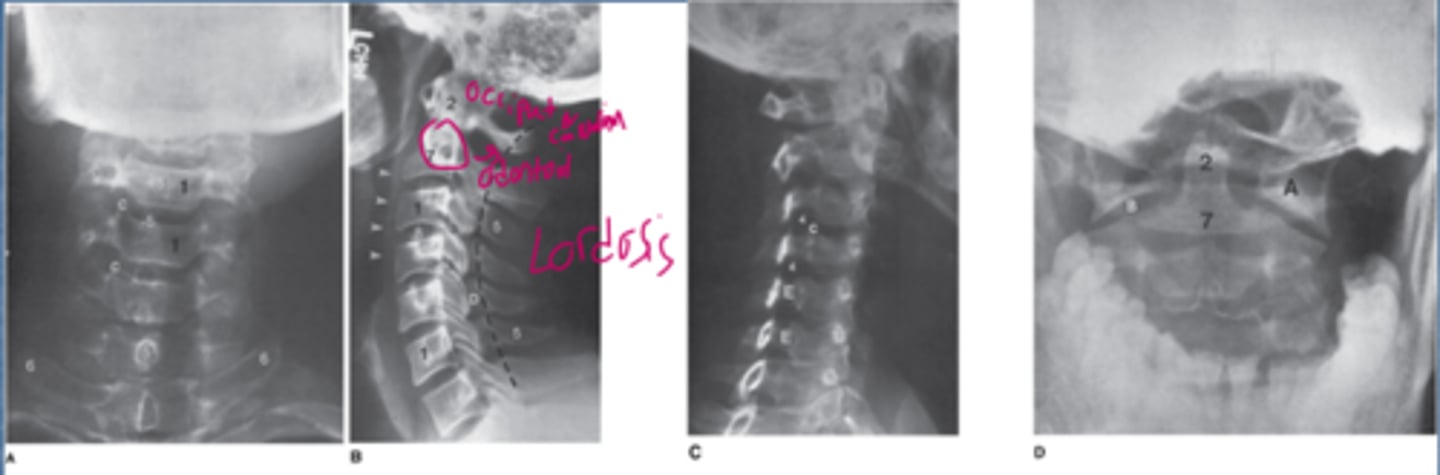
Label the numbers and letters on here
Answers
1=vertebral body
2=odontoid process (dens)
3=articular facet joint
4=intervertebral foramen
5=spinous process
6=transverse process
7=body of axis
8=interverbal disk space
9=anterior arch of atlas
A=lateral mass of atlas
B=atlantoaxial joint
C=uncinate process
D=lamina
E=pedicle
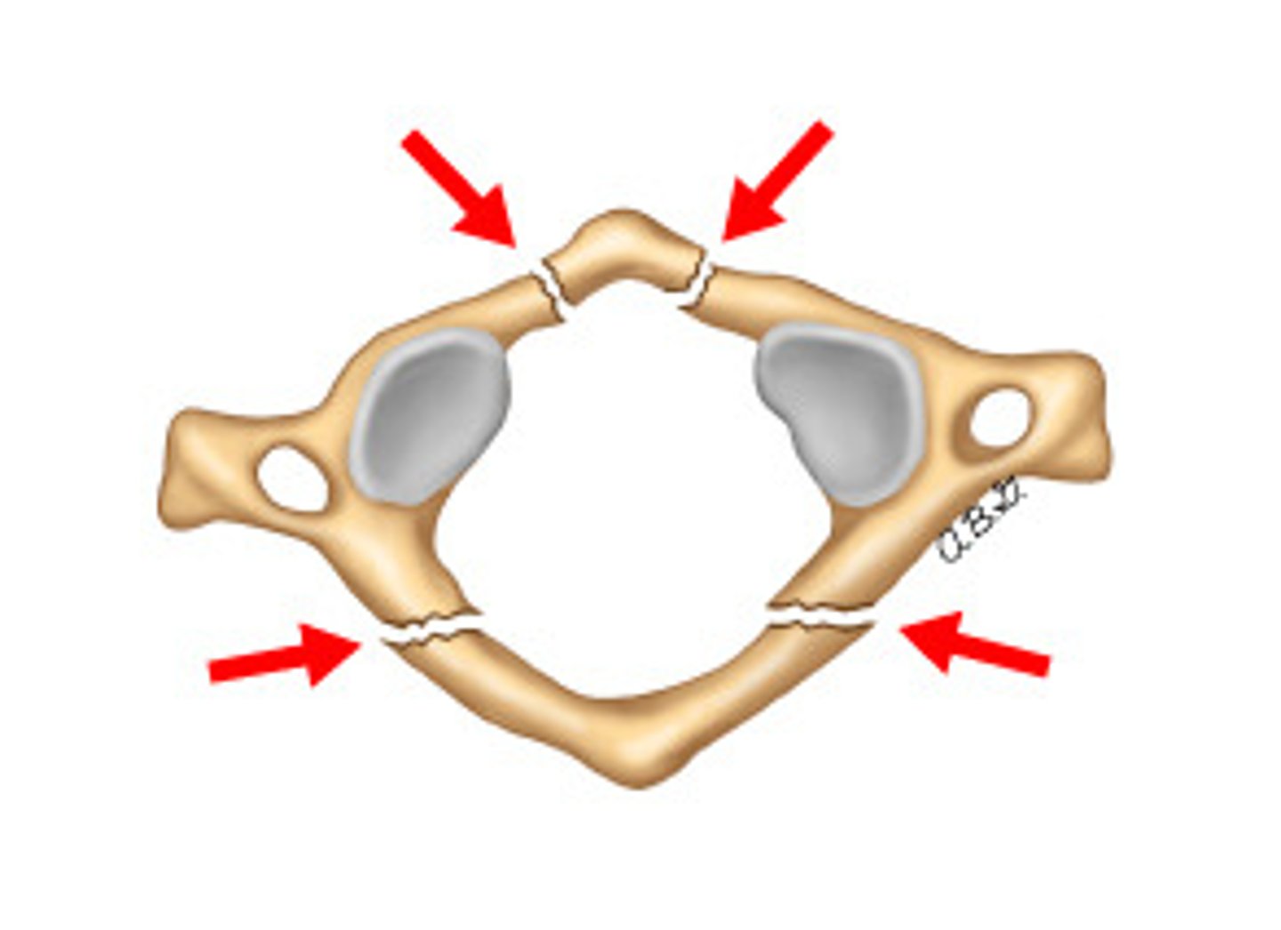
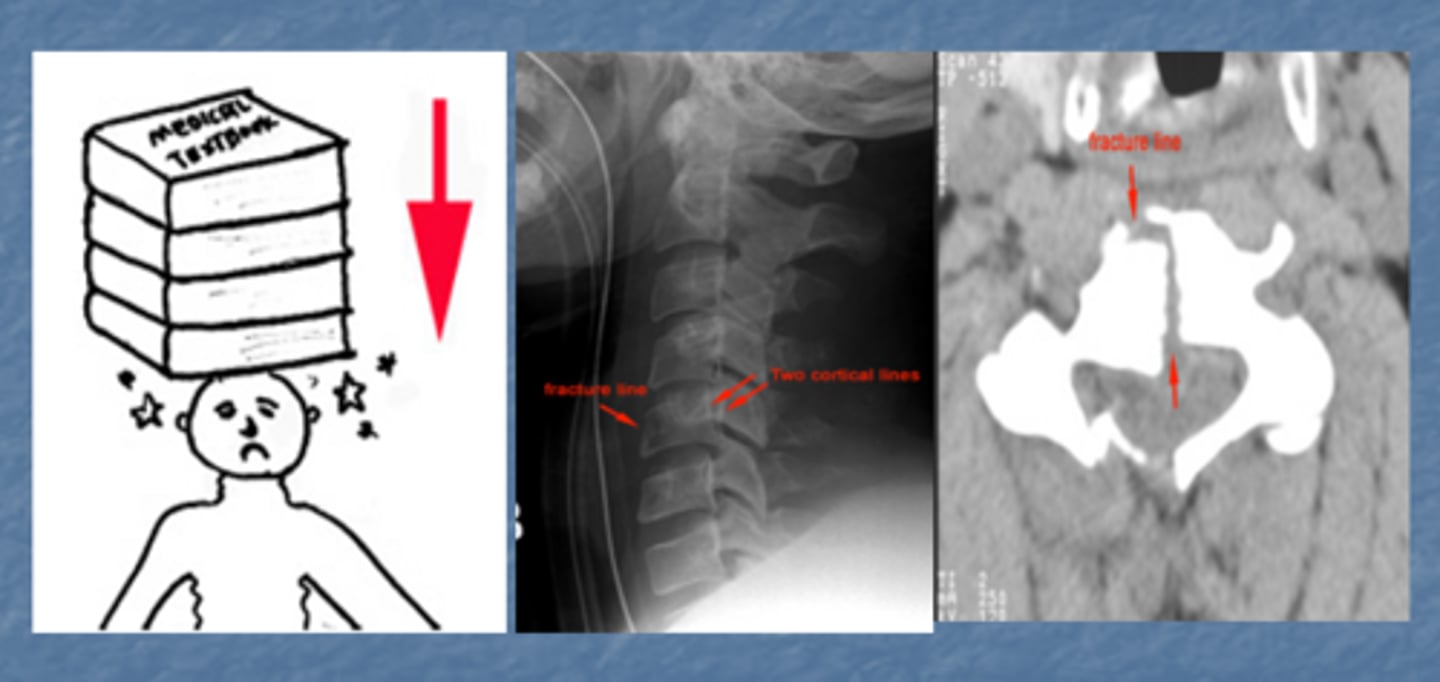
A 14 y/o comes to you with their mom with neck pain after a diving accident. What did they most likely fracture?
C1=jefferson's fracture, extreme axial load most commonly from either diving or you are in an MVA and you hit the top of the car with your head.
What fracture is commonly named after the fracture that occurs in the neck of victims who died by hanging?
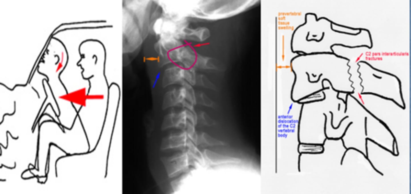
Hangman's fracture=C2. Fracture in the pars interarticularis.
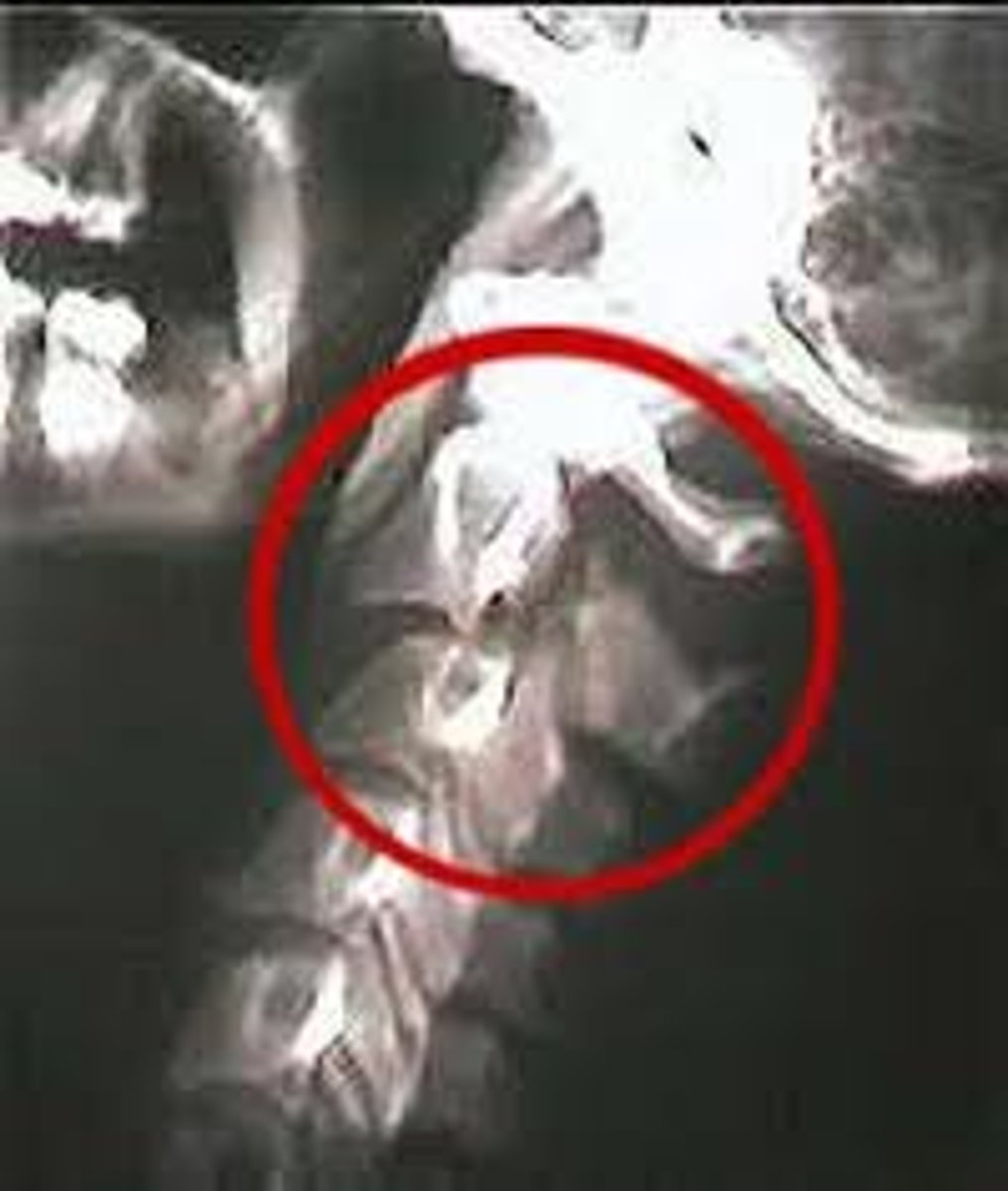
pars interarticularis
Between the superior and inferior articular processes
Normal cervical spine projections
1.C1 and C2 for any widening (atlantoaxial joint >3 mm)
2.First look at the anterior vertebral line then go down the list.
3.I want to see the top of T1 if I can
Plain film of the cervical spine (lateral view) of a 53-year-old woman who presents with neck and right arm pain
The lateral cervical spine plain film suggests that the most likely diagnosis is
degenerative disk disease at C2-3 and C3-4.
neoplastic disease at C4.
degenerative disk disease at C5-6 and C6-7.
traumatic injury.
disk space infection at C5-6 and C6-7.
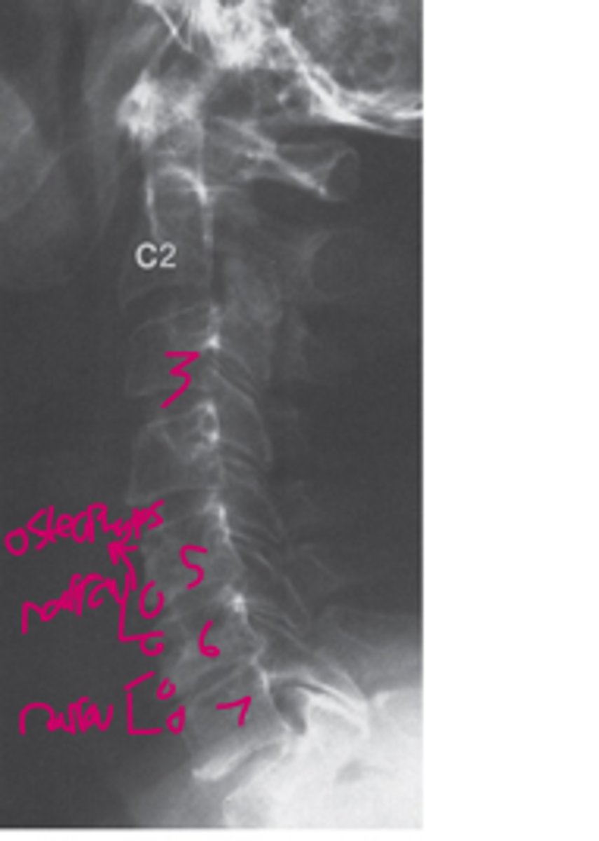
Answer
Degenerative Disk disease at C5-C6 and C6-C7:
-There is disk space narrowing and osteophytes at the C5-C6 and C6-C7 disk spaces
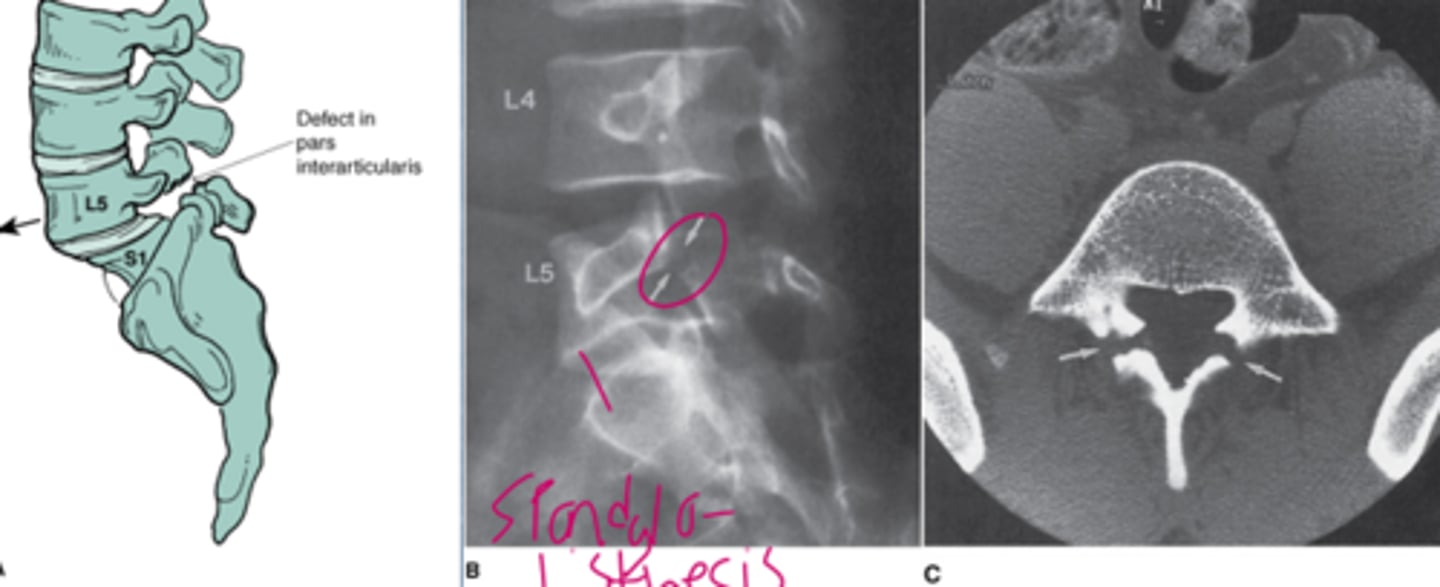
Spondylosis
degeneration of the spine
Spondylolysis
fracture of pars interarticularis
Spondylolisthesis
Slippage of vertebra
Make sure you can recognize this
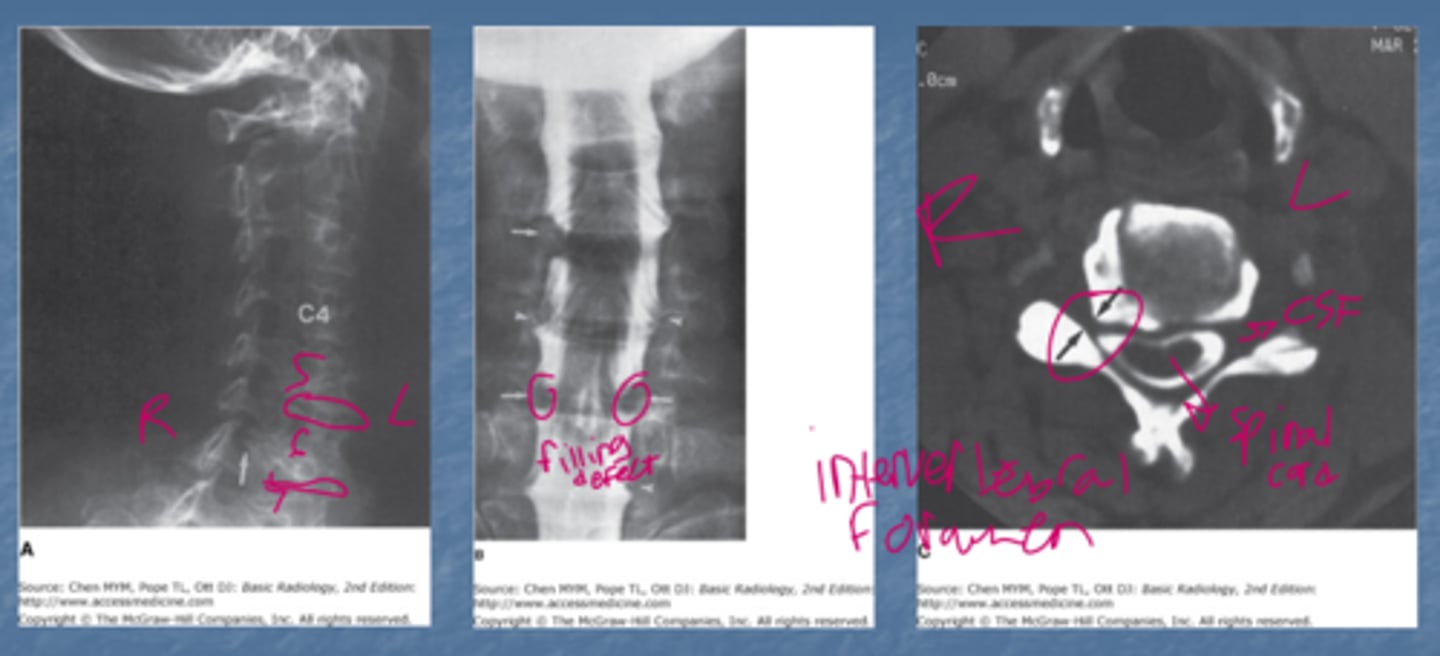
Note that in A there is compromise of the neural foramen by osteophytes. Note the defects where the nerve roots would be on the myelogram.
Burst fracture of the spine
Vertebra loses height both anteriorly and posteriorly and there is often 'retro-pulsion' backwards movement into spinal canal. May be stable or unstable depend on extent of damage
So what does this patient have (Warning: My chicken scratch)?
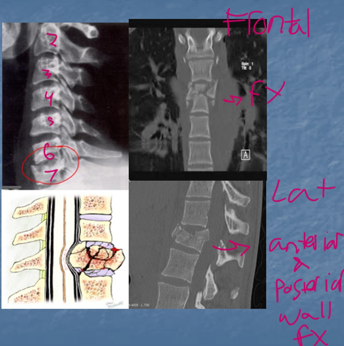
Answer
Burst fracture of C7
Anytime someone has an injury of their spinal cord what image modality should I immediately get?
MRI
What happened to this person?
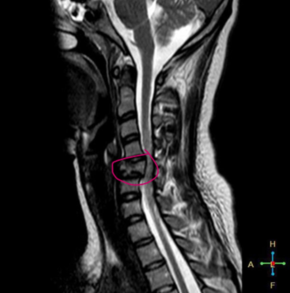
Answer
-C5 is abnormal and is also compressing the spinal cord...thats probably not good.
-This is an anterior compression fracture from a flexion injury of the neck with posterior subluxation
-C5 compression fracture
Scary, what is it?
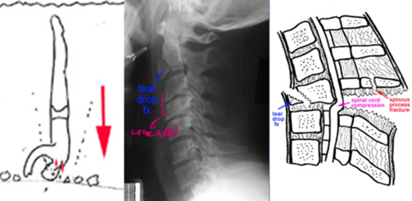
Answer
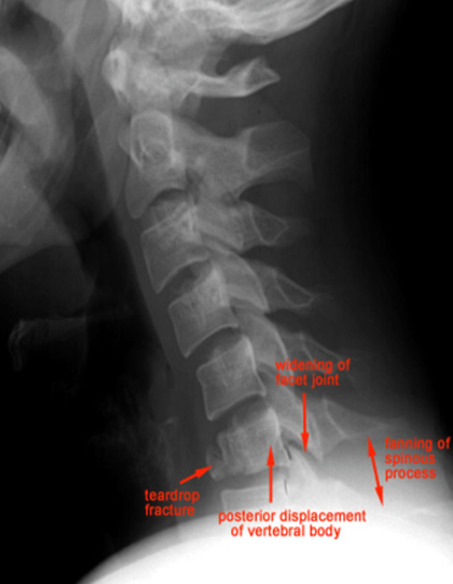
-Tear drop fracture seen with neck flexion.
-Usually significant ligamentous disruption.
-Would follow up with an MRI for ligamentous injury
What does this person have?
Answer
Flexion tear drop fracture:
-C7 is anteriorly subluxated
Oof what do they have?
Answer
Hangman's fracture, C2 through the pars interarticularis region with soft tissue widened
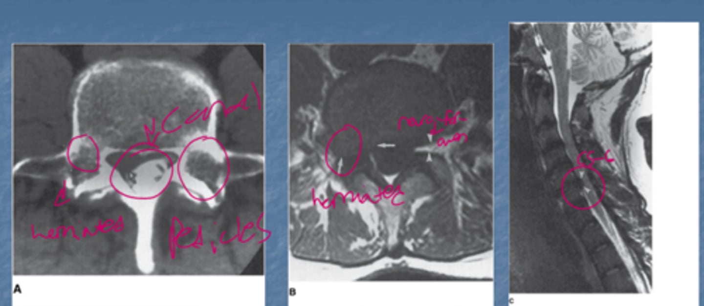
Make sure you can identify that this is herniated.
Note that there is compression at the L4-L5 disk space from a herniated disc
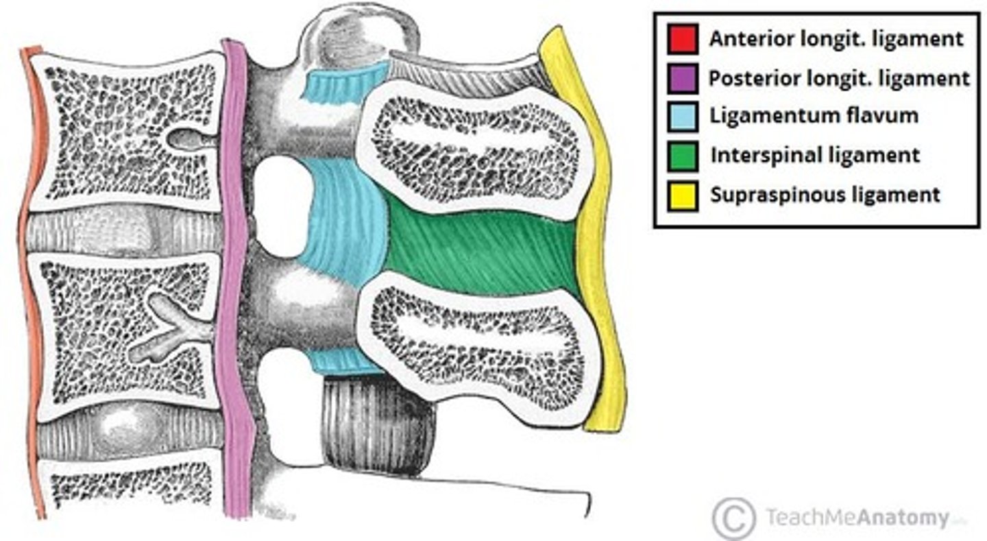
What ligament is a herniated disc pushing up against
PLL
A 35-year-old female with recent onset of paresthesia's in her extremities.
What is the main abnormality visualized?
None
Artifact
Subarachnoid lesion
Intramedullary lesion
Syrinx
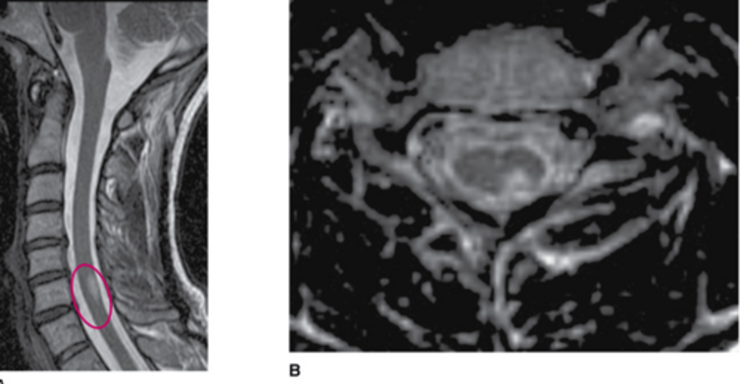
Answer
Intramedullary lesion. its literally in the spinal cord
What could that be?
A syrinx! Can be congenital, can be delayed from trauma, can also be seen with Chiari malformation.
A 44-year-old male with history of intravenous drug use presents to the emergency department with neck pain.
What is the most likely diagnosis?
Spine metastases
Posttraumatic changes
Discitis and osteomyelitis
Epidural abscess
Degenerative changes
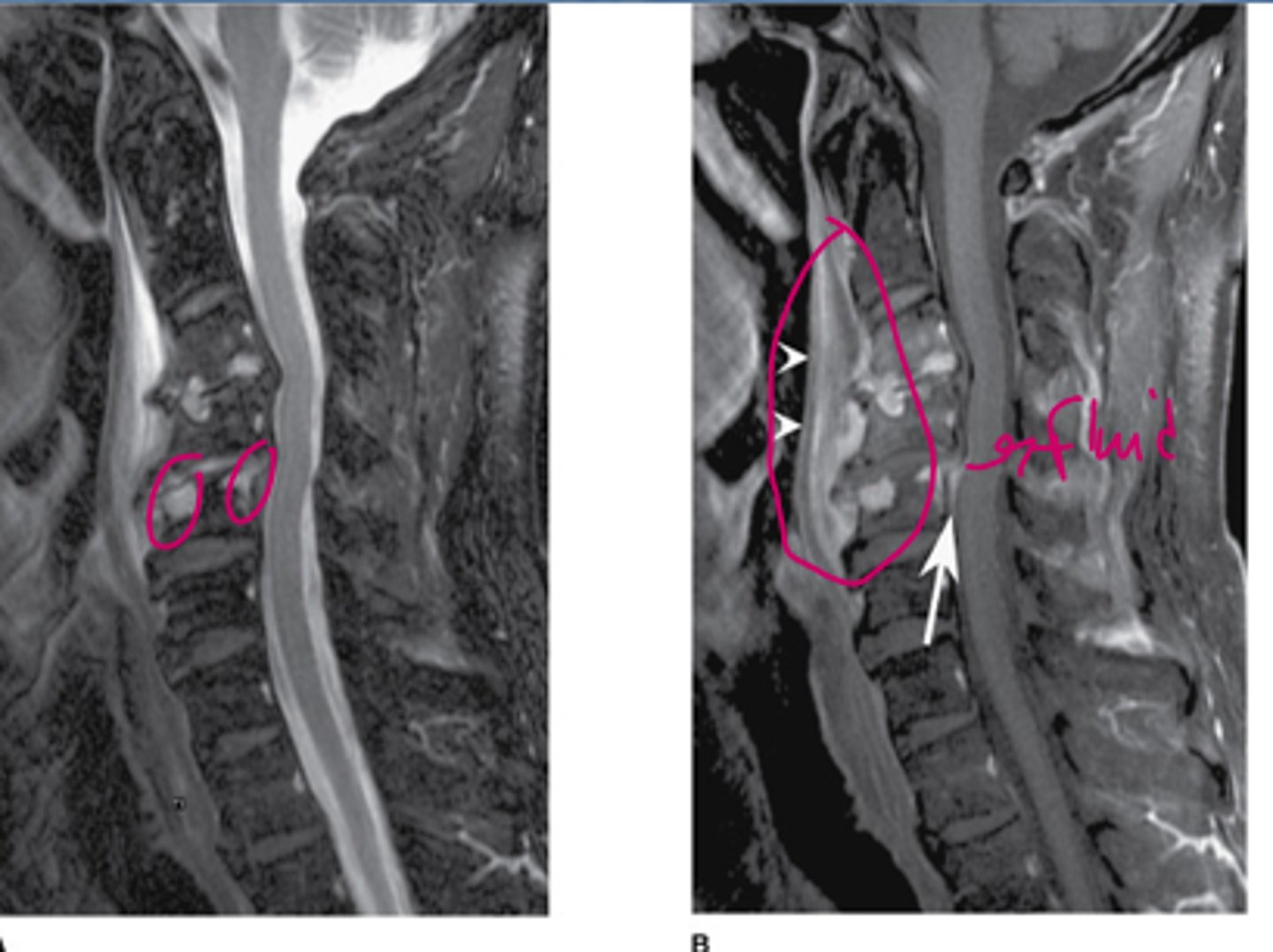
Answer
Discitis and osteomyelitis
A 63-year-old man presents with severe upper neck pain not responding to anti-inflammatory medication.
On the lateral cervical spine radiograph. what is the main radiologic finding?
A lesion of the C7 spinous process
An osteoblastic bony lesion
An abnormality of alignment
A destructive lesion at C2
A fracture
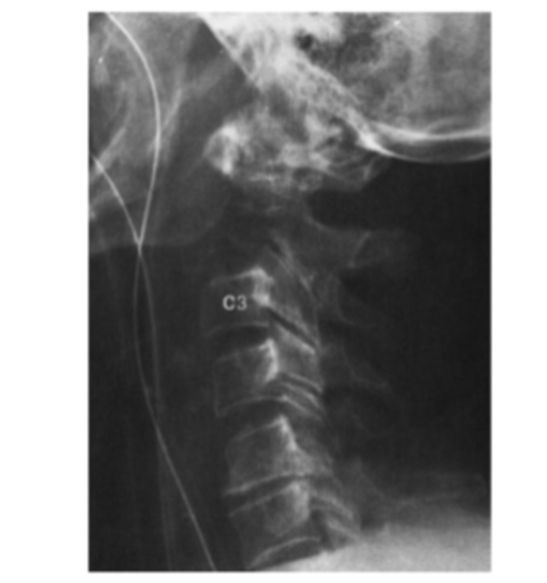
Answer
Destructive lesion at C2:
-This is a lytic lesion most concerning for malignancy
MOLE for osteolytic lesion
-M=Mets
-M=Myeloma
-O=Osteosarcoma (any primary bone sarcoma)
-O=Osteomyelitis
-L=lymphoma
-L=leukemia
-E=ewing
-E=eosinophilic granuloma
This is unfortunately sad: A 21-year-old quadriplegic woman had a motor vehicle accident 4 weeks ago.
Regarding the patient shown, which of the following is true?
The condition probably predated the trauma.
The prospects for a full recovery are good.
Surgical repair will likely be successful.
The patient will probably never have normal neurologic function below C6.
The spinal cord is intact.
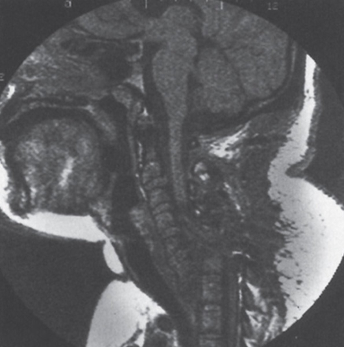
Answer
She will never have normal neurologic function below C6 and will never walk again.
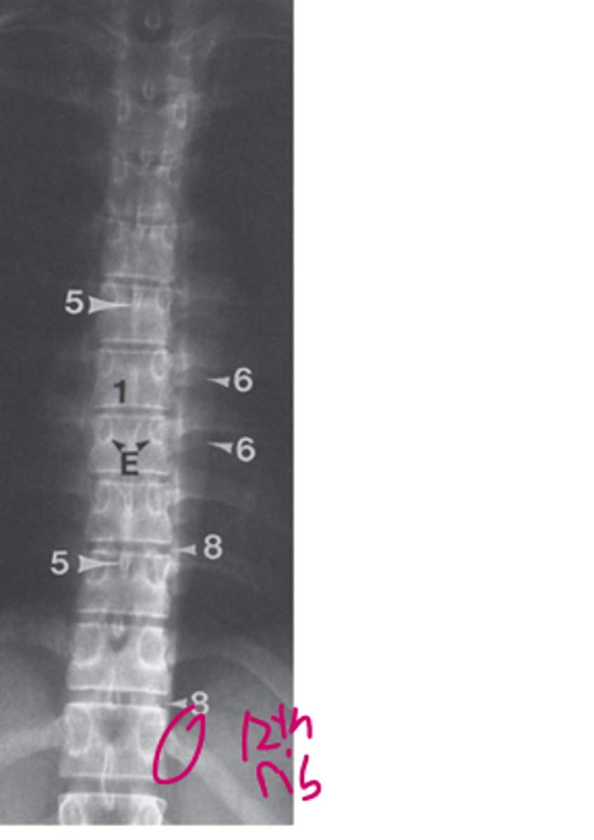
Make sure you can identify the structures on this normal film of the thoracic spine
1=vertebral body
5=spinous process
6=transverse process
8=intervertebral disc space
E=pedicle
A 38-year-old woman presents slowly with progressive upper extremity and upper trunk sensory deficits 3 years after a motor vehicle accident.
The MRI most likely demonstrates
delayed posttraumatic syrinx.
subluxation.
spinal cord tumor.
abnormal bone marrow.
disk abnormality.
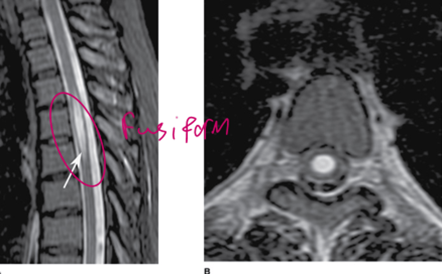
Answer
Delayed posttraumatic syrinx:
-Can happen after trauma
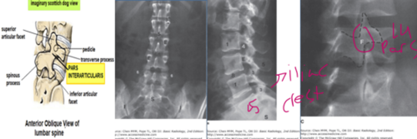
Make sure you can label these
-AP view, lateral view, oblique view
-Scottie dog sign! (my favorite X-ray finding).
-The neck of the Scottie dog represents pars interarticularis.
1=vertebral body
3=articular facet joint
4=intervertebral foramen
5=spinous process
6=transverse process
8=intervertebral disk space
E=pedicle
F=pars interarticularis
S=sacrum
I=sacroiliac joint
Make sure you can label this
A=aorta
D=intervertebral disk
N=neural foramen
P=psoas muscles
arrows=ligamentum flavum
Asterisk on A=thecal sac
Asterisk on B=spinal canal
P=pedicle
B=Vertebral body
T=transverse process
F=facet joint
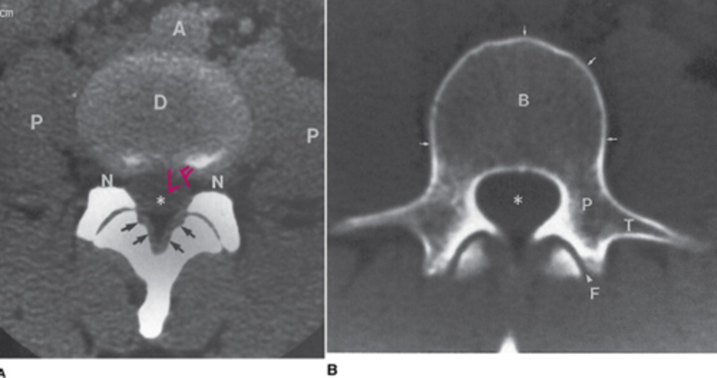
Thick ligamentum flavum + disc + narrowing of neural foramen = what?
Spinal stenosis!
A coned-downed lateral plain film of the lumbar spine in a whiny 45-year-old neuroradiologist who presents with low back pain
What is the abnormality ?
The bones are too dense.
The bones are not dense enough (osteopenia).
There is a destructive bony lesion.
There is an abnormality of alignment(spondylolisthesis
There is a soft-tissue abnormality.
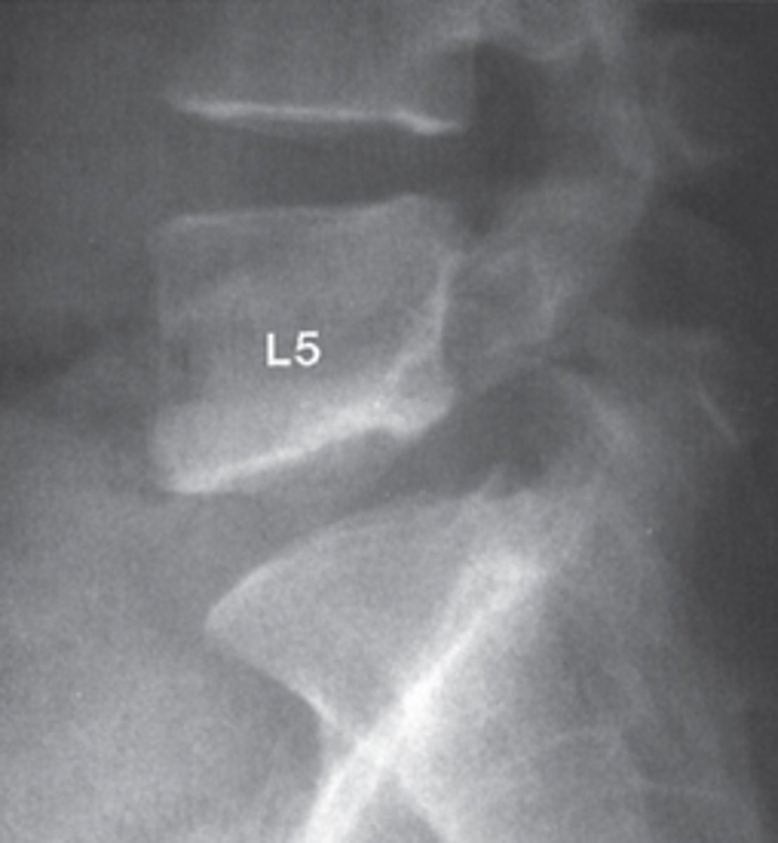
Answer
There is an abnormality of alignment (spondylolisthesis)
What is this?
-Spondylolisthesis
-L5 over S1
A 23-year-old woman was involved in a motor vehicle accident.
What is the most likely diagnosis?
Spinal tumor, aggravated by trauma
Abnormality of bone density
Disruption of facet joints at multiple levels
Subluxation of L4 over L5
L2 compression fracture with kyphotic angulation
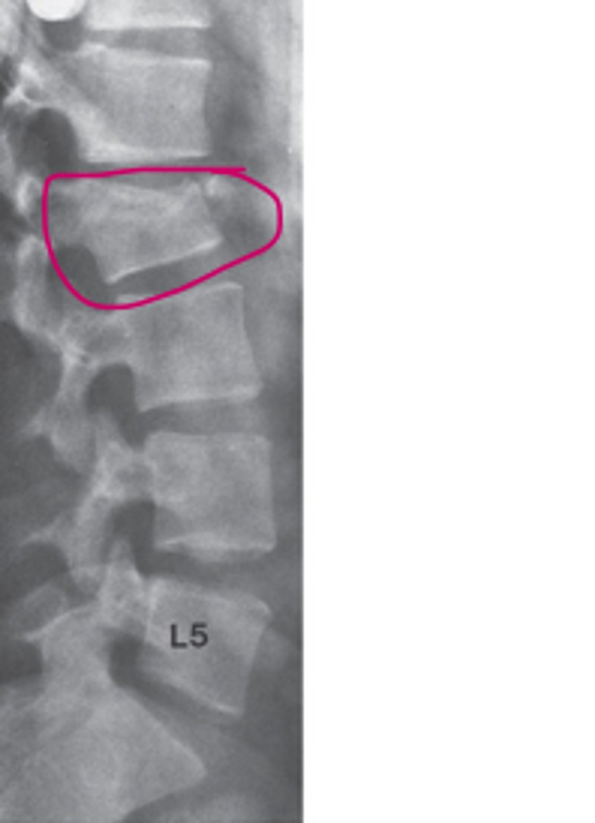
Answer
L2 compression w/ kyphotic angulation.
A 58-year-old man with right-sided L5 radiculopathy. A myelogram was performed, and an oblique view demonstrating the right-sided nerve roots is displayed.
In Case 13-2, the lesion represented by an arrow is most likely to be
a right-sided L4-5 herniated nucleus pulposus.
an extradural tumor.
an epidural abscess.
an intradural mass.
a bony lesion.
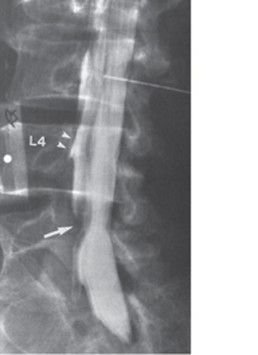
Answer
A right sided L4-:5 herniated nucleus pulposus
Most common fracture site of the lumbar vertebra
T10-L2
Most common compression site of the lumbar vertebra
L4-L5
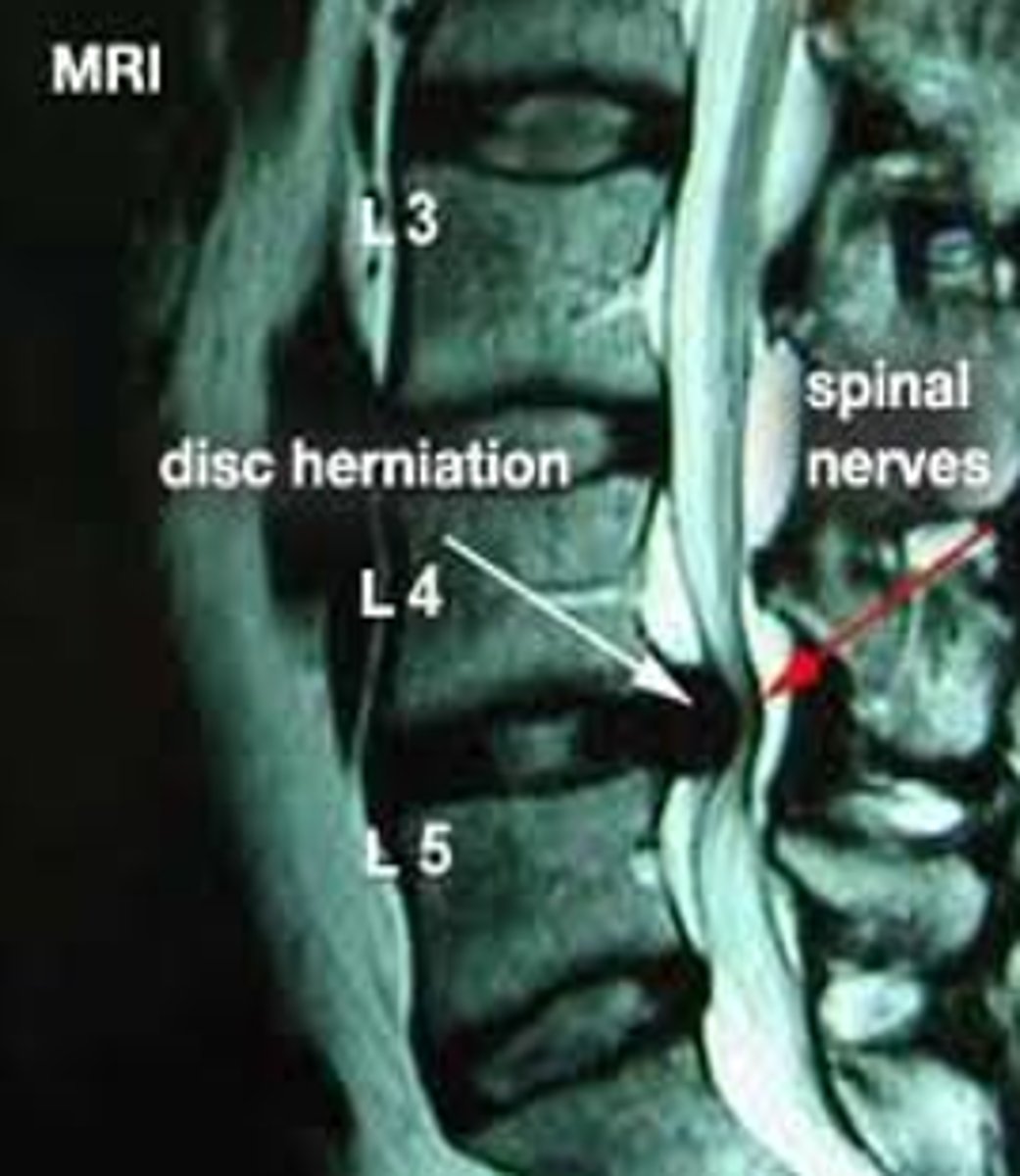
A 22-year-old female presenting with back and leg pain and lower extremity paresthesias.
Whats she got?
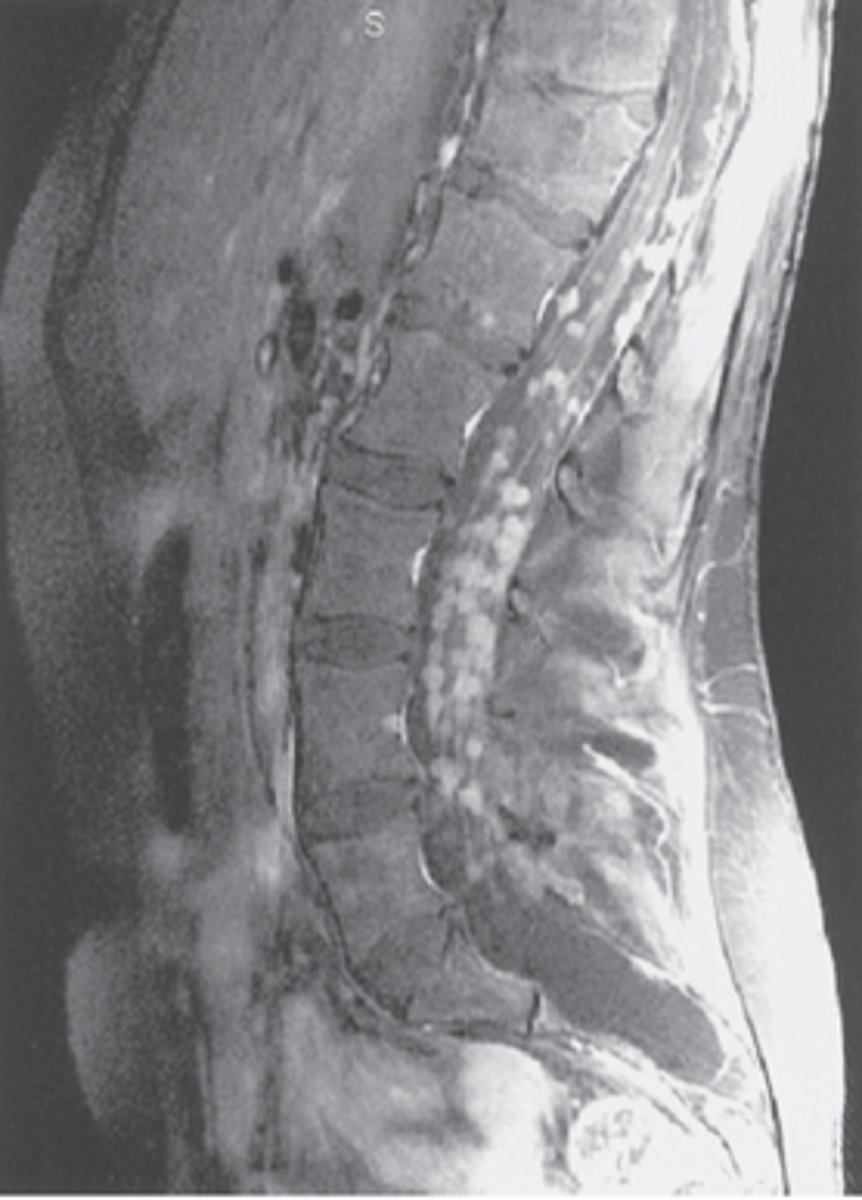
Answer
Metastatic Ewings Sarcoma
A thoracic myelogram in a 39-year-old man who presents with leg pain and weakness. A prior lumbar spine MR examination was normal.
What does this the AP view from a thoracic myelogram show?
A bony abnormality
An extradural mass
An intradural-extramedullary mass
An intramedullary mass
A really big disk herniation
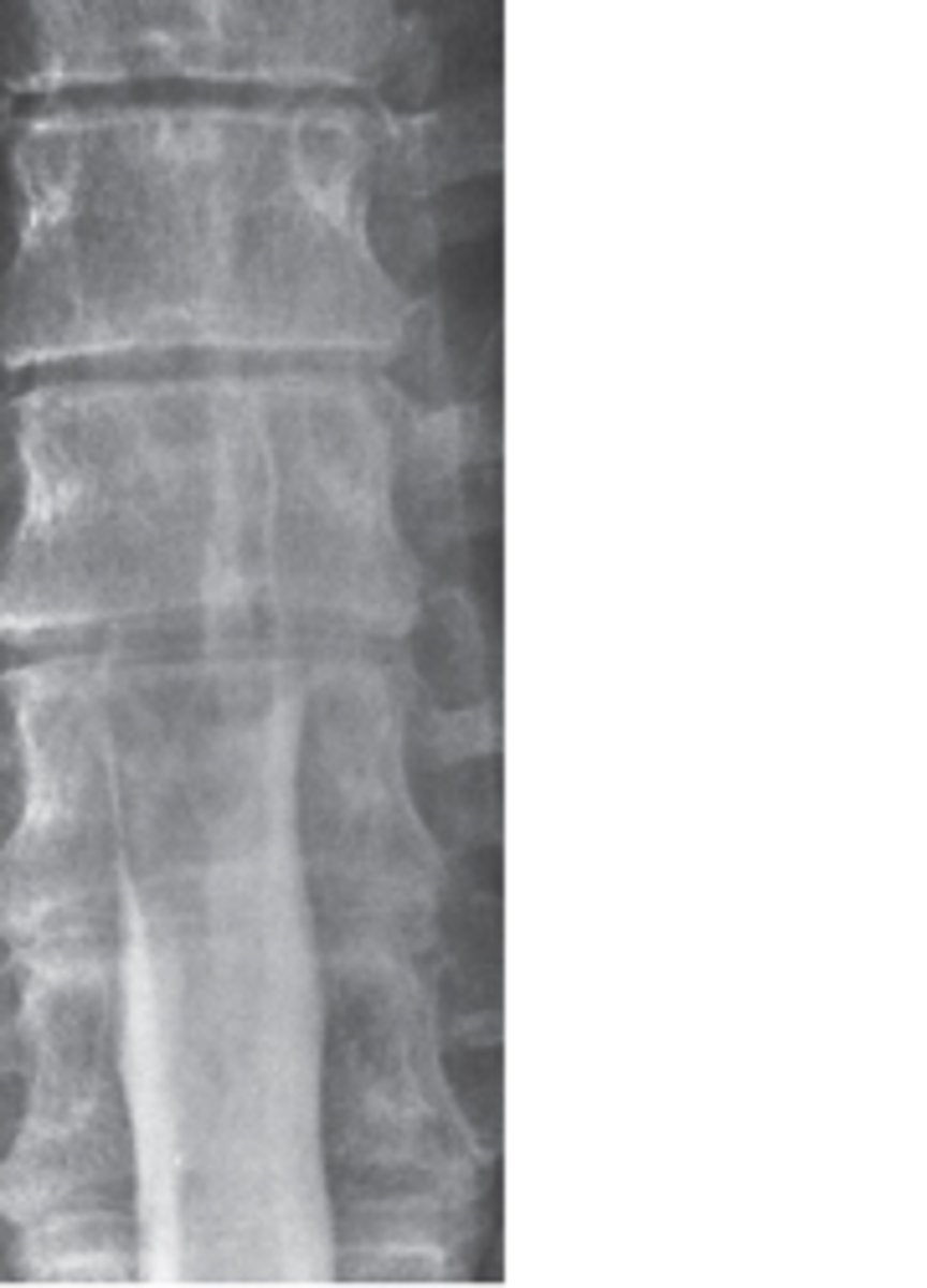
Answer
Intramedullary mass:
-It was a spinal cord astrocytoma but point being its literally in the spinal cord thats why theres a filling defect.
A 70-year-old woman presents with a 5-year history of back pain and recent onset of paresthesia in the groin and inner thighs (saddle distribution).
What is the most likely diagnosis?
Sacroiliitis
A sacral tumor
Constipation
Osteoporosis
Uterine malignancy
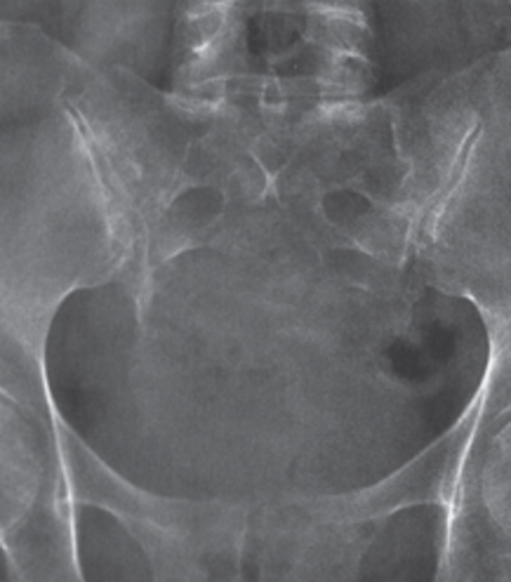
Answer
Sacral tumor:
-Chordoma which is a comon lesion of the sacrum causing those symptoms
A CT bone window in a 65-year-old man who presents with back pain.
What diagnostic possibilities should be most seriously considered?
Congenital or traumatic lesions
Metabolic or endocrine disease
Myeloma or metastatic disease
Infectious or inflammatory disease
Degenerative or inflammatory disease
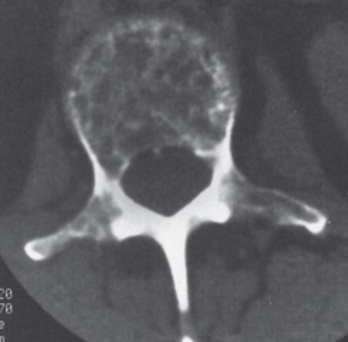
Answer
Myeloma or mets:
-Its lytic
What can MM cause in the vertebra?
Compression fractures
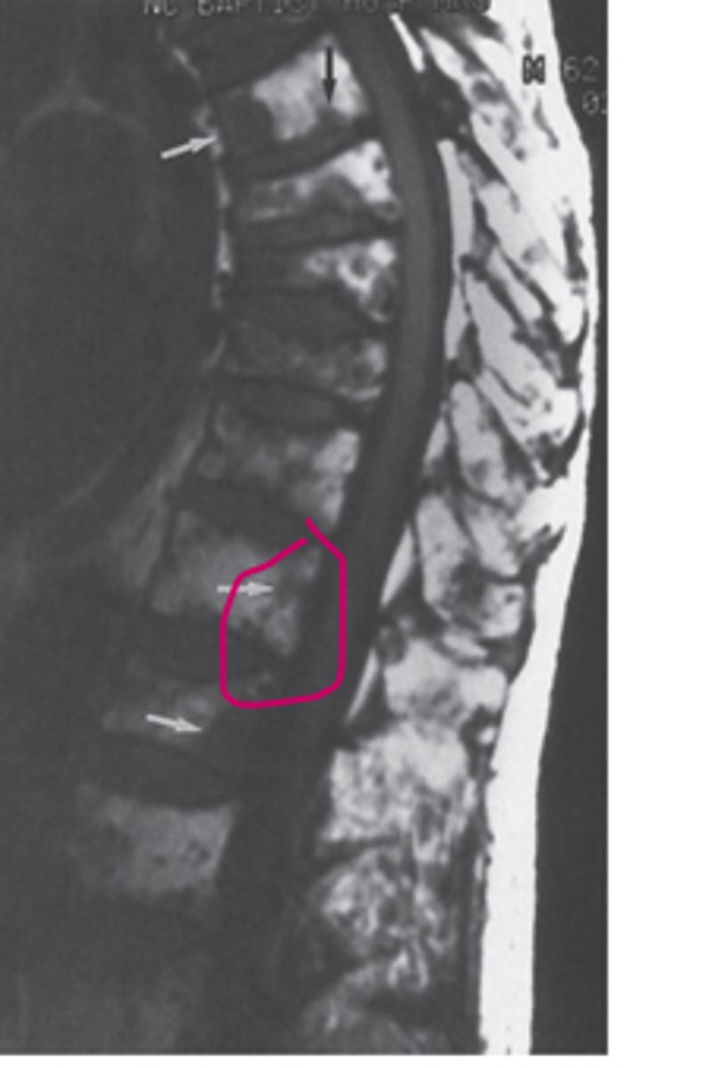
25 y/o with back pain chronically
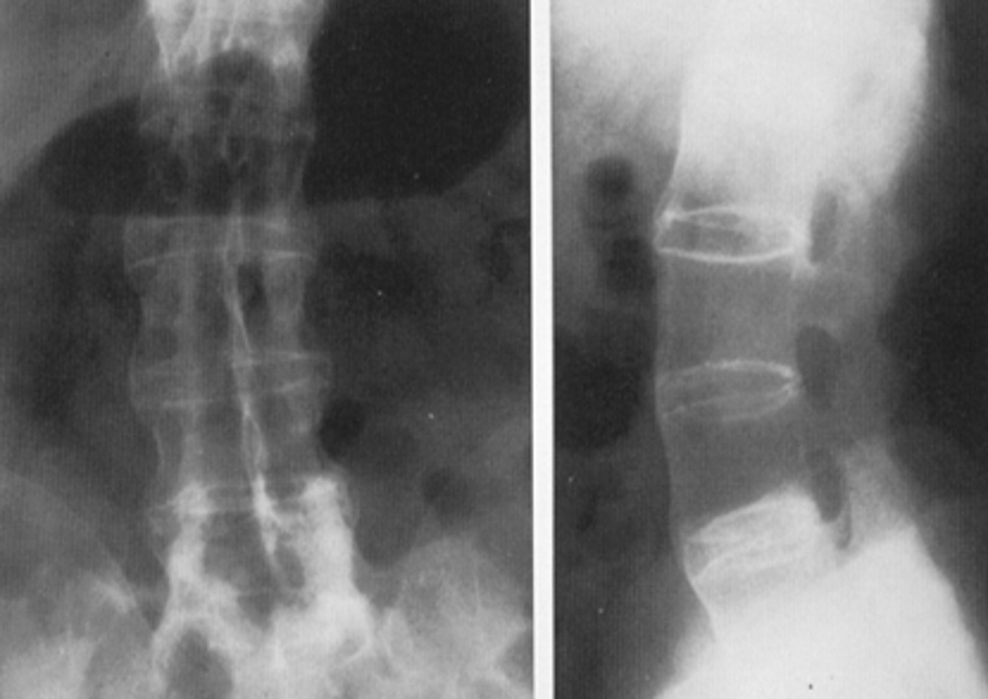
Answer
Ankylosing Spondylitis
Now for textbook stuff
yaaayyy
Decreased bone density
Osteopenia, osteolytic mets
Increased bone density
osteoblastic mets, pagets
What does the open mouth view allow you to look at?
Atlantoaxial articulation with C1 and C2 to look for possible trauma.
What passes through the cervical transverse foramen
Vertebral arteries
Myelography indications
Herniated nucleus pulposus, tumors, cysts, trauma
What level is the conus medullaris at?
L1-L2
What level is the cauda equina at?
L2-S5
Lesions can be what?
-Extradural
-Intradural but extramedullary
-Intramedullary
Indications for a CT of the spine
-Trauma
-Spine tumors
-Degenerative disk disease
Indications for an MRI of the spine
-Mainly anytime we think theres trauma to the spinal cord.
Imaging you should use in trauma
1. Start with the plain film.
2. THen do a CT
Red flags for back pain
- Saddle anesthesia
- Fecal or urinary incontinence
- Focal weakness
- Cancer history
- Recent invasive spinal procedure
- IV drug use
MRI is really good for what?
Herniated disk its the most sensitive
Where do osteophytes like to form?
Facet joints.
Where do nerve roots exit?
Cervical=above the corresponding vertebra
Thoracic and lumbar=below the corresponding vertebra
Then sacral are fused they don't have foramen
Two most common spinal cord tumor
Astrocytomas and ependymomas
Most common primary sacral malignancy
Chordoma
How do spinal infections typically result?
From hematogenous spread, usually in the lumbar spine, common culprit is staph aureus
Congrats now you're an expert
Yay