Lab Practical 3 Study
 Call Kai
 Learn
 Practice Test
 Spaced Repetition
 Match
 Flashcards
 Knowt Play
1/193

There's no tags or description
Looks like no tags are added yet.
194 Terms
What are the two major divisions of the cranium?
Neurocranium and viscerocranium
What is the function of the neurocranium?
It forms the bony case that encloses the brain and its membranous coverings
How many bones make up the adult cranium?
22 bones
How many bones make up the adult neurocranium?
8 bones: four unpaired bones and two paired sets (temporal and parietal)

What is the calvaria?
The dome-like roof or skull cap
What forms the cranial base?
The floor of the cranium
Which bones primarily form the calvaria?
Frontal, parietal, and occipital bones
Which bones contribute substantially to the cranial base?
Sphenoid and temporal bones
How many bones make up the viscerocranium?
15 irregular bones
Which three unpaired bones are included in the viscerocranium?
Mandible, ethmoid, and vomer
Which bones form the paired groups of the viscerocranium?
Maxillae, inferior nasal conchae, zygomatic, palatine, nasal, and lacrimal bones
Which bones house the teeth?
Maxilla and mandible
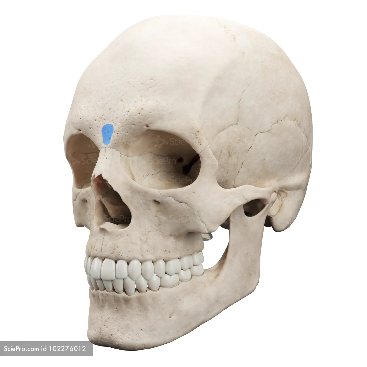
What is the glabella?
The smooth, slightly depressed area between the superciliary arches
What are the zygomatic bones commonly called?
Cheekbones

What is the piriform aperture?
The pear-shaped opening inferior to the nasal bones
What is the zygomatic arch formed by?
The temporal process of the zygomatic bone and the zygomatic process of the temporal bone
What four bones meet at the pterion?
Frontal, parietal, sphenoid, and temporal bones

What is the external acoustic meatus?
The opening that forms the entrance to the ear canal

Where is the mastoid process located?
On the temporal bone

What does the coronal suture separate?
The frontal and parietal bones
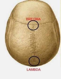
What is the bregma?
The landmark where the sagittal and coronal sutures intersect
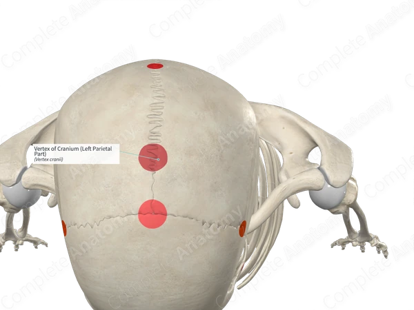
What is the vertex?
The most superior part of the calvaria, near the midpoint of the sagittal suture
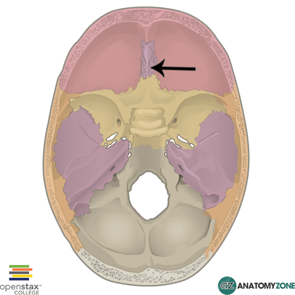
What passes through the cribriform plate?
Olfactory nerve fibers (CN I)

What passes through the optic canal?
Optic nerve (CN II) and ophthalmic artery
Which nerves pass through the superior orbital fissure?
CN III (Oculomotor), CN IV (Trochlear), CN V1 (Ophthalmic), and CN VI (Abducens)

Which division of the trigeminal nerve passes through the foramen rotundum?
Maxillary division (CN V2).

Which division of the trigeminal nerve passes through the foramen ovale?
Mandibular division (CN V3).

What important artery passes through the foramen spinosum?
Middle meningeal artery

Which cranial nerves pass through the jugular foramen?
CN IX, CN X, and CN XI

Which cranial nerve passes through the hypoglossal canal?
Hypoglossal nerve (CN XII)

What structure passes through the foramen magnum?
The medulla continues as the spinal cord; the opening also transmits associated vessels, meninges, and CN XI
What is a depressed calvarial fracture?
A fracture in which bone fragments are pushed inward, potentially compressing or injuring the brain
What is a contrecoup injury?
An injury in which a fracture occurs at the impact site and another occurs on the opposite side of the cranium
What is a craniotomy?
A procedure in which a bone flap is produced and elevated or removed to access the cranial cavity
What is a craniectomy?
Removal of a bone flap that is not replaced; a permanent plate may be used instead
What are the five layers of the scalp, remembered by SCALP?
Skin, Connective tissue, Aponeurosis, Loose areolar tissue, Pericranium
Which scalp layers move together as a unit?
The first three layers: skin, connective tissue, and aponeurosis.

What is the epicranial aponeurosis?
A broad, strong tendinous sheet covering the calvaria and connecting the scalp muscle bellies
What is the function of the loose areolar tissue layer of the scalp?
It allows free movement of the scalp and contains potential spaces that may fill with fluid
What is the pericranium?
Dense connective tissue forming the external periosteum of the calvaria
From which pharyngeal arch do facial expression muscles develop?
The 2nd pharyngeal arch
What is the main function of facial muscles of expression?
They move the skin to change facial expressions

What are the two bellies of the occipitofrontalis?
Frontal belly and occipital belly
What is the action of the frontal belly of occipitofrontalis?
Elevates the eyebrows, wrinkles the forehead, and protracts the scalp
What is the action of the occipital belly of occipitofrontalis?
Retracts the scalp

What is the action of orbicularis oculi?
Closes the eyelids; the palpebral part closes gently and the orbital part closes tightly

What is the action of corrugator supercilii?
Draws the eyebrow medially and inferiorly, creating vertical wrinkles above the nose

What is the action of orbicularis oris?
Closes the mouth and protrudes the lips

What is the action of the buccinator?
Compresses the cheek against the molar teeth and helps keep food between the teeth during chewing

What is the action of zygomaticus major?
Elevates the corner of the mouth during smiling and draws the angle of the mouth superiorly and posteriorly
What is the action of platysma?
Tenses the skin of the neck and assists in depressing the mandible
Which cranial nerve supplies the muscles of facial expression?
Facial Nerve (CN VII)
What are the five terminal motor branches of the facial nerve?
Temporal
Zygomatic
Buccal
Marginal Mandibular
Cervical
What are the three divisions of the trigeminal nerve?
Ophthalmic (V1), Maxillary (V2), and Mandibular (V3)
Which nerve provides the major arterial supply to the face?
Facial artery, a branch of the external carotid artery.
Which arteries supply the scalp from the external carotid artery?
Occipital, posterior auricular, and superficial temporal arteries.
Which arteries supply the scalp from the internal carotid artery?
Supratrochlear and supra-orbital arteries
Where does the facial vein drain?
Into the internal jugular vein
What is a cephalohematoma?
Bleeding between an infant's pericranium and calvaria, often after a difficult birth.
What is trigeminal neuralgia?
A sensory disorder of CN V causing sudden, severe, stabbing or lightning-like facial pain
What structures make up the eye?
The eyeball and optic nerve
What does the orbit contain?
The eyeball and its accessory visual structures
What structures are included in the orbital region?
The upper and lower eyelids and lacrimal apparatus overlying the orbit and eyeball
Which wall of the orbit is strongest and thickest?
The lateral wall
Which bones mainly form the orbital floor?
Maxilla, with contributions from the zygomatic and palatine bones

What is the palpebral conjunctiva?
The transparent mucous membrane lining the inner surface of the eyelids
What does the palpebral conjunctiva become when reflected onto the eyeball?
Bulbar conjunctiva

What does the lacrimal gland secrete?
Lacrimal fluid (tears), which contains the bactericidal enzyme lysozyme
What is the function of lacrimal fluid?
Moistens and lubricates the conjunctiva and cornea and supplies some nutrients and dissolved oxygen to the cornea
Where do the nasolacrimal ducts drain tears?
Into the inferior nasal meatus
What are the three layers of the eyeball?
Fibrous layer, vascular layer (uvea), and inner layer (retina)
What structures make up the fibrous layer?
Sclera and cornea
What structures make up the vascular layer (uvea)?
Choroid, ciliary body, and iris

What is the sclera?
The tough, opaque portion of the fibrous layer of the eyeball

What is the cornea?
The transparent anterior portion of the fibrous layer
Which nerve supplies sensation to the cornea?
Ophthalmic division of the trigeminal nerve (CN V1)
What are the functions of the iris and pupil?
The iris is a contractile diaphragm; its central opening, the pupil, allows light to enter
Which muscle constricts the pupil?
Sphincter pupillae
Which muscle dilates the pupil?
Dilator pupillae
What is miosis?
Pupillary constriction
What is mydriasis?
Pupillary dilation

What is the retina?
The sensory neural layer of the eyeball

What is the optic disc?
The point where optic nerve fibers and vessels enter the eyeball; it has no photoreceptors and is insensitive to light

What is the macula lutea?
The area of the retina lateral to the optic disc
Where is the area of most acute vision?
The fovea centralis, in the center of the macula
What is the function of rods?
They detect shapes and black-and-white vision
What is the function of cones?
They distinguish color
What are the extraocular muscles?
Levator palpebrae superioris; superior, inferior, medial, and lateral recti; superior and inferior obliques
Which cranial nerves control the extraocular muscles?
CN III, CN IV, and CN VI; CN II is the sensory nerve for vision.
Which cranial nerves are involved in the pupillary light reflex?
CN II (sensory input) and CN III (parasympathetic motor output)
What is the normal response in the corneal reflex?
Blinking when the cornea is lightly touched
Which cranial nerves are involved in the corneal reflex?
CN V1 provides the sensory limb and CN VII provides the motor response
What is a blowout fracture of the orbit?
An indirect injury that displaces an orbital wall, commonly involving the medial wall or orbital floor
What is exophthalmos?
Protrusion of the eyeball, which may result from orbital bleeding, pressure, or tumors
What is retinal detachment?
Separation associated with fluid collecting between the neural and pigmented layers of the retina
What is papilledema?
Swelling of the optic disc associated with increased cerebrospinal fluid pressure.
What is glaucoma associated with?
Reduced aqueous humor outflow, causing increased pressure in the anterior and posterior chambers
What are the classic signs of Horner's syndrome?
Ipsilateral miosis, ptosis, vasodilation with increased skin temperature, and anhidrosis
How many cervical vertebrae are there?
Seven (C1-C7)
What are C1 and C2 called?
Atlas (C1) and axis (C2)
