Microm 410 Midterm 1
 Call Kai
 Learn
Learn Practice Test
 Spaced Repetition
 Match
Match Flashcards
 Knowt Play
1/30

There's no tags or description
Looks like no tags are added yet.
Name | Mastery | Learn | Test | Matching | Spaced | Call with Kai | Chat |
|---|
No analytics yet
Send a link to your students to track their progress
31 Terms
Key differences between prokaryotes and eukaryotes?
Eukaryotes have:
nucleus
membrane bond organelles
cytoskeletal elements
much larger
Why was 16S rRNA used?
Found in all cells (18S in eukaryotes)
Performs same function in all organisms
Not subject to horizontal gene transfer
Highly conserved and highly variable regions
Bacteria & Archaea vs Eukaryotes
Genome structure and metabolic diversity
circular DNA vs linear
operons vs single genes
Archaea & Eukaryotes vs Bacteria
Transcription and translation
Bacteria & Eukaryotes vs Archaea
Membrane lipids
ester linked fatty acids vs ether linked isoprenoids
Pathogens infecting animals/plants
many vs none
What is an endosymbiont?
Microbes that live intracellularly within host organisms in often mutually beneficial association
What traits are associated with transition from endosymbiont to organelle?
Reduced genome size with significant gene transfer to host nucleus
Significant loss of metabolic versatility
High numbers of proteins imported from host
Evidence of symbiotic origins of mitochondria and plastids?
Multiply by binary fission
Bacterial ribosomes
Circular DNA genomes
Bacterial envelope structure
Organization of bacterial cytosol?
Genome (single, circular dsDNA) is condensed in nucleoid
High degree of organization and structure
Examples of bacterial sub-cellular structures that have specific functions?
Carboxysome: carbon fixation
Gas vesicle:
Inclusion bodies: storage (nutrients, molecules, proteins)
Magnetosome: makes magnetic particles for movement and direction
Phospholipid composition and effect on permeability?
No unsaturated fatty acids → lower permeability, more stable
Many unsaturated fatty acids → higher permeability, less stable
What fills the gaps in the phospholipid membrane?
Eukaryotes use sterols (cholesterol)
Prokaryotes use hopanoids
How do archaeal phospholipids differ?
L-glycerol, not D
1 phosphate linked, not 3
Branched tails that can cross link and forms rings (inhibits movement → more stability)
Ether linked, not ester linked
*can form monolayer instead of bilayer
Gram staining process
Heat fix cells → stain with crystal violet → add iodine (locks violet in G+) → destain with acetone-ethanol → counterstain with safranin
Make up and purpose of bacterial cell wall?
Made of linked polymers called peptidoglycan
Constrains osmotic expansion of plasma membrane
What is a peptidoglycan polymer sub-unit made of?
*What do some G+ bacteria do that G- don’t?
Peptidoglycan = Peptide + Glycan = D amino acid peptide + NAG,NAM
NAG and NAM disaccharides joined by B-1,4 linkage
Lactic acid group attached to NAM is attached to a peptide w/ D amino acids
Peptide cross-links glycan strands
*Some G+ have glycine bridge to cross-link peptides
Peptidoglycan biosynthesis steps?
Make subunit: add 5 amino acids to NAM → link NAG to NAM pentapeptide
Get across membrane: use lipid carrier bactoprenol to flip subunit into periplasm
Insert into existing PG: Periplasmic transglycosylases link disaccharide to existing glycan strand → transpeptidases (PBPs) cross-link pentapeptide
How are peptidoglycan polymers arranged?
Glycan strands have high tensile strength; peptides have high elasticity
Glycan around circumference; peptides length wise
How does expansion of the peptidoglycan sacculus happen?
Selective formation and then cleavage of peptide cross-links

Gram + cell envelope
Thick PG layer
Teichoic acids (alcohol sugars w/ amino acid/alcohol sugar side group): linked to NAM of PG
Lipoteichoic acids: link membrane lipids and PG

Gram - cell envelope
Outer membrane surrounding cell wall
inner leaflet made of phospholipids
outer leaflet made of LPS
lipid A w/ polysaccharide attached
initial portion are core sugars - same for all
majority is O-antigen - varies
Lipoproteins: connect cell wall to outer membrane
Thin PG layer
Periplasm: between inner and outer membrane containing enzymes and proteins

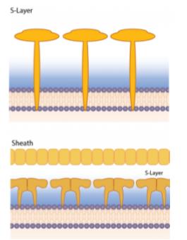
S layers composition and location
Both G± can have S layer
Self assembling array of proteins
G+: in PG G-: in LPS
Glycocalyx definition, forms, function?
Most prokaryotes have outermost external later called glycocalyx made of proteins and polysaccharides
Capsule: compact, discrete glycocalyx
Slime layer: less discrete, loose glycocalyx
Attachment/adherence, protection, essential for biofilms
Archaeal cell envelopes
Most have S layer, some have protein layer sheath
Methanogenic have pseudomurein/PG
NAT instead of NAM
B-1,3 linkage instead of B-1,4
No D amino acids
Bacterial wall elongation
PG synthesis resulting in cell elongation is mediated by scaffolding protein MreB
MreB polymers move in circumferential direction, require active PG synthesis

Bacterial septation and division
Septation: zonal PG synthesis
Scaffolding element is complete ring of FtsZ
FtsZ ring creates large complex of enzymes to form divisome
Inward growth of cell envelope from contraction of FtsZ ring

Polar aging
Cylindrical growth and division leads to aging poles; older pole = increased chance of lysis
Bacterial flagella
Peritrichous: flagella all over cell body
Flagellar filament made of flagellin spins for movement
Hook is flexible to allow filament repositioning
Basal body resembles Type III secretion system
Cap protein passes first and assembles hook and filament (grows from distal end)

Proton motive force
Creates energy to spin flagellum
Chemical and electrical gradient
Can be used for ATP synthesis, solute transport, motility
Archaellum vs flagellum
Basal body resembles Type IV pili
Pili are extended and retracted via polymerization for adherence and twitching motility
No hook
Filament assembled proximally
Rotation powered by ATP hydrolysis
Chemotaxis
Bacteria swim in straight lines (runs) and stop (tumbles) for reorientation
No ligand → CheA autophosphorylates → phosphorylates CheY → promotes tumbling
CheZ phosphatase dephosphorylates CheY
MCP binds ligand → CheA blocked → less CheY → no tumbling → longer runs