A&P1 Lab Exam 1
 Call Kai
 Learn
 Practice Test
 Spaced Repetition
 Match
 Flashcards
 Knowt Play
1/27

There's no tags or description
Looks like no tags are added yet.
28 Terms
Total Magnification
Ocular lens (10X) x Objective lens
Example: 10X x 40X (Obj.) = 400
Microscopy Steps
1. Start on 4x objective
2. Secure slide on stage
3. Use course adjustment knob
to raise stage
4. Use stage control knobs to
move specimen into view
5. Slowly lower the stage until
specimen is in focus
6. Fine tune focus with fine
adjustment knob
7. Move up to 10x objective lens
8. Fine tune focus with fine
adjustment knob
9. Move up to 40x objective lens
10. Fine tune focus with fine
adjustment knob
The life of a cell contains two divisions
Interphase and Cell Division
Cell Division
Includes mitosis and cytokinesis
Interphase
Includes G1,S, and G@
G1 phase
• Normal function
• Division preparation
o ↑ Organelles & cytoplasm
o Centriole replication begins
S phase
DNA replication
G2 phase
Last minute protein synthesis, organization & completion of centriole replication
Epithelial Tissue
Functions
• Protection
• Absorption
• Filtration
• Excretion
• Secretion
• Sensory reception
Characteristics
• Polarity
• Cellularity and specialized contacts
• Supported by connective tissue
• Avascularity
• Regeneration
Connective Tissue
Functions
• Protect other tissues
• Support other tissues
• Bind other tissues
Characteristics
• Rich supply of blood vessels (mostly)
• Composed of many cell types
• Extracellular matrix between cells
o Ground substance
-Interstitial fluid, adhesion proteins & proteoglycans
o Fibers
-Collagen, elastic &/or reticular
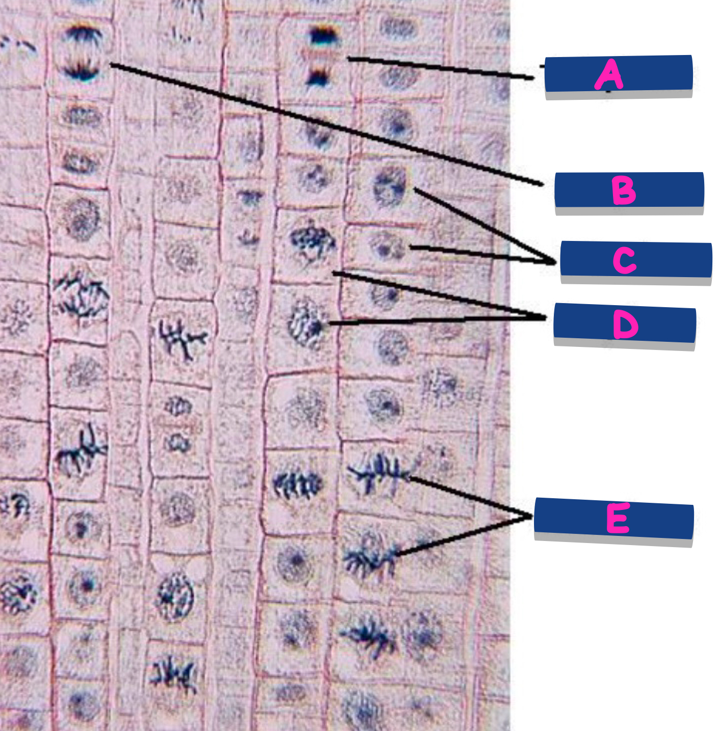
What phase is shown at A?
Telophase
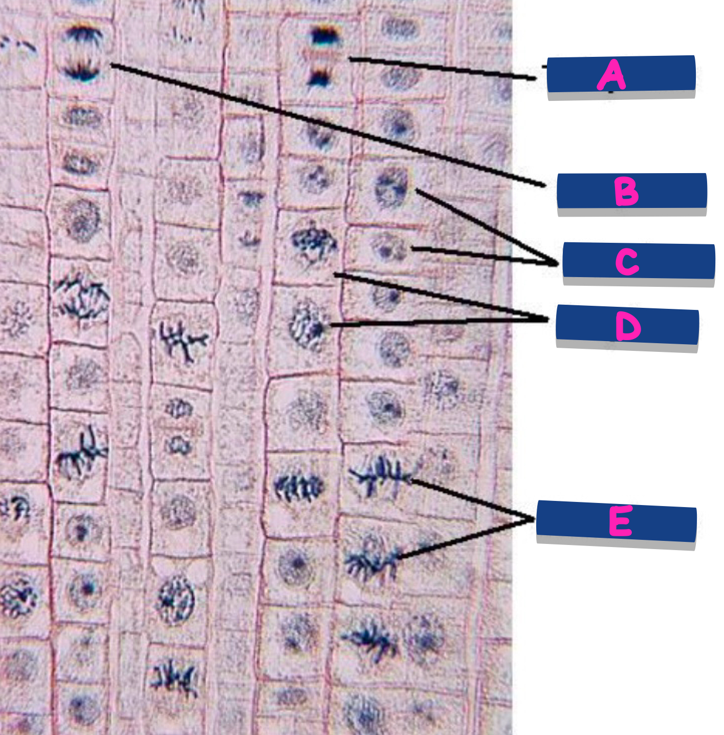
What phase is shown at B?
Anaphase
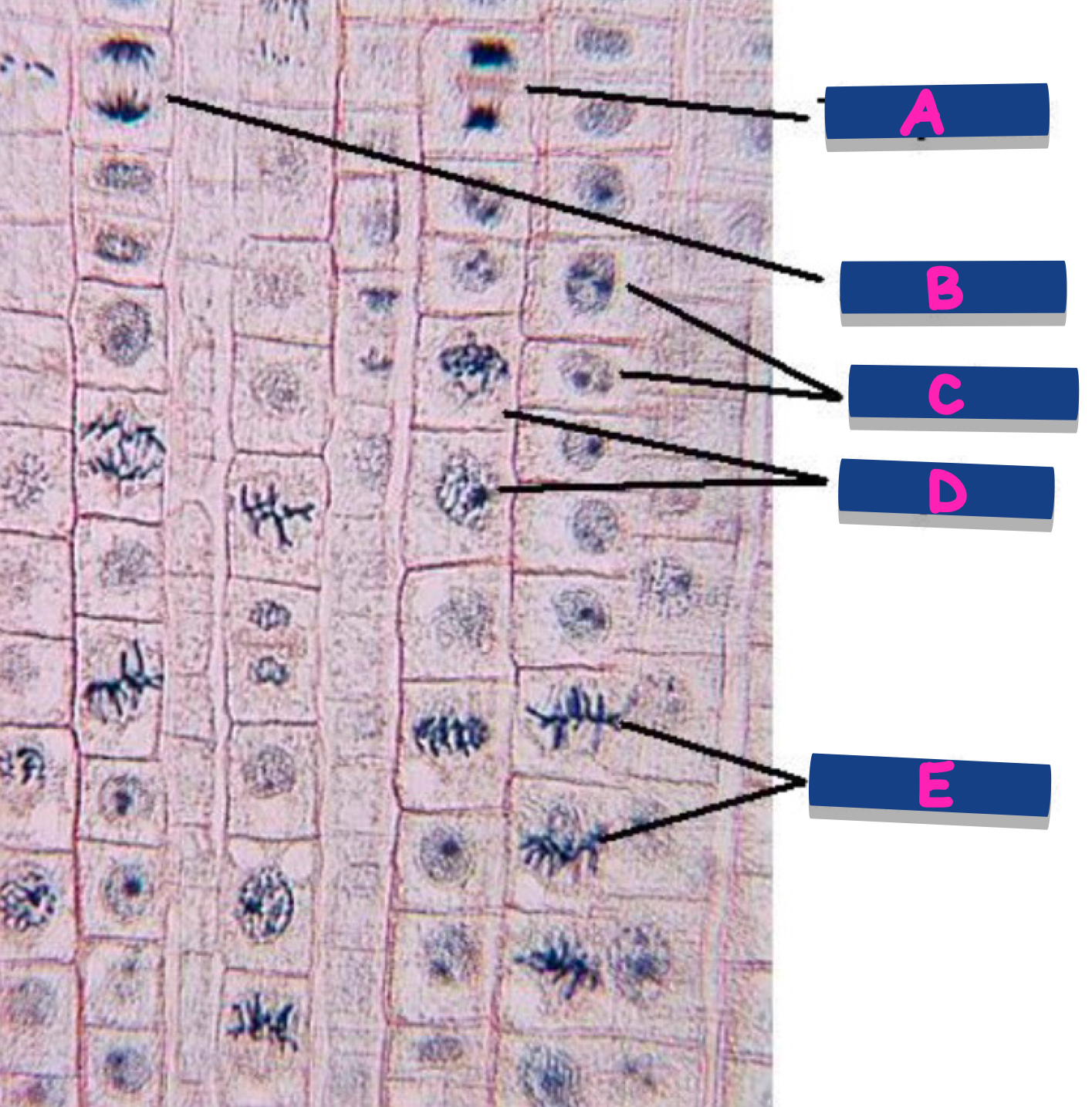
What phase is shown at C?
Interphase
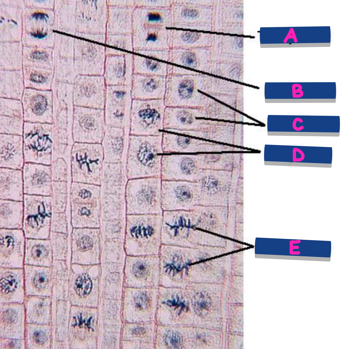
What phase is shown at D?
Prophase
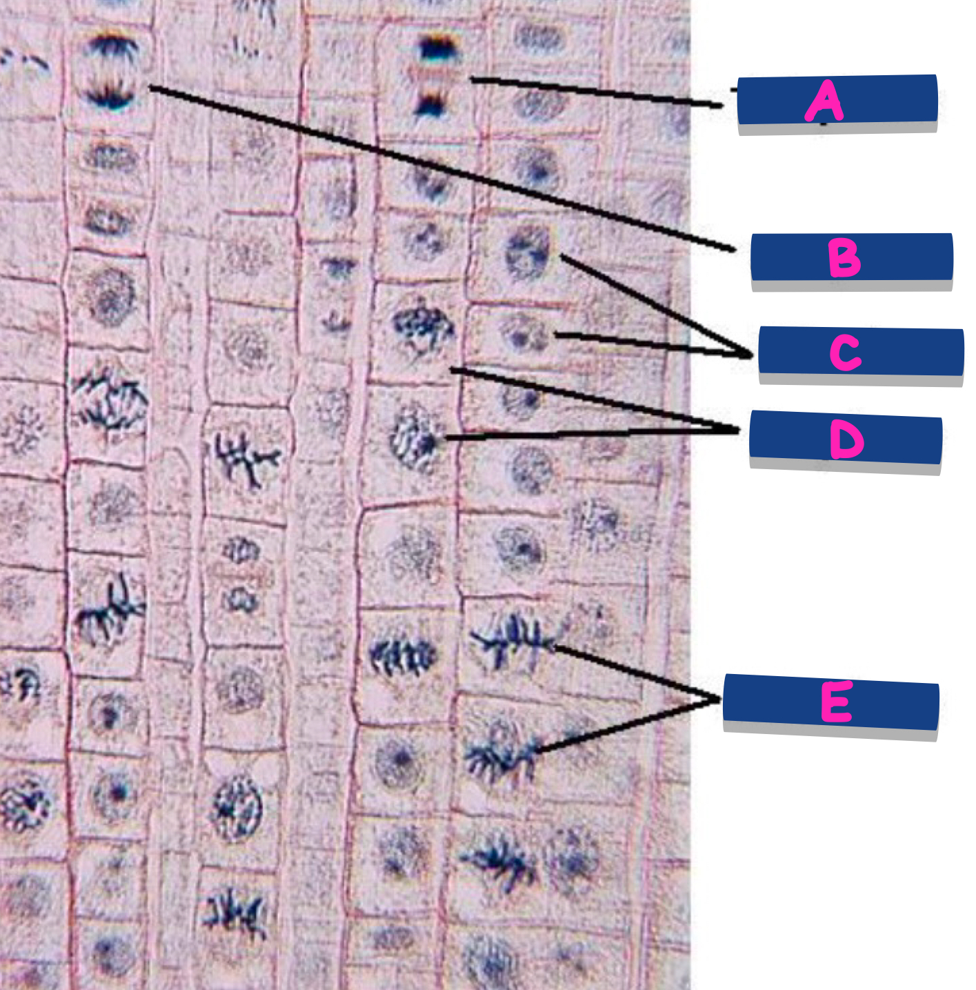
What phase is shown at E?
Metaphase
Simple Squamous
Structure: single layer of this, flat cells; the relatively flat single nucleus of each cell bulges at its center
Function: thinnest possible barrier to allow for rapid diffusion and filtration; secretion in serous membranes
Location: air sacs in lungs (alveoli); lining of lumen of blood vessels and lymph vessels (endothelium); serous membranes if body cavities (mesothelium), segments of kidney tubules
Simple Cuboidal
Structure: single layer of cells about as tall as they are wide; spherical and centrally located nucleus
Function: absorption and secretion; forms secretory tissue of most glands and small ducts
Location: lining of kidney tubules; thyroid gland follicles; surface of ovary; secretory regions and ducts of most exocrine glands
Simple Columnar
Structure: single layer of non ciliated cells taller than they are wide; oval-shaped nucleus oriented lengthwise in basal region of cell; apical surface of cell may have microvilli; may contain goblet cells
Function: absorption and secretion
Location: lining of most of gastrointestinal (digestive) tract (stomach, small intestine, and large intestine)
Ciliated Pseudo stratified Columnar
Structure: single layer of cells with varying heights; all cells connect to the basement membrane, but not all cells reach the apical surface; has goblet cells and cilia
Function: protection; secretion of mucin and movement of mucus along apical surface of epithelium by cilia
Location: lining of the larger airways of respiratory tract, including nasal cavity, part of pharynx, parts of larynx, trachea, and bronchi
Nonkeratinized Stratified Squamous
Structure: multiple cells layers, basal cells are cuboidal or polyhedral, whereas apical (superficial) cells are squamous; all cells are living; each cell contains a nucleus and organelles and lacks keratin; superficial cells kept moist
Function: protection of underlying tissue from abrasion
Location: lining of mouth, part of pharynx, parts of larynx, esophagus, lining of vagina and anus
Keratinized Stratified Squamous
Structure: multiple cells layers, basal cells are cuboidal or polyhedral and alive, whereas apical (superficial) cells are squamous, lack a nucleus and organelles, are filled with the protein keratin, and are dead
Function: protection of underlying tissue from abrasion
Location: epidermis of skin
areolor
Structure: scattered fibroblasts within an abundant viscous ground substance; few elastic and collagen fibers; many blood vessels
Function: protects tissue and organs; binds skin and some epithelia to deeper tissue; provided space for blood vessels and nerves
Location: papillary layer of the dermis (skin); subcutaneous layer (deep to skin); surrounds organs, nerve cells, some muscle cells, and component of blood vessel walls
adipose
Structure: closely packed adipocytes; nucleus pushed to edge of cells by large fat droplet; contains many blood vessels
Function: stores, energy; insulated, cushions, and protects
Location: subcutaneous layer; surrounds and covers some organs
dense regular
Structure: fibroblasts squeezed between densely packed, parallel arrays of collagen fibers; scarce ground substance; limited blood vessels
Function: attach bone to bone (most ligaments) as well as muscle to bone (tendon); resists stress applied in one direction
Location: tendons (attach muscle to bone); ligaments (typically attach bone to bone)
dense irregular
Structure: fibroblasts between densely packed but randomly arranged and clumped collagen fibers; more ground substance than in dense regular connective tissue; extensive blood vessels
Function: withstands stresses applied in all directions; durable
Location: reticular layer of the dermis (skin): epimysium covering skeletal muscle; epineurium covering nerves; periosteum covering bone; perichondrium covering cartilage; some organ capsules
hyaline cartilage
Structure: irregularly arranged chondrocytes in lacunae; glassy- appearing ground substance; collagen fibers are not visible
Function: provides support; forms most of fetal skeleton
Location: tip of nose; trachea; bronchi; most of larynx, costal cartilage; both the epiphyseal (growth) plates and articular ends of long bones; most of fetal skelton
Fibrocartilage
Structure: large chondrocytes in lacunae; numerous, parallel collagen fibers; limited ground substance
Function: weight- bearing cartilage that resists compression; acts as shock absorber in some joints
Location: intervertebral discs; pubis symphysis; menisci of knee joints
elastic cartilage
Structure: closely packed chondrocytes in lacunae; abundant elastic fibers that form weblike mesh; limited ground substance
Function: maintains shape while permitting extensive flexibility
Location: external ear; epiglottis of larynx