Brain Stem
 Call Kai
 Learn
 Practice Test
 Spaced Repetition
 Match
 Flashcards
 Knowt Play
1/37

There's no tags or description
Looks like no tags are added yet.
38 Terms
Medulla Function
sends sensory information from the body to the cerebellum (spinocerebellar tract [inferior cerebellar peduncle])
swallowing (CN 9, CN 10, CN 12)
regulation of cardiovascular function
respiratory and visceral function (CN10, CN 11)
head movement and position relative to gravity (CN 9 and CN 11)
Pons Function
processes motor info from cortex and sends it to cerebellum (pontocerebellar fibers and middle cerebellar peduncle)
CN 5 sensory processing; face, sinus, teeth
Motor control of chewing (CN 5), lateral eye movement (CN 6), and facial expression (CN 7)
Hearing and head position (CN 8)
Midbrain Function
eye movement (CN 3 and 4)
processing visual and auditory info (colliculi)
connects forebrain to hindbrain (vertical tracts)
connects cerebellum to cortex (cerebello-thalamo-cortico fibers [superior cerebellar peduncle])
Brainstem Surface Anatomy: Ventral
medulla
pyramids
decussation of pyramids
olive
inferior olivary nucleus
pons
transverse pontine fibers (pontocerebellar fibers)- go from nuclei in pons → across the pons→ headed into cerebellum
middle cerebellar peduncle
midbrain
mammillary bodies- memory, spatial navigation
crus cerebri, cerebral peduncle, basis pedunculi- carries big motor tracts that start in the cortex

Brainstem Surface Anatomy: Dorsal
Medulla
dorsal columns- carries sensory info to the brain
fasciculis gracilis- more medial, info from legs
fasciculis cuneatus- more lateral, info from arms
1st order sensory neurons
sensory tracts cross in the mid-rostral medulla (higher than the motor tracts on the ventral side)
possible to have a lesion that causes ipsilateral sensory and contralateral motor symptoms
nuclei gracilis and cuneatus
termination of 1st order sensory neurons in dorsal column
continuation of central canal (caudal 2/3)
4th ventricle begins in rostral medulla
lateral recess at pontomedullary junction and corresponds with widest point of 4th ventricle
Pons
rostral 2/3 of 4th ventricle posterior wall is dorsal surface of pons
cerebellar peduncles
superior and inferior make up lateral walls of 4th ventricle
Midbrain
superior colliculi- vision
inferior colliculi- auditory
trochlear nerve

Descending White Matter Tracts Originating in the Cortex 1
crus cerebri (cerebral peduncle)
corticobulbar (nuclear) tract- motor commands from motor cortex to CN motor nuclei on contralateral side; except facial CN nuclei are bilateral
corticospinal tract- biggest motor tract; motor commands from motor cortex to motor neurons in ventral horn of SC on contralateral side (80%)
other 20%- bilateral to motor neurons innervating trunk muscles; half of those also bilateral innervation to extremities
corticopontine fibers- cortex → pons; relaying motor commands to cerebellum on contralateral side via pontocerebellar fibers; redundant info because it informs the cerebellum what the motor command is
L cerebellum gets motor info from R cortex
crus cerebri (cerebral peduncle)

Descending White Matter Tracts Originating in Cortex 2
corticoreticular tract- motor tract that initiates in the BS; motor commands from cortex to pontomedullary BS to initiate corticoreticular tract to support balance, postural control and axial muscle activation
corticotectal tract- connects cortex to tectum in midbrain (superior colliculus) to direct eye gaze
Ascending White Matter Originating in the Cerebellum
cerebello-thalamo-cortical pathway- initiates in BS and terminates in cortex; feedback loop sensory info between cerebellum and cortex involved in movement coordination
both connections are contralateral cortex ←→ cerebellum
lesion between cortex and cerebellum- cerebellum symptoms are contralateral
CN Connection in BS

superior cerebellar peduncle
lateral border of pons; connects cerebellum to the midbrain
primary function- output of cerebellum (cerebello-thalamo-cortical fibers) to inform cortex of real time sensory info as movement in occurring
info going to thalamus and cortex
info crosses to contralateral cortex
Lesion = contralateral impairment of motor coordination (ataxia)
small input from body (spinocerebellar tract) supports real-time bilateral coordination of gait
proprioceptive input from LE
primarily info from ipsilateral body with a bit of contralateral
takes the scenic route: crosses sides to get info about the other side of the brain and crosses back aging though the SCP as it goes into the midbrain
Lesion = impairment of bilateral coordination of LE in gait
middle cerebellar peduncle
located on the pons
primary function- to inform cerebellum on motor commands being sent to muscles
info from cortex → corticopontine tracts → pontine nuclei → pontocerebellar fibers cross → enter contralateral cerebellum
Lesion = contralateral impairment in motor coordination
inferior cerebellar peduncles
located at pontomedullary junction
primary function= input to cerebellum from peripheral sensory receptors (spinocerebellar tract) to inform cerebellum on real-time sensory circumstances during the ongoing movement
info comes from body to spinal cord (peripheral nerve → nerve root → spinal nerve)
at SC, synapse (clarke nucleus or external caudate nucleus)
at SC synapse, spinocerebllar fibers begin and take info to ipsilateral cerebellum
Lesion = ipsilateral impairment in motor coordination
Important Nuclei: Medulla
cuneate and gracilis- sensory info synapses → tracts cross towards the top of the medulla
when fibers cross they come anterior and the head back posteriorly
one exception to motor anterior and sensory posterior rule
olives- on the anterolateral side; motor

Important Nuclei: Pons
pontine nuclei- connect corticopontine to pontocerebellar fibers

Important Nuclei: Midbrain
periaqueductal gray- area around central canal; pain perception
substantia nigra- dopamine production → targets basal ganglia motor
red nucleus- initiation of rubrospinal tract
superior colliculus- vision
inferior colliculus- audition
mammillary bodies- memory
ventral tegmental area (VTA)- dopamine production → targets limbic structures
pedunculopontine nucleus (PPN)- automatic locomotion and arousal
ACh, glutamatergic, GABA
excites arousal system- controls sleep wake cycles
inhibits motor circuits

Reticular Formation
matrix of neurons that extend through the length of BS
long and short axons
origination of reticulospinal tracts
ascending reticular activating system
production and release of slow acting NTs/neuromodulators
influence activity in cortex, cerebellum, BS, and SC

Reticular Formation: VTA
ventral tegmental area (VTA) in midbrain
dopamine
cerebrum (motivation and decision making)

Reticular Formation: PPN
Pedunuclopontine nucleus (PPN) in midbrain
Acetylcholine for arousal function
globus pallidum, subthalamic nucleus, emotion system, Ret SPT (movement)
target of deep brain stimulation in parkinson’s disease

Reticular Formation: Raphe nuclei
in midbrain, pons, and medulla
seretonin
thalamus, hypothalamus, BG, amygdala, hippocampus, cerebellum (modulates sensory, motor, and autonomic activity)

Reticular Formation: Locus Coeruleus
located in pons
norepinephrine
cerebellum, diencephalon, cortex, medulla, and lateral horn of SC (attention, modulating pain signals in STT, CV, respiration)

Ascending Reticular Activating System (ARAS)
consciousness and awareness
alertness, sleep, attention
nuclei project to intralaminar nuclei of thalamus
thalamus projects throughout cerebral cortex

Descending Tracts 1
passing through the BS:
corticospinal tract (CST) from cortex → corona radiata → internal capsule → crus cerebri (cerebral peduncle) → pons → pyramids in medulla → cross at caudal medulla at pyramidal decussation → on its way to motor neurons in ventral horn in SC
terminating in BS:
corticobulbar- same path as CST until level of target CN motor nucleus where they cross to go to target
corticopontine- same path as CST to pontine nuclei
corticoreticular- same path as CST to pontomedullary reticular formation
corticotectal- visual cortex and FEF → internal capsule → superior colliculus
Descending Tracts: corticobulbar tracts
UMNs of CNs originate from precentral gyrus (primary motor cx)
neurons descend as the CBT down to midbrain
synapse as
CN 3- oculomotor nucleus
CN 4- trochlear nucleus
CN 6- abducens nucleus
→ cerebral peduncles → midbrain → pons
synapse as
CN 5- motor trigeminal nucleus
CN 7- facial nucleus
pons → medulla
synapse as
CN 9- Glossopharyngeal
CN 10- vagus
CN 11- accessory
CN 12- hypoglossal
neurons to lower face have contralateral control, upper half have bilateral conrol

Descending Tracts: motor tracts originating in BS, Rubrospinal
arises from red nucleus in midbrain → crosses immediately in midbrain → influences large muscle and fine movements primarily of UE
Descending Tracts: motor tracts originating in BS, Reticulospinal
arises from reticular nuclei
medullary (lateral)- crosses in medulla, influences limb flexion, supports rudimentary gait
pontine (medial)- uncrossed from pons- does receive input from bilateral cortex
biases extensors
Descending Tracts: motor tracts originating in BS, Vestibulospinal
arises from medial and lateral vestibular nuclei
lateral- uncrossed, influences proximal limb musculature
medial- crossed and uncrossed, influences neck and axial muscles
Descending Tracts: motor tracts originating in BS, RAS
RAS tracts- target SC interneurons from RAS
raphespinal- raphe nucleus in throughout the BS releases seretonin
ceruleospinal locus coeruleus in pons releases norepinephrine
Tracts within the BS
pontocerebellar tract- pontine nuclei to contralateral cerebellum
medial longitudinal fasciculus- 2-way superhighway; linking CN nuclei for eue movement with vestibular nucleus
Ascending Tracts: initiating from cerebellum
cerebello-thalamo-cortico feedback look- updating cortex on real time sensory circumstances
initiates in deep nuclei of cerebellum → travels out of cerebellum in sup. cerebellar peduncle → crosses to thalamus and cortex
Ascending Tracts: initiating from SC
dorsal columns/medial lemniscus
proprioception and discriminative touch and body
comes into SC via sensory spinal nerve
synapses in dorsal horn of SC and ascends as dorsal column
ascends to medulla and decussates in mid to rostral medulla at nucleus gracilis (leg) and cuneatus (arm) and continues ascending to thalamus as medial lemniscus
called the medial meniscus after it crosses and makes its final ascent to thalamus
spinal thalamic tract (STT)
pain, temp., gross touch from body
comes into SC via sensory spinal nerve
enters edge of dorsal horn → travels up 2 spinal segments and crosses to contralateral side of SC
ascends in SC and through BS as the STT (part of anterolateral system) to thalamus
only one that crosses before it gets to the brainstem


BS vasculature
Midbrain- Basilar
posterior cerebral a. (PCA)
superior cerebellar a. (SCA)
Pons
anterior inferior cerebellar a. (AICA)
pontine branches of basilar
Medulla
posterior inferior cerebellar a. (PICA)
caudal medulla at SC/BS junction (posterior spinal artery)
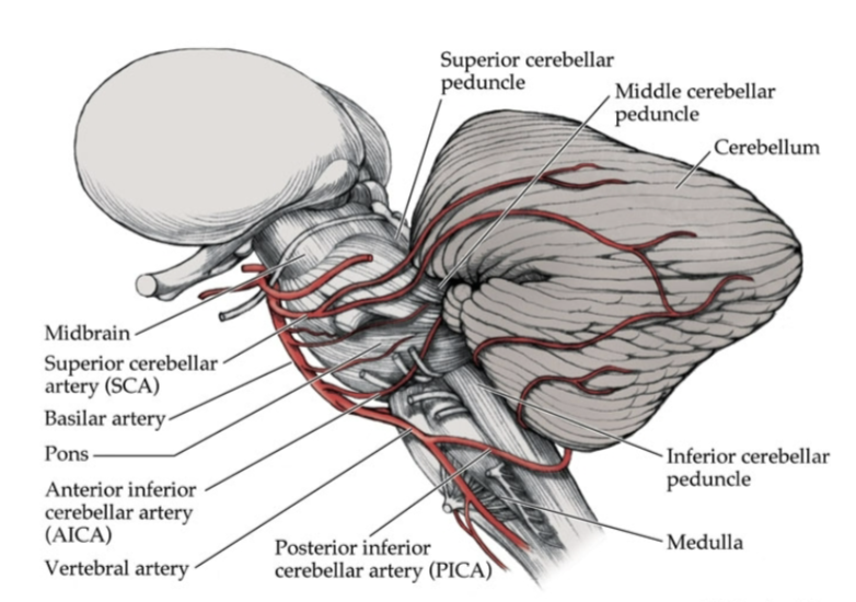
Common BS Strokes- PICA
PICA- lateral medulla (wallenberg’s syndrome or lateral medullary syndrome)
structures:
CN 8, 9, 10 12
spinocerebllar tract
lateral cuneate nucleus
inf. cerebellar peduncles
spinal tract and nucleus of trigeminal
spinothalamic tract
descending sympathetic fibers

Common BS Strokes- AICA
lateral inferior pons
structures: CN 7 and 8
descending sympathetic tract
spinal tract and nucleus of trigeminal nerve
spinocerebellar tract
inf. cerebellar peduncle
spinothalamic tract
Common BS Strokes- Basilar a.
anteromedial midbrain
structures:
CN 3
trigemeinal lemniscus
cerebellothalamic axons
red nucleus
corticopontine tract
Common BS Strokes- PCA at midbrain
structures:
CN 3
CST
CBT
substantia nigra
edinger-westphal nucleus
horner’s syndrome
damage to descending sympathetic tract in BS
ipsilateral symptoms
drooping eye lid
constricted pupil
skin vasodilation
absence of sweating
locked-in syndrome
pons level total occlusion of basilar a. system
paralysis of all muscles except eye
conscious
cognition- can communicate through eye blink