LS plexus, LL arteries, ant-med thigh, hip muscles
 Call Kai
 Learn
Learn Practice Test
 Spaced Repetition
 Match
Match Flashcards
 Knowt Play
1/26

There's no tags or description
Looks like no tags are added yet.
Name | Mastery | Learn | Test | Matching | Spaced | Call with Kai | Chat |
|---|
No analytics yet
Send a link to your students to track their progress
27 Terms
nerve roots + region/action for compartments of lower limb
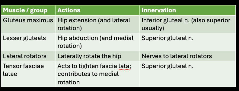
*table summary of drawing + actions

gluteal region action + innervation
arteries of lower limb
-external iliac → inguinal l → femoral (ant thight) → deep femoral (post thigh) + popliteal a (adductor hiatus, in adductor magnus)
-internal iliac → obturator int (med thigh)
-popliteal → tibial → ant (ant leg) and post (post leg) tibial a
-post tibial a → fibular a (lat post leg and lat leg, though not actually in lateral compartment?)

foot arteries
Dorsalis pedis
-Distal to extensor retinaculum
-Terminal branch of anterior tibial a.
Dorsal pedis → Arcuate a.
-Gives off dorsal metatarsal aa.
Plantar
-post tibial → lat and medial plantar arteries

table: blood supply + innervation lower limb


identification: ant thigh superficial


identification: ant thigh deep

Iliopsoas parts
-combine past inguinal ligament
-action=flexion of hip and trunk (depends on position/other muscles activated/if fixed on ground)
*Psoas major: last TV and all LV
*Ilioposas: postural muscle

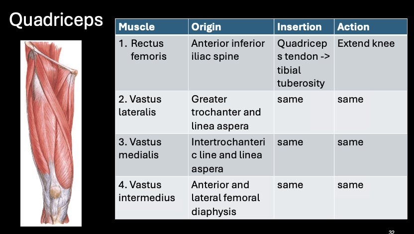
quadriceps muscel tabel
“knee cap”
platella
-largest sesamoid bone in body=lives in a tendon
-”patellar ligament” below bc bone → bone


identificatin: medial thigh muscles superficial


identification: medial thigh muscles deep

pes anserinus
-multiple tendons insert; “foot of goose”
-unique in that the 3 muscles of 3 diffferent: compartments, actions, and innervations
Sartorius: femoral n; adduct and LR
Gracilis: obt n; adduct
ST: tibial n; ext hip and flex knee

adductor brevis and obturator nerve

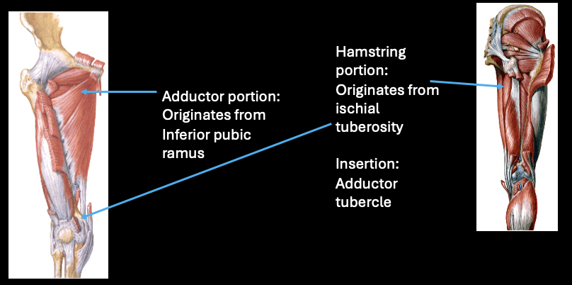
adductor magnus: 2 parts
-Adductor portion: Originates from Inferior pubic ramus; Insertion=Linea aspera; obturator n
-Hamstring portion: Originates from ischial tuberosity; Insertion= Adductor tubercle; tibial n
forms adductor hiatus (where femoral a → popliteal a)
adductor hiatus
femoral a → popliteal a

femoral triangle
-3 Boundaries: Inguinal l., Sartorius, Adductor longus
*can get a femoral hernia here; IL is most inf part of abdominal wall
Contents from lateral to medial: “NAVL”
1. Femoral n. (*broom apperance)
2. Femoral a.
3. Femoral v.
4. Lymph nodes (superficial inguinal)
deep femoral artery
-femoral (ant thigh) → deep femoral (post thigh)
-deep femoral → lateral and medial (loop around proximal femur) circumflex femoral a


muscle identification
1.Gluteus maximus
2.Gluteus medius
3.Piriformis
4.Gluteus minimus
5.Superior gemellus
6.Obturator internus (tendon)
7.Inferior gemellus
8.Quadratus femoris

muscle identification
1.Iliopsoas
2.Sartorius
3.Tensor fasciae latae
4.Rectus femoris
5.Vastus medialis
6.Vastus lateralis
7.Pectineus
8.Adductor longus
9.Gracilis
10.Vastus intermedius
11.Gluteus medius
12.Adductor brevis
13.Adductor magnus
14.Obturator externus

muscle identifcation
1.Semitendinosus
2.Semimembranosus
3.Biceps femoris long head
4.Vastus lateralis
5.Adductor magnus
6.Biceps femoris short head
Hip
-2 bony components
-type of joint
-actions
-Head of femur + Acetabulum of os coxa (innominate)
-ball and socket
-Flexion; Extension; Abduction; Adduction; Medial Rotation; Lateral Rotation; Circumduction (A combination of the above)
muscle table: hip flexion

muscle table: hip extension

muscle table: hip abduction and adduction

muscle table: hip MR

muscle table: hip LR
