Biliary system
 Call Kai
 Learn
 Practice Test
 Spaced Repetition
 Match
 Flashcards
 Knowt Play
1/141

There's no tags or description
Looks like no tags are added yet.
142 Terms
BILIARY SYSTEM consists of and contributes to ..
•Consists of gallbladder and ducts that drain the liver of bile
•Contributes to digestive process along with liver and pancreas
Biliary duct function
allow bile to flow to gallbladder (tansports bile)
Gallbladder functions
acts as reservoir for BILE (stores and concentrates bile)
GB location
mostly intraperitoneal and situated on the posterior surface of the liver
Cystic artery arises from and supplies
arises from the RT hepatic artery & supplies the GB and hepatic ducts
Function of the Biliary System
•Hepatocytes make bile
•Drain the liver of bile
•Store the bile until it is needed to aid digestion
Biliary System Anatomy
Intimately associated with the liver and pancreas
Consists of:
• Gallbladder
• Intra and Extra hepatic bile ducts
Biliary Tree Anatomy
•Common hepatic duct joins the cystic duct to form the common bile duct
•Common bile duct empties into the duodenum
Indication for Exam (WO)
•Pain
•Right upper quadrant (RUQ) pain
•Right shoulder pain / scapula (with inflammation)
•Positive Murphy’s sign
•Nausea
•Vomiting
•Intolerance to fatty foods and dairy products
•Jaundice or abnormal liver function tests (LFTs)
•Loss of appetite
Patient preparation
NPO 6-8 hrs
Transducer selection
Curved, linear phased, higher frequency when possible
Patient positioning
supine, LDEC, RDEC to use liver as a “backdrop”. Can appreciate small stones and shadows which sometimes are not appreciated due to bowel gas
Image optimization
DEPTH, SECTION WIDTH and FOCAL ZONE!!!!! harmonics, compression, intercostal, anterior artifact is often present due to reverb from abdominal wall and adipose. Do not clean up too much as you may miss sludge.
Examination protocol
probe at inferior rt liver at costal margin
Scan superior to inferior and side to side
look carefully in the neck for small stones
Examination limitations
•Bowel gas
•Artifact (reverberation)
•Pt unable to hold breath
•Pt ate
•NPO 6-8hrs
(suspected Cholecystitis ? – do it)
•Obesity
gallbladder wall should not be more than …
2-3mm in thickness (3mm is considered abnormal)
Normal measurements
k

Associated Tests
•Oral cholecystogram
•Nuclear medicine (HIDA scan)
•Computed axial tomography (CT scan)
•Cholangiography
•Endoscopic retrograde cholangiopancreatography (ERCP)
•Hepatobiliary scintigraphy
•Endoscopic ultrasonography (EUS)
Gallbladder Anatomy
•Variety of shapes
• Neck is superior, fundus is inferior
Where is bile made and how does it get to the hapetocytes ? (WO)
Bile is produced by liver (HEPATOCYTES) and carried to the gastrointestinal system by the biliary tract
Sphincter of Oddi, in the duodenum, regulates passage of bile into the duodenum, and at the same time prevents reflux of gastrointestinal fluids into the biliary system.
When closed, the sphincter of Oddi forces the gallbladder to fill with bile
When fats and amino acids are ingested, the duodenal mucosa releases cholecystokinin (CCK)
Where is bile more concentrated ?
Bile in the gallbladder is much more concentrated than hepatic bile
What is bile composed of ?
mostly water (82%) and bile acids (12%)
Remaining constituents include cholesterol, bilirubin (bile pigment), proteins, electrolytes, and mucus
What supplies the GB?
Cystic artery supplies GB
What supplies the gallbladder and liver ?
Proper/ Right hepatic artery supplies the gallbladder and liver
Where does the cystic artery originate ?
may originate directly from the common hepatic artery or, rarely, from the gastroduodenal artery
Venous drainage of the gallbladder is by way of the
hepatic portal system
What is the only O2 supply to biliary system?
CYSTIC ARTERY IS ONLY O2 SUPPLY TO BILIARY SYSTEM.
•GB wall layers
Serosal: outer most layer, connective tissue
Subserosa Muscularis: middle layer, muscular
Mucosa: innermost layer, epithelial cells with folds
Rokitansky-Aschoff sinuses
Rugae or pocket-like invaginations of the epithelium into the muscularis
GB Wall 3 or more mm…
•Rokitansky-Aschoff sinuses rugae
Adenomyomatosis
Wall thickness measurement
Wall should be measured on the anterior border when the wall is perpendicular to the transducer.
GB Location
• Intraperitoneal cavity
• Anterior*
• Superior & Anterior to RK
• Relationship to liver & gut
bile reservoir
on posteroinferior surface of liver
Divided into:
•Fundus
•Body
•Neck
•1. FUNDUS -
1. Is the bottom of the pouch (inferior)
2. Most distal and inferolateral portion of GB
3. Projects below the inferior margin of liver
4. Close to hepatic flexure of colon & duodenum
5. Lateral to duodenum & pancreatic head
5. Can be palpated if GB is inflamed or enlarged
•2. BODY -
1. Portion between neck & fundus
2. Is in contact with the visceral surface of liver
•3. NECK -
1. Narrowing area leading to the cystic duct
2. Is continuous with cystic duct
3. Orientated posteromedially towards the porta hepatis
Laboratory Values
•White Blood Count (WBC)
•Serum bilirubin
•Serum alkaline phosphatase (ALP)
•Liver Function Tests (LFTs)
•1) Aspartate aminotransferase (AST)
•2) Alanine aminotransferase (ALT)
•3) Prothrombin time (PT)
GB protocols
•AIUM & ACR GUIDELINES
Sonography of Gallbladder
•Anechoic lumen with thin smooth walls
•Sonolucent oblong ANTERIOR structure located anterior to RK, lateral to head of panc & duodenum.
•Fundus is slightly more anterior. This is appreciated on sagittal scans.
•Main lobar fissure can be used to identify the GB. It is located between the GB and RPV. GB is inferior to MLF.
•Hyperehoic wall <3mm
What is palpatation of GB used to evaluate for ?
Murphy’s Sign
Anterior Reverberation Artifact Versus Sludge
Scanning intercostal can eliminate this artifact, scan inferior to see fundus well.
use a higher frequency probe to inspect the fundus, also helping to eliminate the artifact.
The fundus is ….
ANTERIOR and INFERIOR
Gallbladder Fossa
Indentation located on the posteroinferior portion of the right lobe of the liver where the gallbladder is situated
Main Lobar Fissure
•Thin hyperechoic line between the portal vein and gallbladder
•Landmark for locating the gallbladder and gallbladder fossa
HARTMANN’S POUCH
out pouching, forms when the neck of GB folds on itself
PHRYGIAN CAP
forms when the fundus of the GB bends/folds on itself.
JUNCTIONAL FOLD
forms when the GB folds on itself in the region where the neck & the body meet
DUPLICATION DOUBLE GB
rare (1/ 4000), hard to diagnose on US.
Appears as two parallel cystic structures in the GB fossa
SEPTATED
septations seen within GB
other normal varients of the GB
Rare agenesis, hypoplasia (underdeveloped), opening directly into duodenum, left sided with situs inversus
Common Normal Variants
•Phrygian cap at fundus
•Junctional Fold
•Hartmann's pouch

1) junctional fold
2) phrygian cap
BILE DUCTS
•Transports bile: liver to GB & GB to duodenum
•Surrounded by a CT sheath, which gives hyperechoic appearance
RIGHT & LEFT BILE DUCTS
completely enclosed by the liver and considered INTRAHEPATIC
COMMON BILE DUCT, CYSTIC DUCT & PART OF COMMON HEPATIC DUCT
are not enclosed by the liver and considered EXTRAHEPATIC
Bile Ducts sonography
avascular, anechoic lumens which vary in size
Intrahepatic ducts are normally not seen.
Bright/Hyperechoic walls
Intrahepatic Right and Left Ducts sonography
• Normally not seen
• <2mm or <40% of PV
• Adjacent to PV (linear structures)
Which structure is not routinely seen on US ?
Cystic duct is not routinely seen.
What structures are routinely seen on US ?
CHD & CBD are routinely seen
The common hepatic duct is the union of….
Rt and Lt (intra and extra) hepatic ducts
Cystic Duct
Joins the CHD
Extrahepatic
Bi-directional bile flow

spiral valves of heister
Common Bile Duct
Union of CHD & CD
Extrahepatic
Hepatoduodenal ligament

common bile duct
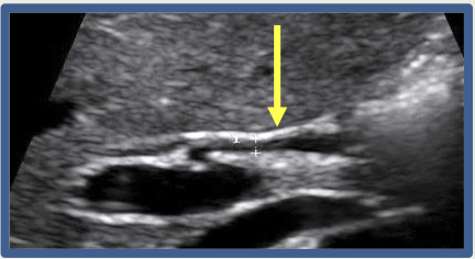
common bile duct
Portal triad
CBD forms Portal Triad along with portal vein & hepatic artery.
MICKEYS SIGN:
PV=face, HA=Lt ear, CBD=Rt ear

distal CBD
measuring CBD
follow any dilatation to the obstruction

distal CBD
Right and left INTRA hepatic ducts diameter
0.1 - 0.2 mm in diameter
Common bile duct measurement
5-15cm in length
0.1-0.7 cm in diameter
Intrahepatic ducts
•Formed from canaliculi, which drain into the right & left hepatic ducts
•2mm or less
•Should not be more than 40% of the diameter of the adjacent portal vein.
•not normally seen
Rt & Lt hepatic ducts
drain bile from rt & lt lobes and bring bile to GB through cystic duct
run parallel & anterior to Rt & Lt Portal Vein
The Common Hepatic Duct is the union of
Rt & Lt intrahepatic bile ducts
Common Hepatic Duct
• 2 mm in diameter
•Is slightly intra & extra hepatic
Common hepatic duct is formed by the union of …
rt and lt intrahepatic ducts
Common hepatic duct joins with the cystic duct to form….
extrahepatic common bile duct

common hepatic duct
CYSTIC DUCT
•3-4 cm long (average), EXTRAHEPATIC
•1-4 mm in diameter
•S shape
•Usually not seen on ultrasound
The cystic duct connects
the GB neck to CHD
Bile from the hepatocytes flows through ___ to get to the ___
Bile from the Hepatocytes flows through cystic duct to get to the GB
Spiral valves of heister
mucosal folds in the cystic duct
What forms the common bile duct ?
the union of the cystic duct and the common hepatic duct
SPIRAL VALVES OF HEISTER
prevent the duct from over distending folding & kinking on itself….does not control flow of bile. Bile flows freely in both directions in cystic duct. Pressures in the biliary along with GB contraction control the flow of bile.
These valves may cause shadowing and should not be confused with stones.
COMMON BILE DUCT
•CBD AP measurements ranging upper limits of normal 1-7mm.
•Union of cystic duct & common hepatic duct
•Located anterolateral to Main Portal Vein
•Range of 6 to 11.5 cm in length.
COMMON BILE DUCT Courses through ….
the Hepatoduodenal Ligament
AMPULLA OF VATER aka
hepatopancreatic ampulla.
AMPULLA OF VATER
point where the CBD and PANCREATIC DUCT unite and join the duodenum
SPHINCTER OF ODDI
muscle sheathe surrounding the end of the CBD and pancreatic ducts (ampulla of vater)
regulates the bile and pancreatic juices into the duodenum
CBD transverses…
the POSTERIOR aspect of the head of the pancreas and uncinate process
Cholecystectomy
removal of GB
•Post cholecystectomy the diameter of the CBD can exceed 10 mm / 1cm.
Loss of GB =
loss of contraction & relaxation. Sphincter of Oddi loses its tonus, remains open for several months after cholecystectomy & extra hepatic bile ducts may dilate and concentrate bile - substituting as a one of the function lost from the GB removal. If the common & hepatic ducts start to concentrate bile they are then susceptible to stone formation by precipitation of biliary solids.
CHOLEDOCHAL CYSTS
(3 TYPES) can be seen as anechoic masses connected to the CBD or CHD (porta hepatis - to doudenal insertion)
Congenital Cystic Dilatation
most common is a localized dilatation of the CBD. A “fusiform” dilatation.
Intraduodenal (choledochocele)
an entangled or herniated CBD enters the duodenum
Congenital diverticulum
outpouching of the wall of the CBD, may be seen as separate or loosely attached structure to the CBD

choledochal cysts
Cholelithiasis
gb stone
Choledocholithiasis
stone in the bile duct
Cholecystitis
inflamed gb