Hemostasis I - Dr. Thomas - Wk 6
 Call Kai
 Learn
Learn Practice Test
 Spaced Repetition
 Match
Match Flashcards
 Knowt Play
1/64

There's no tags or description
Looks like no tags are added yet.
Name | Mastery | Learn | Test | Matching | Spaced | Call with Kai | Chat |
|---|
No analytics yet
Send a link to your students to track their progress
65 Terms
⭐ 1. Q: What are the microscopic features of healthy, non-activated platelets?
Healthy platelets may be round, ovoid, or elongate and normally contain fine pink-to-purple granules.
🔎 What this means:
Pseudopods, loss of granules, or aggregation can appear when platelets become activated, including during sample collection.

⭐ 2. Q: Why may platelets lack granules, form pseudopods, or appear clumped on a blood smear?
These changes can occur because the platelets became activated during blood collection.
🔎 What this means:
Activation can cause platelets to:
extend pseudopods,
centralize or release granules,
and aggregate with one another.
These changes are not automatically evidence of disease in the patient.
⭐ 3. Q: What is the significance of finding platelet clumps on a blood smear?
Platelet clumps can make the measured or estimated platelet concentration falsely low, so the reported platelet concentration should be considered a minimum value.
🔎 What this means:
The platelets are present, but some are stuck together rather than being counted individually.
A clump caused during collection can occur even in a healthy animal.
Q: How can traumatic venipuncture promote platelet activation and clumping?
Traumatic venipuncture can expose tissue factor and collagen, leading to thrombin generation and platelet activation; activated platelets also release ADP and thromboxane A2, which recruit additional platelets.
🔎 What this means:
Several activation signals can be accidentally created in the sample during a difficult blood draw.
⭐ 🧠 LECTURE QUIZ 5. Q: Where is the primary site of platelet production in healthy mammals?
The bone marrow is the primary site of platelet production in healthy mammals.
🔎 What this means:
If decreased production is suspected, the bone marrow is the tissue you would evaluate.
⭐ 🧠 LECTURE QUIZ 6. Q: What cell type produces mammalian platelets?
Megakaryocytes produce mammalian platelets by releasing cytoplasmic fragments.
🔎 What this means:
When investigating inadequate platelet production, you would examine the bone marrow for megakaryocytes.

⭐ 🧠 LECTURE QUIZ 7. Q: What is the major regulator of platelet production, and where is it produced?
Thrombopoietin, or TPO, is the major regulator of platelet production and is produced primarily by the liver.
🔎 What this means:
TPO increases when platelet and megakaryocyte numbers decrease.
⭐ 🧠 LECTURE QUIZ 8. Q: What is the approximate platelet lifespan, and how does it compare with RBCs and neutrophils?
Platelets circulate for about 5–10 days, which is shorter than the lifespan of red blood cells but longer than the lifespan of neutrophils.
🔎 What this means:
Platelets have an intermediate circulating lifespan.
For neutrophils, think 1-2 days
For platelets, think 1-2 weeks
For RBCs, think months
🧠 LECTURE QUIZ 9. Q: How does platelet concentration normally compare with RBC and neutrophil concentrations?
Platelets are less numerous than red blood cells but more numerous than neutrophils.
🔎 What this means:
Think of the rough ranking:
RBCs most numerous → platelets → neutrophils.
For neutrophils think thousands
For platelets, think hundreds of thousands
For RBCs, think millions
⭐ 10. Q: What are the major functions of platelets?
Platelets form the primary hemostatic plug, provide a surface for secondary hemostasis, and promote vascular repair and revascularization.
🔎 What this means:
Platelets do more than physically plug a hole: their activated membrane also helps the coagulation system work.
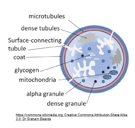
⭐ 11. Q: What major platelet structures support platelet function?
Platelets have membrane receptors, phosphatidylserine, an open canalicular system, a cytoskeleton, and granules that support adhesion, activation, secretion, spreading, and coagulation.
🔎 What this means:
Membrane receptors bind collagen, fibrinogen, and von Willebrand factor.
Open canalicular system transports substances and increases surface area.
Cytoskeleton enables shape change.
Granules store substances used in hemostasis and repair.
⭐ 12. Q: What happens to circulating platelets during their normal lifespan?
Healthy platelets circulate in a non-activated state, some reside in the spleen, aging platelets are likely removed by the liver, and platelets are consumed when they form hemostatic plugs.
🔎 What this means:
“Consumed” means platelets incorporated into a plug are no longer freely circulating.
As much as one-third of the blood platelet population may be in the spleen in health.
13. Q: What is the overall purpose of normal hemostasis?
Normal hemostasis keeps blood fluid in healthy vessels while allowing rapid, localized clot formation after vessel injury and clot removal after healing.
🔎 What this means:
Hemostasis must balance prothrombotic forces that promote clotting with antithrombotic forces that inhibit unnecessary clotting.
Prothrombotic = promotes clot formation.
Antithrombotic = inhibits or limits clot formation.
Q: What are the major components of hemostasis?
The major components are the vessel wall, primary hemostasis, secondary hemostasis, and tertiary hemostasis or fibrinolysis.
🔎 What this means:
Vessel wall responds to injury.
Primary hemostasis forms a platelet plug.
Secondary hemostasis adds fibrin.
Fibrinolysis limits and later removes the thrombus.
⭐ 15. Q: How does the vessel wall participate in hemostasis after injury?
Vessel injury causes vasoconstriction and changes or removes the normally antithrombotic endothelium, exposing prothrombotic subendothelial tissue.
🔎 What this means:
Healthy endothelium keeps blood away from clot-promoting structures underneath it.
Injury exposes those structures and helps initiate platelet and coagulation responses.
Prothrombotic = promotes clot formation.
Antithrombotic = inhibits or limits clot formation.
⭐ 🧠 LECTURE QUIZ 16. Q: What forms first after vascular injury: fibrin, a thrombus, the primary plug, or the secondary plug?
The primary hemostatic plug forms first after vascular injury.
🔎 What this means:
Platelet plug formation occurs before fibrin has fully stabilized the site.
⭐ 17. Q: What is the overall sequence of primary hemostasis?
Primary hemostasis proceeds through platelet adhesion, platelet activation and recruitment, and platelet aggregation to form the primary hemostatic plug.
🔎 What this means:
Adhesion: platelets are captured at the injured wall.
Activation: platelets change shape and recruit others.
Aggregation: platelets bind one another through fibrinogen.
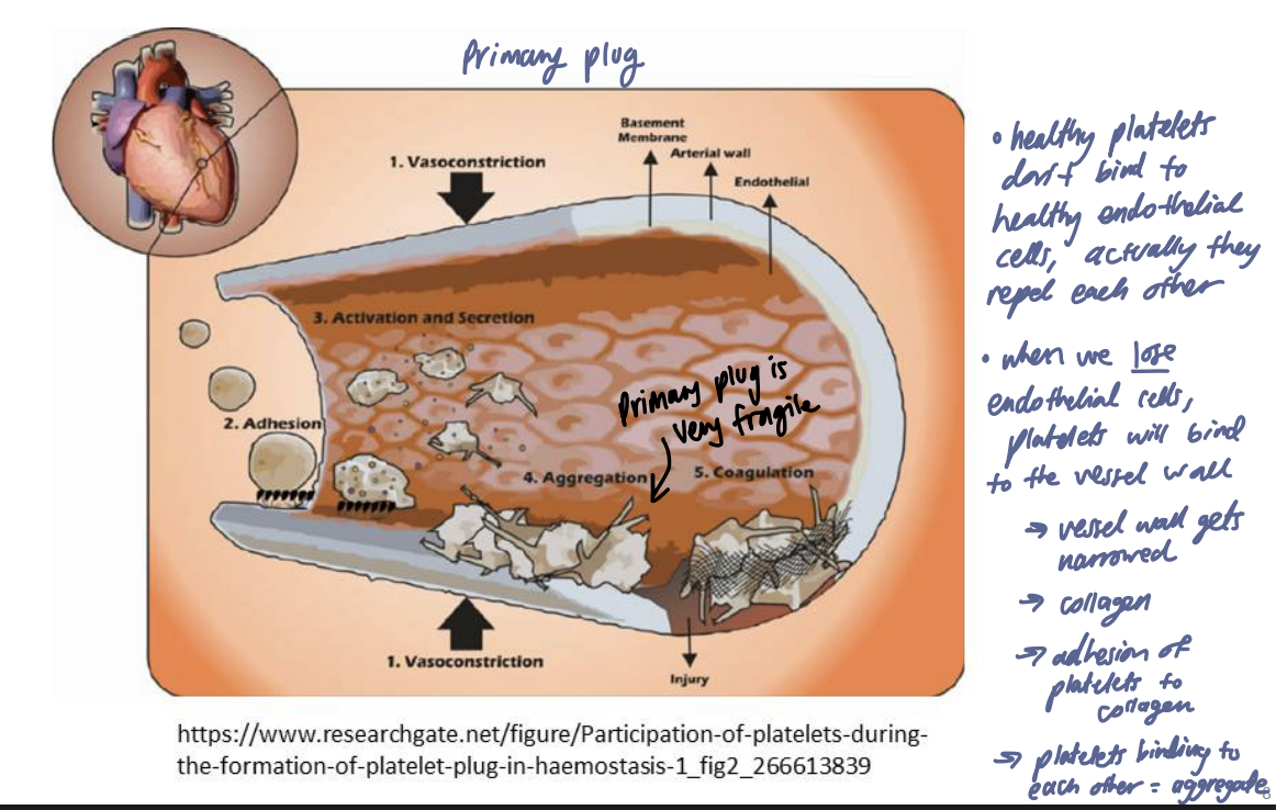
⭐ 🧠 LECTURE QUIZ 18. Q: How do von Willebrand factor and collagen promote platelet adhesion and activation?
Von Willebrand factor binds exposed collagen and tethers platelets to the vessel wall, after which platelet binding to collagen activates the platelets.
🔎 What this means:
Von Willebrand factor, abbreviated VWF, acts like a tether.
The sequence is:
injury exposes collagen → VWF binds collagen → VWF captures platelet → platelet binds collagen → platelet activates.
Platelets do not bind to vWF when not activated. Think of vWF as the thing that pulls platelets out of a fast moving river (aka capillaries)

⭐ 19. Q: Why is VWF especially important for platelet adhesion in vessels with high shear?
VWF rapidly tethers flowing platelets to the injured vessel wall, which is particularly important when high blood-flow forces would otherwise make direct platelet adhesion inefficient.
🔎 What this means:
At slow flow and low shear, direct interaction with exposed collagen is easier.
At high shear, platelets need VWF to grab and tether them quickly because blood is moving past the injured area more forcefully, the platelet may get swept away before it can attach well.
Shear means the dragging force created when layers of blood move at different speeds past each other and past the vessel wall.
A simple way to picture it:
Blood in the center of the vessel tends to move faster.
Blood right next to the vessel wall moves more slowly because of friction.
That difference in speed creates a kind of sliding/dragging force between layers of blood. That force is called shear.
⭐ 🧠 LECTURE QUIZ 20. Q: Which platelet products recruit and activate additional platelets after activation?
Activated platelets release stored ADP and produce thromboxane A2, and both recruit and activate additional platelets.
🔎 What this means:
ADP is stored in dense granules.
Thromboxane A2, or TxA2, is produced from platelet membrane lipids.
This distinction also fulfills the learning objective asking for the source and role of TxA2.
⭐ 🧠 LECTURE QUIZ 21. Q: What membrane change allows activated platelets to aggregate?
Platelet activation exposes fibrinogen-binding sites on the platelet membrane, allowing fibrinogen to bind and bridge adjacent platelets together aka “aggregate”
🔎 What this means:
Resting platelets have the fibrinogen-binding site hidden. After activation, the site becomes exposed.
Fibrinogen then binds receptors on two neighboring platelets, linking them together and causing platelet aggregation.
⭐ 22. Q: How does fibrinogen help form the primary hemostatic plug?
Fibrinogen binds exposed fibrinogen-binding sites on adjacent activated platelets and bridges the platelets together.
🔎 What this means:
This platelet-to-platelet binding is called aggregation.
💡 Fibrinogen = bridge between activated platelets.

⭐ 🧠 LECTURE QUIZ 23. Q: Where is phosphatidylserine in a resting platelet, what happens to it during activation, and why is that important?
Phosphatidylserine is normally on the inner membrane leaflet, but activation moves it to the outer surface, where it provides the major platelet phospholipid surface supporting coagulation.
🔎 What this means:
Phosphatidylserine is abbreviated PS.
The platelet changes from simply being part of the primary plug to becoming a surface on which secondary hemostatic reactions can occur.
we don’t need to know where in the cascade it is needed, we just need to know what role it plays in the cascade
⭐ 24. Q: How do the primary and secondary hemostatic plugs differ?
The primary plug is a relatively unstable platelet aggregate linked mainly by fibrinogen, whereas the secondary plug contains fibrin that reinforces the platelet plug and makes it more stable.
🔎 What this means:
Primary plug → platelet-rich, quick, easily dislodged
Secondary plug → fibrin-reinforced, stronger, more stable
25. Q: What defects can disrupt primary hemostasis, and what bleeding pattern can result?
Primary hemostasis can fail because of marked thrombocytopenia, dysfunctional platelets, or inadequate VWF, producing petechial, mucosal, ecchymotic, or prolonged bleeding.
🔎 What this means:
Thrombocytopenia: too few platelets.
Thrombopathy: dysfunctional platelets.
von Willebrand disease: inadequate VWF.
🧠 LECTURE QUIZ 26. Q: Which routine diagnostic assay listed in the lecture directly assesses primary hemostasis?
Platelet concentration directly assesses one major component of primary hemostasis.
🔎 What this means:
Other primary hemostasis tests discussed include VWF concentration and buccal mucosal bleeding time.
PT and PTT evaluate secondary hemostasis, not primary hemostasis.
Q: What is buccal mucosal bleeding time, and what are its limitations?
Buccal mucosal bleeding time measures how long bleeding continues after a standardized buccal incision, but the test is relatively insensitive and operator-dependent.
🔎 What this means:
BMBT can be prolonged with substantial platelet or VWF dysfunction and is independent of fibrin formation.
🧠 CASE QUESTION 28. Q: Why did Belle's hematuria and widespread petechiae point toward a hematologic rather than purely urinary problem?
The combination of mucosal bleeding and petechiae is characteristic of a primary hemostatic defect rather than an isolated urinary tract disorder.
🔎 What this means:
Primary hemostatic problems commonly cause bleeding from mucosal surfaces and small pinpoint hemorrhages.
Q: What caused Belle's immune-mediated thrombocytopenia?
Belle produced antibodies against her own platelets, causing macrophages to remove platelets faster than the bone marrow could replace them.
🔎 What this means:
This represents increased platelet destruction, not simply inadequate production.
⭐ 30. Q: What is secondary hemostasis?
Secondary hemostasis is a cascade of coagulation reactions that generates thrombin and fibrin to reinforce and stabilize the primary platelet plug.
🔎 What this means:
Inactive coagulation proteins are activated in sequence until fibrin is produced.
Q: What do the terms zymogen, cofactor, and the suffix “a” mean in coagulation?
A zymogen is an inactive enzyme precursor, a cofactor is a nonenzymatic helper, and the suffix “a” indicates that a coagulation factor is activated.
🔎 What this means:
For example:
Factor II = prothrombin
Factor IIa = thrombin
⭐ 32. Q: What initiates the intrinsic, or contact-activated, coagulation pathway?
The intrinsic pathway is initiated when blood contacts negatively charged surfaces, such as exposed subendothelial collagen or the surface of a collection tube.
🔎 What this means:
This pathway is especially important for understanding in vitro coagulation testing
⭐ 33. Q: What is the factor sequence of the intrinsic coagulation pathway?
The intrinsic pathway proceeds through factors XII (12), XI (11), and IX (9), with factor VIII (8) serving as a nonenzymatic cofactor for activated factor IX (9).
🔎 What this means:
XIIa = enzyme
XIa = enzyme
IXa = enzyme
VIIIa = cofactor
💡 Professor's mnemonic: $12 advertised as $11.98 → XII, XI, IX, VIII.
⭐ 34. Q: What initiates the extrinsic coagulation pathway, and where is the initiator normally found?
Exposure of tissue factor initiates the extrinsic pathway; tissue factor is widely expressed on extravascular cells (cells outside of the blood) and can also be expressed by activated endothelium and white blood cells.
When a vessel is damaged, blood can contact those tissue-factor-bearing cells and help start the extrinsic coagulation pathway.
🔎 What this means:
Tissue factor is also called factor III (3).
In health, circulating blood is normally separated from extravascular tissue factor
⭐ 35. Q: Which coagulation factor works with tissue factor in the extrinsic pathway?
Factor VII (7) works with tissue factor in the extrinsic coagulation pathway.
🔎 What this means:
Activated VII (7) forms the TF–VIIa complex, which helps initiate coagulation.
⭐ 36. Q: What is the factor sequence of the common coagulation pathway, and which components are enzymes?
The common pathway involves factor X, factor V, factor II, and factor I; activated X and activated II are enzymes, factor V is a nonenzymatic cofactor, and factor I is fibrinogen.
🔎 What this means:
X + V → II → I
Factor IIa is thrombin

⭐ 37. Q: What are the four major roles of thrombin in hemostasis?
Thrombin cleaves fibrinogen into fibrin, activates platelets, activates factor XIII for fibrin cross-linking, and amplifies coagulation by activating factors XI, VIII, and V.
🔎 What this means:
Thrombin is not merely the enzyme that makes fibrin.
It also makes the coagulation response bigger and more stable.
🧠 LECTURE QUIZ 38. Q: Besides cleaving fibrinogen to fibrin, what other direct role does thrombin play in hemostasis?
a) cross-link fibrin
b) activate X to Xa
c) Activate V to Va
d) Activate XII to XIIa
c) Thrombin directly activates factor V to factor Va.
🔎 What this means:
Do not choose “cross-links fibrin”: thrombin activates factor XIII, and XIIIa performs the cross-linking.
⭐ 39. Q: What are the two major roles of fibrinogen in hemostasis?
Fibrinogen bridges activated platelets during primary hemostasis and is cleaved by thrombin to form fibrin during secondary hemostasis.
🔎 What this means:
Primary: fibrinogen connects platelets.
Secondary: fibrinogen becomes fibrin.
This is why fibrinogen participates in both phases.
⭐ 🧠 LECTURE QUIZ 40. Q: Which organ produces most coagulation factors?
Hepatocytes in the liver produce most coagulation factors.
🔎 What this means:
Liver dysfunction can therefore affect secondary hemostasis.
⭐ 41. Q: What role does calcium play in coagulation, and why does hypocalcemia usually not cause a hemostatic disorder?
Calcium ions are required for several coagulation reactions, but only a small amount is needed, so severe hypocalcemia causes serious muscular problems before calcium becomes low enough to impair hemostasis.
🔎 What this means:
You need to know that calcium is required, not every individual calcium-dependent step.
⭐ 42. Q: Which coagulation factors are vitamin K dependent, and which pathways contain them?
Factors II (2), VII (7), IX (9), and X (10) require vitamin K; factor VII (7) is in the extrinsic pathway, factor IX (9) is in the intrinsic pathway, and factors X (10) and II (2) are in the common pathway.
🔎 What this means:
💡 Professor's memory trick:
2 + 7 = 9, and one more is 10.
⭐ 43. Q: How is coagulation primarily initiated in vivo?
In vivo coagulation is primarily initiated by tissue factor binding factor VIIa (7a) and generating a small initial amount of thrombin.
🔎 What this means:
This is the initiation phase.
Factor XII (7) activation is not required for normal in vivo initiation.
⭐ 44. Q: How is thrombin generation amplified in vivo, and where do the reactions occur?
The initial thrombin activates platelets and factors V (5), VIII (8), and XI (11), producing much more thrombin on phosphatidylserine-bearing cell surfaces such as activated platelets.
🔎 What this means:
The lecture describes two important cellular surfaces:
a tissue factor-bearing cell for initiation,
a phosphatidylserine-bearing cell, such as an activated platelet, for amplification.
⭐ 🧠 LECTURE QUIZ 45. Q: Which enzyme cross-links fibrin to stabilize the thrombus?
Activated factor XIII (13), or XIIIa (13a), cross-links fibrin to stabilize the thrombus.
🔎 What this means:
Thrombin activates XIII (13) → XIIIa (13a) performs the actual cross-linking.

🧠 LECTURE QUIZ 46. Q: Which factor or cell does not play a significant role in secondary hemostasis: plasmin, platelets, thrombin, or tissue factor?
Plasmin does not play a significant role in forming the secondary hemostatic plug.
🔎 What this means:
Plasmin belongs to the fibrinolytic system, which breaks down thrombi rather than forming them.
🧠 LECTURE QUIZ 47. Q: Which coagulation factor is not essential for coagulation in vivo?
Factor XII (12) is not essential for coagulation in vivo.
🔎 What this means:
The tissue factor pathway is the major physiologic initiator of coagulation.
Factor XII (12) remains important for the in vitro intrinsic pathway used in PTT testing.
Q: Why should the traditional intrinsic/extrinsic pathway diagram not be treated as a perfect description of coagulation in a living animal?
The traditional intrinsic, extrinsic, and common pathways are based largely on in vitro testing, whereas in vivo coagulation is primarily initiated through tissue factor and proceeds on cell surfaces.
🔎 What this means:
The two models are useful for different purposes:
Traditional pathways → understand PT/PTT
Cell-based model → understand what actually occurs in vivo
⭐ 🧠 LECTURE QUIZ 49. Q: What blood tube should be used for coagulation testing, how does it prevent clotting, and why is proper filling important?
Coagulation samples are collected in a blue-top trisodium citrate tube; citrate reversibly binds calcium, and underfilling causes excess citrate relative to blood, which can falsely prolong clotting times.
🔎 What this means:
The citrate-to-blood ratio matters.
Too little blood → relatively too much citrate → too much calcium binding → PT/PTT may look longer than they truly are.
Q: Why are collection and sample handling especially important for coagulation testing?
Traumatic collection can expose tissue factor and activate coagulation, and plasma should be separated promptly because some coagulation factors are labile (unstable/easily degraded).
🔎 What this means:
The lecture recommends discarding a coagulation sample if venipuncture was not clean.
Plasma can be refrigerated for up to 24 hours; otherwise it should be frozen.
⭐ 51. Q: What does prothrombin time measure, and which pathways does it assess?
Prothrombin time measures the time until fibrin forms after tissue factor, calcium, and phospholipid are added, and it assesses the extrinsic and common pathways.
🔎 What this means:
PT → extrinsic + common.
Because tissue factor is added during the test, PT does not test the patient's own tissue factor.
⭐ 🧠 LECTURE QUIZ 52. Q: What does partial thromboplastin time measure, and which pathways does it assess?
Partial thromboplastin time measures the time until fibrin forms after a factor XII (12) activator, calcium, and phospholipid are added, and it assesses the intrinsic and common pathways.
🔎 What this means:
Partial thromboplastin time is abbreviated PTT or aPTT.
PTT → intrinsic + common.
It was also the correct test among the lecture quiz choices for assessing secondary hemostasis.
⭐ 53. Q: What does a prolonged PT with a normal PTT indicate?
A prolonged PT with a normal PTT indicates an extrinsic pathway defect, which in this pathway model points to factor VII (7) deficiency.
🔎 What this means:
PT abnormal → extrinsic/common problem possible.
PTT normal → common pathway must still be functioning adequately.
Therefore → extrinsic only → factor VII
⭐ 54. Q: What does a prolonged PTT with a normal PT indicate?
A prolonged PTT with a normal PT indicates an intrinsic pathway defect involving factor XII (12), XI (11), IX (9), or VIII (8).
🔎 What this means:
PTT abnormal → intrinsic/common problem possible.
PT normal → common pathway is not significantly affected.
Therefore → intrinsic pathway defect.
⭐ 🧠 LECTURE QUIZ 55. Q: What does prolongation of both PT and PTT suggest, and which single factor deficiency could cause both?
Prolongation of both PT and PTT suggests a common-pathway defect or multiple factor defects, and factor X deficiency is one single-factor defect that can prolong both tests.
🔎 What this means:
Both PT and PTT depend on the common pathway.
The lecture quiz specifically uses factor X deficiency as the single-factor example
Q: Why can PT and PTT be normal despite a defect in factor XIII-mediated fibrin stabilization?
PT and PTT measure the time until fibrin forms, so they do not assess the later cross-linking of fibrin by factor XIIIa (13a).
🔎 What this means:
The clot can form on time but still fail to become normally cross-linked afterward
🧠 LECTURE QUIZ 57. Q: Which assay is used to measure fibrinogen for hemostasis testing?
Thrombin time is the assay used to measure fibrinogen for hemostasis testing in this course.
🔎 What this means:
Thrombin time, abbreviated TT, adds excess thrombin and measures the time required for fibrin to form
⭐ 58. Q: What clinical findings are expected with a defect in secondary hemostasis?
Secondary hemostatic defects can cause excessive bleeding after trauma or surgery, ecchymoses, hematomas, and hemorrhage into body cavities or joints.
Ecchymoses: bruises aka bleeding under the skin
Hematoma: a localized pocked of blood within tissue (caused by hemorrhage)
Hemorrhage: bleeding, where blood escapes from damaged blood vessels into or outside of the body
🔎 What this means:
These defects prevent normal fibrin stabilization of the hemostatic plug.
Q: How do typical bleeding patterns differ between primary and secondary hemostatic defects?
Primary defects commonly cause petechiae and mucosal bleeding, whereas secondary defects more characteristically cause hematomas and bleeding into body cavities; ecchymoses and excessive post-traumatic bleeding can occur with either.
🔎 What this means:
Do not memorize the distinction as an absolute rule.
A better pattern is:
Primary → small-vessel/mucosal bleeding
Secondary → deeper, larger-volume bleeding
Q: How do anticoagulant rodenticides such as d-CON interfere with coagulation?
They prevent regeneration of reduced vitamin K, impairing carboxylation of factors II, VII, IX, and X and thereby reducing functional factor activity and fibrin formation.
🔎 What this means:
Vitamin K-dependent carboxylation is required for these factors to bind calcium properly.
Less functional II, VII, IX, X → less effective coagulation.
🧠 CASE QUESTION 61. Q: Why were both PT and PTT prolonged in the dog with anticoagulant rodenticide toxicity?
Multiple vitamin K-dependent factors were impaired across the extrinsic, intrinsic, and common pathways, so both PT and PTT were prolonged.
🔎 What this means:
VII (7) → affects PT
IX (8) → affects PTT
II (2) and X (10) → affect the common pathway and therefore both tests
This is an acquired multiple-factor defect, not a single isolated factor deficiency.
Q: What happens if the normal hemostatic balance shifts too far toward or away from clot formation?
Excess clot formation or inadequate clot removal promotes thrombosis, whereas inadequate clot formation or excessive clot removal promotes bleeding.
🔎 What this means:
Hemostasis is a balance, not simply “more clotting is better.”
Q: What diagnostic tests were useful in evaluating the case with suspected secondary hemostatic failure?
The case used a CBC together with coagulation testing, including PT, PTT, and fibrinogen measurement, to evaluate bleeding and coagulation abnormalities.
🔎 What this means:
The CBC assessed anemia and platelets, while PT/PTT assessed the coagulation pathways
⭐ 63. Q: What are the coagulation pathways in one compact comparison?
The intrinsic pathway uses XII (12), XI (11), IX (9), and VIII (8); the extrinsic pathway uses tissue factor and VII (7); both converge on the common pathway involving X (10), V (5), II (2), and I.
🔎 What this means:
Intrinsic: XII → XI → IX + VIII
12 → 11 → 9 + 8
Extrinsic: TF + VII
TF + 7
Common: X + V → II → I
10 + 5 → 2 → 1
Then thrombin activates XIII (13), and XIIIa (13a) cross-links fibrin.