Brain cells
 Call Kai
 Learn
 Practice Test
 Spaced Repetition
 Match
 Flashcards
 Knowt Play
1/43

There's no tags or description
Looks like no tags are added yet.
44 Terms
Golgi: Reticular theory
Revealed two parts of neurons:
Cell body
Neurites e.g. axons and dendrites
Cajal: Neuron Doctrine
Neurons are distinct cellular units
Can be studied through ‘double-impregnation’ with silver nitrate
Neuron
Receives, integrates and transmits information through electrical and chemical signalling
Senses detailed environmental changes
Communicates detailed changes to other neurons
Coordinates bodily response
Around 85 billion in an adult human
Soma
Around 20 micrometres
Metabolic centre of the neuron
Consists of cytoplasm, cytosol and organelles e.g. nucleus, reticuli and mitochondria
Cytoplasm
Everything inside the cell membrane including organelles, excluding the nucleus
Cytosol
Intracellular fluid inside a cell
Salty, potassium-rich solution
Organelles
Same as found in other eukaryotic cells
e.g. nucleus, mitochondria, rough endoplasmic reticulum, smooth endoplasmic reticulum
Neural DNA sequence
Transcription → mRNA → Transcription → Protein
e.g. receptors and ion channels are proteins influencing neuronal signalling
Neurons differ from other cells due to a specific subset of genes
Genetic basis of many diseases of the nervous system e.g. Huntingtons
Endoplasmic reticular differences
Rough ER - Protein production and processing
Smooth ER - Lipid production and calcium regulation
Translation
mRNA bind to ribosomes to translate mRNA into proteins
Many ribosomes are attached to the rough ER
Golgi apparatus - proteins and lipids made in the ER are modified and sorted
Gene editing in brain research
Important tool for brain research and understanding and treating nervous system disorders
Basic utilities:
Correcting inherited disorders
Halting disease progression
Simulating diseases for scientific discovery
Overcoming delivery challenges
Gene editing: Correcting inherited disorders
Many brain diseases are monogenic (caused by a mistake in a single gene)
By using precision genome editing tools like CRISPR, researchers can cut out toxic insertions or repair mutations that destroy neurons
e.g. repeat mutations in Huntington’s disease
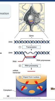
Transcription
DNA - Contains building plan for all cells in the body
DNA acts as a blueprint for RNA → like a photo negative
mRNA exits nucleus, carrying coding information
Cells express different combinations and levels of genes as a result
Gene editing: Halting disease progression
Traditional medications only manage the symptoms of brain diseases (e.g. reducing tremors or eassing pain)
Gene targeting goes for the biological root cause, allowing doctors to potentially stop progressive degeneration before permanent brain damage occurs
Gene editing: Simulating diseases for scientific discovery
Scientists use genetic engineering to create animal models that mimic human brain diseases
By introducing specific mutations into these models, researchers can closely observe how diseases like AD unfold in living tissue and test new therapies safely
Gene editing: Overcoming delivery challenges
Brain protected by the blood-brain barrier, blocking most standard drugs from entering
Precision genetic technologies allow scientists to engineer harmless viruses or specialised nanoparticles to carry therapeutic DNA changes directly to affected brain cells

Soma
Consists of mitochondria, cytoskeleton and neuronal membrane
Mitochondria
Responsible for cellular membrane, generating ATP
ATP is the energy currency of the cell
Cytoskeleton
‘cell skeleton’
Cell shape
Correct positioning of organelles - not static
Tracks for in-cell transportation
Neuronal membrane
Barrier to enclose the cytoplasm inside the neuron
Protein composition varies depending on the location (soma, axon, dendrites)
Structure of discrete membrane regions influences neuronal functions
Axon
Consists of axon hillock (beginning), axon proper (middle) and axon terminal (end)
Only found in neurons, each only having one axon
Highly specialised for information transfer (electrical signals) over distances (electrical cables)
No rough ER, hardly any ribosomes - little protein synthesis in axon, most of proteins in axon come from soma
Axons can extend <1mm to 1m long
Axon hillock
Region where the axon originates
Neurofilaments in the axon hillock become clustered together as fascicles
Axoplasmatic transport
Ensures necessary material travels from the soma to the axon terminal (anterograde transport) and back (retrograde transport)
Differences between axon and soma
ER does not extend into axon
Unique protein composition
Terminal bouton
Region where axon contacts with other neurons
No microtubules in the terminal
Synaptic vesicles - contain important chemicals for communication (neurotransmitters)
Numerous mitochondria due to high energy demand
Synapse
Electrical signal → chemical signal → electrical signal
Synaptic transmission dysfunction
Brain and muscle disorders
Action site for toxins and psychoactive and therapeutically useful drugs
Dendrite
‘Antennae’ of neurons
Dendritic tree
Synapse-receptors
Postsynaptic membrane
Full with receptors - special proteins that bind to NTs
Take up chemical signals from pre-synaptic neuron and carry on electrical signals
Dendritic spines
Small protrusions of membrane that receive signals from the axon terminal.
Increase possible synaptic contact sites between neurons
Effective synapses that can quickly change in appearance
Important for plasticity and learning (new spines can grow, existing spines can become larger or smaller)
Brainbow - Imaging technique
Labels each nerve cell a different colour to identify and track axons and dendrites over long distances
Scientists can use light microscopy to image branching patterns and connections of all axons within a region of the NS, expressing a number of different proteins in individual neurons
Neural classification
Neurons differ from other cells due to being a subset of genes
Genetic basis og many diseases in the NS
Classification can be based on gross anatomy, dendritic morphology, or number of neurites
Classification: Neurites
Classification based on number of neurites
Unipolar: One neurite
Bipolar: Two neurites
Multipolar: More than two neurites
Neurite = axons + dendrites
Uni- and bipolar neurons most often sensory as they take up a smaller amt of info, leading to greater precision
Classification: Morphology
Stellate cells (star shaped) and pyramidal cells (pyramid shaped)
Cerebral and cerebellar cortex have different types of stellate cells
Spiny or aspiny
Stellate cells
Tight network of dendrites
Perfect for interconnection and info exchange between neighbouring neurons
Pyramidal cells
Dendrite tree, similar shape to a pyramid, with a long axon
Perfect for transfer of info over long distances
Classification: Gross anatomy
CNS connections: primary sensory neurons, motor neurons and interneurons
Axonal length: Golgi type I - projection neurons, Golgi type II - local circuit neurons
intrinsic physiology: Intrinsic physiology roughly similar, physiological details differ
Glial cells
Regulate the environment
Provide metabolic and immune support
Influence neuronal communication
Glial cells: Astrocytes
Most abundant neuroglia in the brain
Found in gaps between neurons, influencing neurite growth
Regulate chemical content in extracellular space (K+)
Restrict spread of NTs from synaptic cleft
Actively remove NTs from synaptic cleft
Trigger electrical and biochemical events inside the glial cell
Microglia
10% of brain cells
Immune first responders
Housekeepers of the brain
Act as phagocytes, removing debris left by dead or degenerating cells in both healthy and diseased brains
Conduct synaptic pruning
Reactive gliosis and neuroinflammation are symptoms of Alzheimer’s DIsease
Oligodendrocytes
Myelinate segments of several axons
Located in the CNS
Schwann cells
Wraps one segment of the axon
Located in the PNS
Role of myelin
lipid-rich sheath wrapped around axons, insulates and increases conduction speed
When it is damaged, nerve conduction becomes slower or may fail
Multiple Sclerosis: CNS changes
Immune-mediated inflammation damages CNS myelin and may injure axons
Lesions can occur in the brain, optic nerves and spinal cord
MS can involve axonal injury and neurodegeneration , not only myelin loss
Symptoms of MS
Visual disturbance
Weakness or altered sensation
Balance and coordination difficulties
Fatigue and cognitive difficulties
Symptoms depend on which CNS pathways are affected