Artificial Ventilation
 Call Kai
 Learn
 Practice Test
 Spaced Repetition
 Match
 Flashcards
 Knowt Play
1/62

There's no tags or description
Looks like no tags are added yet.
63 Terms
who is artificial ventilation for?
people who can’t breathe on their own
what does artificial ventilation provide?
adequate ventilation and oxygenation
access to airway/lungs for pulmonary suctioning
connection to positive pressure ventilation
goals of artificial ventilation
maintain patent (unobstructed) airway
reduce potential for aspiration
eliminate airway obstruction
what % of patients with a tracheostomy on ventilators aspirate?
50%
when is mechanical ventilation indicated?
when the patient is unable to spontaneously maintain adequate oxygenation or ventilation
stats for indication of mechanical ventilation
O2 less than 90%
CO2 too high (not expelling enough)
what is important for long term mechanical ventilation?
to try and time swallowing to the expiratory phase of mechanical ventilation
you can teach the patient to swallow on the expiratory event
what are the two types of artificial airways
endotracheal intubation and tracheostomy

endotracheal intubation
through the mouth
requires weaning
tolerated well if patient remains sedated
when is endotracheal intubation used?
surgeries and acute trauma

tracheostomy
ventilation occurs through a stoma
less irritating
more feeding/voice opportunity
when is a tracheostomy used?
disease
long term respiratory
where is a tracheostomy inserted
incision below the cricoid muscle into tracheal ring
indications a tracheostomy is needed
airway obstruction at or above the vocal folds, upper airway obstruction, etc.
tracheostomy: physiological changes
loss or change in airway resistance
inability to generate subglottal pressure during swallow → reduced ability to produce cough/normal phonation
loss of sense of smell - no transnasal airflow
reduced mucosal sensitivity
tracheostomy side effects
globus sensation, disruption of respiratory/swallowing coordination
ventilation of __ ______ results in a trach
27 hours
is it better to trache early?
the earlier you trache, the shorter the hospital stay
is trach meant to be long term or temporary
temporary
tracheostomy etiologies
trauma related
transplants (ex. lung/heart)
head and neck cancer/ENT
burns
trauma related tracheostomy etiologies
gunshot wounds, congestive heart failure, MVA (car accidents), damage to VFs during surgery or intubation
tracheostomy tube anatomy: neck plate/flange
allows tube to be secured to the neck; stabilization

tracheostomy tube anatomy: outer and inner cannula
outside wall of tracheostomy tube provides basic structure
*inner cannula has to be changed more often than outer
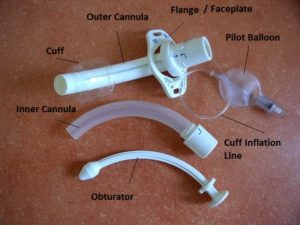
tracheostomy tube anatomy: inner tube
fit snugly inside outer cannula and removed for changing or cleaning
tracheostomy tube anatomy: obturation
only used during placement to guide surgeon in placing the tube through the stoma
tracheostomy tube anatomy: cuffs
surrounds the bottom portion of the inner cannula to prevent air escape
*not present in all trach tubes

tracheostomy tube cuff purpose
ensure delivery of a pre-set tidal volume from the ventilator to the lungs
decreases amount of secretions that enter the lungs
what happens if the trach cuff is deflated?
air comes out around the tube
who do trach cuff patients receive constant monitoring from and what for?
respiratory therapy; constant suctioning and careful management
side effects of tracheostomy cuffs
granulomas can develop in lower airways
reduced tracheal sensations (due to no airflow)
stenosis
overall desensitization of airway activities
cuff side effect: granuloma
a small, tightly packed cluster of immune cells (primarily macrophages) that forms a tiny lump in the body when the immune system tries to wall off and isolate something harmfu
cuff side effect: tracheostomal stenosis
abnormal narrowing of the tracheostoma (the surgical opening made in the neck for a tracheostomy
tracheostomy airway pressure changes
reduced airway pressure
laryngeal elevation is reduced
adverse effects for airway protection and opening of UES
pharyngeal transit
tracheostomy airway pressure changes: reduced airway resistance
impacts normal pressure gradients in oropharyngeal mechanism
open tube disrupts pressure generation
tracheostomy airway pressure changes: reduced laryngeal elevation
significant contributor to dysphagia
tracheostomy airway pressure changes: pharyngeal transit
restricted ROM
increased transit time
prolongation of bolus transit increased risk of pre-swallow spill and pen/asp
trach tubes: cuffed vs. uncuffed (who doesnt need?)
sometimes patients who will definitely not need mechanical ventilation
cystic fibrosis
crohn’s disease
effects of cuffs on swallow
research has found no difference on cuff vs. uncuffed
found pos results with one way valve
what should you never do with a speaking valve/cuff?
never place a one-way valve with the cuff inflated
*the cuff seals the airway off from the outside- patient can’t breathe
checking the cuff (if inflated/deflated inside…)
if inflated, the cuff inside is inflated
if deflated outside, cuff inside is deflated - you can try some talking with the speaking valve
before you place any valve, what should you do?
check if there is a cuff
check if the cuff is deflated or inflated
cap it, finger occlude
if you are going to deflate the cuff, what should you always do?
ask the nursing staff if the patient is appropriate and if they want the cuff re-inflated or remain deflated
tracheostomy tubes: when you place the speaking valve and deflate the cuff…
a lot of secretions will begin to develop around the cuff (sensory receptors activated)
observe to see if they can still produce phonation or swallow
may ask them to cough
speaking valves and their use in dysphagia
decannulation or even tracheostomy tube occlusion using a finger enhances swallowing function in a patient with tracheostomy
expiratory speaking valve
restores subglottic pressure during swallow
removable one-way valve: air comes in, but cannot come out
opens to permit inhalation then travels up: can speak, cough, etc.
closes during expiration to divert the airflow through the larynx
there are some valves that can be used while a patient is on a ventilator (rare)
benefits of a speaking valve
useful in ped and adult populations
patients have a right to communicate and this provides that
kids with MD who have difficulty ventilating generally may have this with a one way valve to help them ventilate
may take the valve off when they get fatigued to get better ventilation
if a patient is unconscious, can you place a speaking valve?
no
when is a speaking valve not appropriate?
severe behavior problems
cognitive disorders
severe medical instability
pulmonary failure
tracheal stenosis
edema
insufficient passage of air around tube with cuff inflated and deflated
thick and copious secretions
airway obstruction above tube before expiration
pt unable to breathe
edema
abnormal buildup of excess fluid trapped in the body's tissues
tracheal stenosis
air won’t be able to move in the right direction
speaking valves: total laryngectomy or laryngotracheal separation
no larynx, no way for air to travel superiorly, not appropriate
tracheostomy care
SLPs can suction to maintain airway patency and keep free of mucous buildup
*oral hygiene; can only be done a certain # of times a day so need to talk with other providers
reduces rate of infection
maintain skin integrity at trach
who does SLP work closely with for trach patients?
respiratory therapist, physician, and nurse
assessment of swallowing in patients with trachs and vents
medical clearance needed
read note but don’t assume something won’t have changed
clinical bedside assessment
assessment of swallowing in patients with trachs and vent: clinical bedside assessment
collecting thorough history from patient can be difficult if patient is semi-sedated
check w/ caregiver and nursing
may deflate the cuff and do some swallowing trials
ice chips
mix blue dye with food, will show on trach/stoma if they aspirate
FEES vs MBS for trach/vent patients
FEES- better for mechanically ventilated patients who can’t transport
*ex. patient post MVA w/ unilateral VF paralysis
VFS- good for tracheotomized patients
assessment of swallowing in patients with trachs and vent: things to consider
onset of swallowing problem (before or after trach/etiology)
methods of trach occlusion
gloved finger
speaking valve placement
make swallowing judgements while watching other stats
voluntary swallow/cough
bolus swallow/cough
change in vocal quality
monitor anxiety- they may be cautious
have suctions ready
weaning process/decannulation
work alongside respiratory therapist
when someone is mechanically vented or trached they generally have a larger tube in the trachea/stoma which can be reduced in size
*some patients with chronic respiratory disease mau require permanent trach and ventilator dependence
Best practices with artificial ventilation: use it or lose it
historians used to think you should wait until full weaning from mech vent before dysphagia intervention
this is NOT TRUE
can lead to muscle weakness of digestive tract
Best practices with artificial ventilation: make the connection
tracheostomy creates a disconnect between the upper and lower airways
aphonia
disrupted laryngeal/pharyngeal sensation
loss of subglottic airflow and pressure
reduced smell and taste
reconnection is best achieved with cuff deflation and placement of a one way valve
Best practices with artificial ventilation: team up
reconnection requires a team approach with respiratory therapy
Best practices with artificial ventilation: size matters
a trach tube that fills the trachea prohibits airflow through the upper airway and placement of a speaking valve
1 week ideal for downsizing (sometimes hospitals way 2)
advocate for your patients
Best practices with artificial ventilation: speaking valves are not just for speaking
restores sensation with redirected airflow